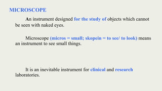
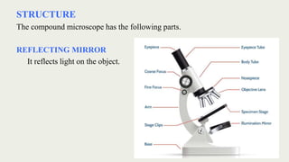
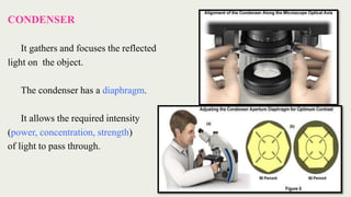
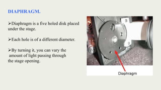
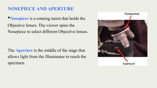
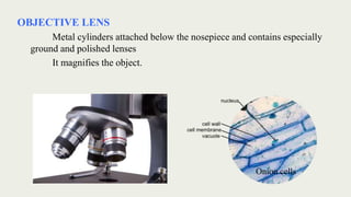
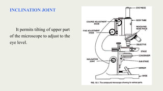
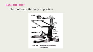
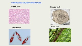
during this ppt of microscopes we will be able to know
INTRODUCTION
DEFINITION
HISTORICAL BACKGROUND
VARIABLES USED IN MICROSCOPY
VARIOUS TYPES OF MICROSCOPES
COMPOUND MICROSCOPE - Structure and Function
USE OF MICROSCOPE
CARE OF MICROSCOPE
defintion
A microscope (Greek: micron = small and scopos = aim)
MICROSCOPE - An instrument for viewing objects that are too small to be seen by the naked or unaided eye
MICROSCOPY - The science of investigating small objects using such an instrument is called microscopy
Slides includes all details about Dark field Microscopy.
useful for MTech, pharmacy student which dealing with microbiology. also for reference to study Dark Field Microscopy. includes principle, instrumentation, working, uses etc.
This presentation is about the introduction of microscopy, its history, parts of a microscope and different types of microscopes along with a brief discussion of their working principles.
1. MICROSCOPY - introduction + principle (Basics)Nethravathi Siri
Basics only
Microscopy is the technical field that uses microscopes to observe samples which are
not in the resolution range of the normal-unaided eye.
Microscope is a scientific-instrument consisting of magnifying lens that enables an
observer to view the minute features distinctly.
In greek, micro = small
skopein = to view.
The microscope has evolved a lot from the time of Leeuwenhoek. This presentation gives a brief overview about the types of microscope their principle of function and application.
BRIGHT FIELD MICROSCOPY by SIVASANGARI SHANMUGAM
bRIGHT FIELD MICROSCOPY is also called a compound microscope. The name bright - field is derived from the fact that the specimen is dark and contrasted by the surrounding bright viewing field.
LIGHT MICROSCOPY by SIVASANGARI SHANMUGAM
The optical microscope, The functions of a light microscope is based on its ability to focus a beam of light through, which is very small and transparent, to produce an image.
DARK FIELD MICROSCOPY by SIVASANGARI SHANMUGAM
Dark-field microscopy is ideally used to illuminate unstained samples causing them to appear brightly lit against a dark background.
This type of microscope contains a special condenser that scatters light and causes it to reflect off the specimen at an angle
Introduction
History
Compound microscope
Variants of microscopes
Dark field microscope
Phase contrast microscope
Fluorescent microscope
Polarising microscope
Electron microscope
A Microscope is an instrument for viewing objects that are too small to be seen by the naked/ unaided eyes.
In Greek micron= small
skopien=to look at
The science of investigating small object using such an instrument is called microscopy
The term microscopic means minute or very small, not visible with the eye unless aided by a microscope
From ancient times, man wanted to see things for smaller than could be perceived with the naked eye.
This led to the construction in the 16th century, of a magnifier composed of a single convex lens, and this in turn led to the eventual development of the microscope.
The most famous early pioneers in the history of microscope are Digges of England and Hans & Zcharias Janssen of Holland
It was Antony Van Leeuwenhoek who became the man to make and use a real microscope.
Leeuwenhoek microscope was called as single lens microscope because it had convex lens attached to metal holder and was focused using screws
Microscopy - Magnification, Resolving power, Principles, Types and ApplicationsNethravathi Siri
Magnification, Resolving power, Principles and Applications of Simple, Compound, Stereozoom, Phase contrast, Fluorescent and Electron microscopes (TEM & SEM).
Microscopy is the technical field that uses microscopes to observe samples which are not in the resolution range of the normal-unaided eye.
INTRODUCTION
HISTORY
TYPES OF MICROSCOPE
Compound Microscope
Dissection Microscope
Scanning Electron Microscope (SEM)
Transmission Electron Microscope (TEM
CARE
PARTS AND FUNCTION
FOCUSING
CONCLUSION
REFERANCE
A **bright field microscope** is a type of compound light microscope that illuminates the background against a stained specimen ¹². It is commonly used in practical labs to study organisms' behavior and characteristics such as size, shape, and arrangement ². The microscope uses light rays to produce a dark image against a bright background ¹. It is specially designed with magnifying glasses known as lenses that modify the specimen to produce an image seen through the eyepiece ¹. The bright field microscope is made up of various parts, including the eyepiece, objective lenses, focusing knobs, and stage ¹.
I hope this helps!
Source: Conversation with Bing, 7/11/2023
(1) Brightfield Microscope (Compound Light Microscope)- Definition .... https://microbenotes.com/brightfield-microscope/.
(2) Bright Field Microscopy - Biology Reader. https://biologyreader.com/bright-field-microscopy.html.
(3) Bright-field microscopy - Wikipedia. https://en.wikipedia.org/wiki/Bright-field_microscopy.
(4) Bright Field Microscope: Definition, Parts, Working Principle, Application. https://microbiologynote.com/bright-field-microscope-definition-parts-working-principle-application/.
Microscopy is the technique of using microscopes to observe and analyze objects that are too small to be seen by the naked eye. Microscopes are instruments that magnify and resolve the details of objects, allowing scientists and researchers to study the structure, composition, and behavior of materials and specimens at a microscopic level
Microscopy is the technical field of using microscopes to view objects and areas of objects that cannot be seen with the naked eye. There are three well-known branches of microscopy: optical, electron, and scanning probe microscopy, along with the emerging field of X-ray microscopy.
History of Microbiology Discovery Era, Transition Era, Golden Era, Modern Era, Louis Pasteur, Antony Van Leevenhoek, Pasteurization, Alexander Fleming, fermentation, Agar,
Preservation of industrially important microorganisms, methods of preservation, periodic transfer, storage in saline suspension, storage in sterile soil, cryopreservation
Introduction to AI for Nonprofits with Tapp NetworkTechSoup
Dive into the world of AI! Experts Jon Hill and Tareq Monaur will guide you through AI's role in enhancing nonprofit websites and basic marketing strategies, making it easy to understand and apply.
Biological screening of herbal drugs: Introduction and Need for
Phyto-Pharmacological Screening, New Strategies for evaluating
Natural Products, In vitro evaluation techniques for Antioxidants, Antimicrobial and Anticancer drugs. In vivo evaluation techniques
for Anti-inflammatory, Antiulcer, Anticancer, Wound healing, Antidiabetic, Hepatoprotective, Cardio protective, Diuretics and
Antifertility, Toxicity studies as per OECD guidelines
Safalta Digital marketing institute in Noida, provide complete applications that encompass a huge range of virtual advertising and marketing additives, which includes search engine optimization, virtual communication advertising, pay-per-click on marketing, content material advertising, internet analytics, and greater. These university courses are designed for students who possess a comprehensive understanding of virtual marketing strategies and attributes.Safalta Digital Marketing Institute in Noida is a first choice for young individuals or students who are looking to start their careers in the field of digital advertising. The institute gives specialized courses designed and certification.
for beginners, providing thorough training in areas such as SEO, digital communication marketing, and PPC training in Noida. After finishing the program, students receive the certifications recognised by top different universitie, setting a strong foundation for a successful career in digital marketing.
Unit 8 - Information and Communication Technology (Paper I).pdfThiyagu K
This slides describes the basic concepts of ICT, basics of Email, Emerging Technology and Digital Initiatives in Education. This presentations aligns with the UGC Paper I syllabus.
Acetabularia Information For Class 9 .docxvaibhavrinwa19
Acetabularia acetabulum is a single-celled green alga that in its vegetative state is morphologically differentiated into a basal rhizoid and an axially elongated stalk, which bears whorls of branching hairs. The single diploid nucleus resides in the rhizoid.
Embracing GenAI - A Strategic ImperativePeter Windle
Artificial Intelligence (AI) technologies such as Generative AI, Image Generators and Large Language Models have had a dramatic impact on teaching, learning and assessment over the past 18 months. The most immediate threat AI posed was to Academic Integrity with Higher Education Institutes (HEIs) focusing their efforts on combating the use of GenAI in assessment. Guidelines were developed for staff and students, policies put in place too. Innovative educators have forged paths in the use of Generative AI for teaching, learning and assessments leading to pockets of transformation springing up across HEIs, often with little or no top-down guidance, support or direction.
This Gasta posits a strategic approach to integrating AI into HEIs to prepare staff, students and the curriculum for an evolving world and workplace. We will highlight the advantages of working with these technologies beyond the realm of teaching, learning and assessment by considering prompt engineering skills, industry impact, curriculum changes, and the need for staff upskilling. In contrast, not engaging strategically with Generative AI poses risks, including falling behind peers, missed opportunities and failing to ensure our graduates remain employable. The rapid evolution of AI technologies necessitates a proactive and strategic approach if we are to remain relevant.
1. MICROSCOPE
R. NITHYA M. Sc., M. Phil., (Ph. D)
ASSISTANT PROFESSOR IN BIOTECHNOLOGY
SRI ADI CHUNCAHNAGIRI WOMEN’S COLLEGE, CUMBUM,
THENI DT, TAMIL NADU, INDIA
2. MICROSCOPE
An instrument designed for the study of objects which cannot
be seen with naked eyes.
Microscope (micros = small; skopein = to see/ to look) means
an instrument to see small things.
It is an inevitable instrument for clinical and research
laboratories.
4. MICROSCOPY
The field of designing and using microscopes to view objects that
cannot be seen with unaided eyes is known as microscopy.
Microscopy has three distinct branches. They are
Optical microscopy
Electron microscopy
Scanning probe microscopy.
5. TYPES OF MICROSCOPE
The microscopes are classified into three types
based on the source of illumination.
They are
Light microscope
Electron microscope
X-ray microscope
6. LIGHT MICROSCOPE
In light microscopes, light is the source of illumination.
They are subdivided into following types.
Simple microscope
Compound microscope
Binocular microscope
Phase contract microscope
Interference microscope
Polarizing microscope
Dark field microscope
Ultraviolet microscope
Fluorescence microscope
Cinematography
7. Compound Microscope
It is an optical microscope.
It is formed by the combination of two simple microscopes (a simple
microscope is formed of only one lens).
Principle
The lenses magnify objects.
It has light source, a diaphragm, an object, an objective lens and an eye
piece.
The light passes through the diaphragm.
The diaphragm gathers the light on the object.
The objective lens produces a real (actual), inverted (upturned – upside
down) magnified image of the object.
The magnified image acts as an object for the eye piece.
The eyepiece produces a virtual (effective, actual) inverted and magnified
image of the object.
8. MAGNIFICATION POWER
The ratio of magnified image to that formed in retina of unaided eye is called
magnification.
Magnification power = Size of retinal image seen with a microscope
Size of retinal image seen with unaided eye
The magnifying power of a compound microscope is the ratio of size of the
final image to the size of the object.
M = Size of final image/ Size of the object
11. RESOLVING POWER
The ability of the microscope to distinguish two very small and
closely spaced objects as separate entities is called resolving power of the
microscope.
Resolving power of microscopes depends on the wavelength of rays
and numerical aperture (NA) of objective lens.
The RP of a microscope can be calculated by using the formula –
Resolving power (RP) = ʎ
2 x NA
12.
13. NUMERICAL APERTURE
The resolving power of microscope
can be increased by increasing the size
of numerical aperture.
The NA is the function of the
objective lens in relation to its focal
length.
It can be calculated by using the formula
Numerical aperture (NA) = ƞ sin Ɵ
14.
15. SOURCE OF ILLUMINATION
These two microscopes mainly differ in the source of light they use.
One uses a mirror to converge sunlight
while other uses directly an illuminator.
17. CONDENSER
It gathers and focuses the reflected
light on the object.
The condenser has a diaphragm.
It allows the required intensity
(power, concentration, strength)
of light to pass through.
18. DIAPHRAGM.
Diaphragm is a five holed disk placed
under the stage.
Each hole is of a different diameter.
By turning it, you can vary the
amount of light passing through
the stage opening.
19. NOSEPIECE AND APERTURE
•Nosepiece is a rotating turret that holds the
Objective lenses. The viewer spins the
Nosepiece to select different Objective lenses.
The Aperture is the middle of the stage that
allows light from the Illuminator to reach the
specimen.
20. OBJECTIVE LENS
Metal cylinders attached below the nosepiece and contains especially
ground and polished lenses
It magnifies the object.
Onion cells
21. TYPES OF OBJECTIVE LENSES
LPO / Low Power Objective
Gives the lowest magnification, usually 10x
HPO / High Power Objective
Gives higher magnification usually 40x or 43x
OIO / Oil Immersion Objective
Gives the highest magnification, usually 97x or 100x, and is used cedar wood
oil or synthetic oil for better resolution.
23. BODY TUBE
It is a tube with the objective lens at the lower end and the eye piece lens at
the upper end.
24. COARSE ADJUSTMENT
It moves the body tube up and
down rapidly to correct the distance
from the object to get focusing.
FINE ADJUSTMENT
It moves the body tube up
and down slowly to make exact
focusing.
25. SPECIMEN STAGE
It is a platform with a hole in the centre.
The light falls on the object through the hole.
The slide is placed on the stage.
30. USES OF COMPOUND LIGHT MICROSCOPE.
• Compound Microscope is used in pathology labs to identify diseases.
• In Forensic laboratories, Compound Light Microscopes are used to
identify presence of minerals or metals in human cells so as to solve
criminal cases.
• Forensic Experts can also find out the origin of a drug by viewing its
component particles under a Microscope.