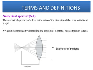
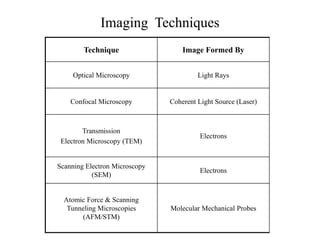
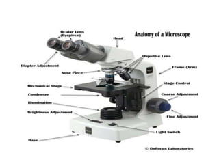
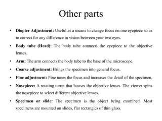
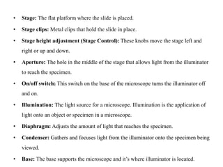
SlideShare a Scribd company logo
Microscopy
Microscopy
• Microscope is used to view objects or specimens that are too small to be
seen with just the human eye.
• Microscopy is a technical field that involves the use of Microscopical
components such as microscopes or microscope objectives to obtain
greater detail of examined samples.
• Definition-A microscope is a high precision optical instrument that uses a
lens or a combination of lenses to produce highly magnified images of
small specimens or objects especially when they are too small to be seen
by the naked (unaided) eye.
• A light source is used (either by mirrors or lamps) to make it easier to see
the subject matter.
TERMS AND DEFINITIONS
Principle
Microscopy is to get a magnified image, in which structures may be resolved
which could not be resolved with the help of an unaided eye.
Magnification
•It is the ratio of the size of an object seen under microscope to the actual size
observed with unaided eye.
•The total magnification of microscope is calculated by multiplying the
magnifying power of the objective lens by that of eye piece.
Resolving power
•It is the ability to differentiate two close points as separate.
•The resolving power of human eye is 0.25 mm
•The light microscope can separate dots that are 0.25µm apart.
•The electron microscope can separate dots that are 0.5nm apart.
TERMS AND DEFINITIONS
Limit of resolution
•It is the minimum distance between two points to identify them separately.
•It is calculated by Abbé equation.
Working distance
•It is the distance between the objective and the objective slide.
•The working distance decreases with increasing magnification.
TERMS AND DEFINITIONS
Numerical aperture(NA)
The numerical aperture of a lens is the ratio of the diameter of the lens to its focal
length.
NA can be decreased by decreasing the amount of light that passes through a lens.
Diameter of the lens
Imaging Techniques
Technique Image Formed By
Optical Microscopy Light Rays
Confocal Microscopy Coherent Light Source (Laser)
Transmission
Electron Microscopy (TEM)
Electrons
Scanning Electron Microscopy
(SEM)
Electrons
Atomic Force & Scanning
Tunneling Microscopies
(AFM/STM)
Molecular Mechanical Probes
History of microscope
• In 1590 F.H Janssen & Z.Janssen constructed the first simple compound light
microscope -10x to 30x .
• In 1665 Robert Hooke developed a first laboratory compound microscope.
• Later, Kepler and Galileo developed a modern class room microscope.
• In 1672 Anton Von Leeuwenhoek developed a first simple microscope with a
magnification of 200x – 300x.
• In 1674, Anton was the first to see and describe bacteria, yeast, plants, and life in a drop
of water- He is called as Father of microscopy.
• The term microscope was coined by Faber in 1623.
• In the early 1930’s the first electron beam microscopes were developed which were a
breakthrough in technology as they increased the magnification from about 1000x or so
up to 250,000x or more.
Compound Microscope
• Common type of microscope.
• High power microscope- The magnification (power) 40x to 1000x.
• Compound refers to the fact that in order to enlarge an image - a single
light path passes through a series of lenses in a line where each lens
magnifies the image over the previous one.
• In the standard form – 2 lenses
• an objective lens (closest to the object or specimen)
• an eyepiece lens (closest to the observers’ eye)
• Uses light to illuminate the specimen
• The objective lens usually consists of three or four lenses.
• The most used light method is trans-illumination.
• At 400x much detail can be seen at the cellular level of biological
specimens.
• Applications: Learn about cells and microorganisms in both medical and
science field.
Objective lenses
• One of the most important parts of a compound microscope, as they
are the lenses closest to the specimen.
• A standard microscope has three, four, or five objective lenses that
range in power from 4X to 100X.
• Objectives vary in power from 1x to 160x in compound microscopes
but the most common power range is from 4x to 100x.
• Most compound microscopes have three or four (occasionally five)
objectives usually of 4x, 10x, 40x, and 100x (oil immersion) which
revolve on a nosepiece (turret) to give different magnifying powers.
Numerical Aperture
• NA of a microscope objective is a measure of its ability to gather light
• The more light (higher NA) the better the resolving power of the lens
– Better resolution
• The N.A. will be marked on the objective and the typical N.A. for the
following are;
• 4x=0.10,
• 10x=0.25
• 40x=0.65
• 100x=1.25.
Ocular Lens or Eye piece
• The eyepiece consists of a series of lenses mounted in a tube (barrel) at the upper
end of the microscope.
• Its basic function is to look at the focused, magnified image projected by the
objective lens and magnify that image a second time before your eye looks at the
image of the specimen.
• The eyepieces are usually 10x but also come in 5x, 12.5x, 15x, and 20x. The “x”
refers to the amount of magnification (power) that this lens adds as a multiplier to
the magnification of the objective.
• For special applications, eyepieces can have scales, pointers, crosshairs, markers,
etc. on them.
• The eyepoint is the location (or position) of the eye from the eyepiece which allows
for the best possible viewing of the image.
Other parts
• Diopter Adjustment: Useful as a means to change focus on one eyepiece so as
to correct for any difference in vision between your two eyes.
• Body tube (Head): The body tube connects the eyepiece to the objective
lenses.
• Arm: The arm connects the body tube to the base of the microscope.
• Coarse adjustment: Brings the specimen into general focus.
• Fine adjustment: Fine tunes the focus and increases the detail of the specimen.
• Nosepiece: A rotating turret that houses the objective lenses. The viewer spins
the nosepiece to select different objective lenses.
• Specimen or slide: The specimen is the object being examined. Most
specimens are mounted on slides, flat rectangles of thin glass.
• Stage: The flat platform where the slide is placed.
• Stage clips: Metal clips that hold the slide in place.
• Stage height adjustment (Stage Control): These knobs move the stage left and
right or up and down.
• Aperture: The hole in the middle of the stage that allows light from the illuminator
to reach the specimen.
• On/off switch: This switch on the base of the microscope turns the illuminator off
and on.
• Illumination: The light source for a microscope. Illumination is the application of
light onto an object or specimen in a microscope.
• Diaphragm: Adjusts the amount of light that reaches the specimen.
• Condenser: Gathers and focuses light from the illuminator onto the specimen being
viewed.
• Base: The base supports the microscope and it’s where illuminator is located.
Microscopy.ppt Microscopy.pptvMicroscopy.pptMicroscopy.pptMicroscopy.pptMicroscopy.ppt

More Related Content

Similar to Microscopy.ppt Microscopy.pptvMicroscopy.pptMicroscopy.pptMicroscopy.pptMicroscopy.ppt

microscope.pptx
microscope.pptxmicroscope.pptx
microscope.pptx
Dinraj Kulkarni
 
microscopy ppt.pptx
microscopy ppt.pptxmicroscopy ppt.pptx
microscopy ppt.pptx
Ravipatel149335
 
History of microscopy i
History of microscopy iHistory of microscopy i
History of microscopy i
Pharmacy Universe
 
Microscopy
MicroscopyMicroscopy
Microscopy
YhanzieCapilitan
 
light microscopy seminar.pptx
light microscopy seminar.pptxlight microscopy seminar.pptx
light microscopy seminar.pptx
Ahalya40
 
Microscope
MicroscopeMicroscope
Microscope
MUKESH SINGH
 
Introduction to microscope
Introduction to microscopeIntroduction to microscope
Introduction to microscope
TasmiaZeb1
 
microscope- lecture 2.pptx
microscope- lecture 2.pptxmicroscope- lecture 2.pptx
microscope- lecture 2.pptx
OsmanAli92
 
Compound microscope
Compound microscopeCompound microscope
Compound microscope
meghashridhar
 
Microscopy use this.ppt
Microscopy use this.pptMicroscopy use this.ppt
Microscopy use this.ppt
ShantanuDharekar
 
Microscopy and staining techniques details
Microscopy and staining techniques detailsMicroscopy and staining techniques details
Microscopy and staining techniques details
JyotirmoyPanda1
 
Light Microscope and types
Light Microscope and typesLight Microscope and types
Light Microscope and types
SuganyaPaulraj
 
Microscope(1).pptx
Microscope(1).pptxMicroscope(1).pptx
Microscope(1).pptx
AshleyGarnica
 
History of microbiology and types of microscope
History of microbiology and types of microscopeHistory of microbiology and types of microscope
History of microbiology and types of microscope
sanjithkumarsanjith
 
History of microbiology and types of microscope
History of microbiology and types of microscopeHistory of microbiology and types of microscope
History of microbiology and types of microscope
sanjithkumarsanjith
 
Microscopy - Study
Microscopy - StudyMicroscopy - Study
Microscopy - Study
AyushiSharma843565
 
Microscopy - Study
Microscopy - StudyMicroscopy - Study
Microscopy - Study
AyushiSharma843565
 
microscope
microscopemicroscope
microscope
AireenDiamsimMalto
 
MICROSCOPY.pptx
MICROSCOPY.pptxMICROSCOPY.pptx
MICROSCOPY.pptx
Saraswathi29574
 
Compound microscope
Compound microscopeCompound microscope
Compound microscope
Komal Nikumbh
 

Similar to Microscopy.ppt Microscopy.pptvMicroscopy.pptMicroscopy.pptMicroscopy.pptMicroscopy.ppt (20)

microscope.pptx
microscope.pptxmicroscope.pptx
microscope.pptx
 
microscopy ppt.pptx
microscopy ppt.pptxmicroscopy ppt.pptx
microscopy ppt.pptx
 
History of microscopy i
History of microscopy iHistory of microscopy i
History of microscopy i
 
Microscopy
MicroscopyMicroscopy
Microscopy
 
light microscopy seminar.pptx
light microscopy seminar.pptxlight microscopy seminar.pptx
light microscopy seminar.pptx
 
Microscope
MicroscopeMicroscope
Microscope
 
Introduction to microscope
Introduction to microscopeIntroduction to microscope
Introduction to microscope
 
microscope- lecture 2.pptx
microscope- lecture 2.pptxmicroscope- lecture 2.pptx
microscope- lecture 2.pptx
 
Compound microscope
Compound microscopeCompound microscope
Compound microscope
 
Microscopy use this.ppt
Microscopy use this.pptMicroscopy use this.ppt
Microscopy use this.ppt
 
Microscopy and staining techniques details
Microscopy and staining techniques detailsMicroscopy and staining techniques details
Microscopy and staining techniques details
 
Light Microscope and types
Light Microscope and typesLight Microscope and types
Light Microscope and types
 
Microscope(1).pptx
Microscope(1).pptxMicroscope(1).pptx
Microscope(1).pptx
 
History of microbiology and types of microscope
History of microbiology and types of microscopeHistory of microbiology and types of microscope
History of microbiology and types of microscope
 
History of microbiology and types of microscope
History of microbiology and types of microscopeHistory of microbiology and types of microscope
History of microbiology and types of microscope
 
Microscopy - Study
Microscopy - StudyMicroscopy - Study
Microscopy - Study
 
Microscopy - Study
Microscopy - StudyMicroscopy - Study
Microscopy - Study
 
microscope
microscopemicroscope
microscope
 
MICROSCOPY.pptx
MICROSCOPY.pptxMICROSCOPY.pptx
MICROSCOPY.pptx
 
Compound microscope
Compound microscopeCompound microscope
Compound microscope
 

Recently uploaded

AKS UNIVERSITY Satna Final Year Project By OM Hardaha.pdf
AKS UNIVERSITY Satna Final Year Project By OM Hardaha.pdfAKS UNIVERSITY Satna Final Year Project By OM Hardaha.pdf
AKS UNIVERSITY Satna Final Year Project By OM Hardaha.pdf
SamSarthak3
 
J.Yang, ICLR 2024, MLILAB, KAIST AI.pdf
J.Yang,  ICLR 2024, MLILAB, KAIST AI.pdfJ.Yang,  ICLR 2024, MLILAB, KAIST AI.pdf
J.Yang, ICLR 2024, MLILAB, KAIST AI.pdf
MLILAB
 
ML for identifying fraud using open blockchain data.pptx
ML for identifying fraud using open blockchain data.pptxML for identifying fraud using open blockchain data.pptx
ML for identifying fraud using open blockchain data.pptx
Vijay Dialani, PhD
 
Planning Of Procurement o different goods and services
Planning Of Procurement o different goods and servicesPlanning Of Procurement o different goods and services
Planning Of Procurement o different goods and services
JoytuBarua2
 
Architectural Portfolio Sean Lockwood
Architectural Portfolio Sean LockwoodArchitectural Portfolio Sean Lockwood
Architectural Portfolio Sean Lockwood
seandesed
 
Standard Reomte Control Interface - Neometrix
Standard Reomte Control Interface - NeometrixStandard Reomte Control Interface - Neometrix
Standard Reomte Control Interface - Neometrix
Neometrix_Engineering_Pvt_Ltd
 
The Benefits and Techniques of Trenchless Pipe Repair.pdf
The Benefits and Techniques of Trenchless Pipe Repair.pdfThe Benefits and Techniques of Trenchless Pipe Repair.pdf
The Benefits and Techniques of Trenchless Pipe Repair.pdf
Pipe Restoration Solutions
 
weather web application report.pdf
weather web application report.pdfweather web application report.pdf
weather web application report.pdf
Pratik Pawar
 
power quality voltage fluctuation UNIT - I.pptx
power quality voltage fluctuation UNIT - I.pptxpower quality voltage fluctuation UNIT - I.pptx
power quality voltage fluctuation UNIT - I.pptx
ViniHema
 
English lab ppt no titlespecENG PPTt.pdf
English lab ppt no titlespecENG PPTt.pdfEnglish lab ppt no titlespecENG PPTt.pdf
English lab ppt no titlespecENG PPTt.pdf
BrazilAccount1
 
NO1 Uk best vashikaran specialist in delhi vashikaran baba near me online vas...
NO1 Uk best vashikaran specialist in delhi vashikaran baba near me online vas...NO1 Uk best vashikaran specialist in delhi vashikaran baba near me online vas...
NO1 Uk best vashikaran specialist in delhi vashikaran baba near me online vas...
Amil Baba Dawood bangali
 
Design and Analysis of Algorithms-DP,Backtracking,Graphs,B&B
Design and Analysis of Algorithms-DP,Backtracking,Graphs,B&BDesign and Analysis of Algorithms-DP,Backtracking,Graphs,B&B
Design and Analysis of Algorithms-DP,Backtracking,Graphs,B&B
Sreedhar Chowdam
 
ASME IX(9) 2007 Full Version .pdf
ASME IX(9)  2007 Full Version       .pdfASME IX(9)  2007 Full Version       .pdf
ASME IX(9) 2007 Full Version .pdf
AhmedHussein950959
 
ethical hacking-mobile hacking methods.ppt
ethical hacking-mobile hacking methods.pptethical hacking-mobile hacking methods.ppt
ethical hacking-mobile hacking methods.ppt
Jayaprasanna4
 
Runway Orientation Based on the Wind Rose Diagram.pptx
Runway Orientation Based on the Wind Rose Diagram.pptxRunway Orientation Based on the Wind Rose Diagram.pptx
Runway Orientation Based on the Wind Rose Diagram.pptx
SupreethSP4
 
MCQ Soil mechanics questions (Soil shear strength).pdf
MCQ Soil mechanics questions (Soil shear strength).pdfMCQ Soil mechanics questions (Soil shear strength).pdf
MCQ Soil mechanics questions (Soil shear strength).pdf
Osamah Alsalih
 
Nuclear Power Economics and Structuring 2024
Nuclear Power Economics and Structuring 2024Nuclear Power Economics and Structuring 2024
Nuclear Power Economics and Structuring 2024
Massimo Talia
 
AP LAB PPT.pdf ap lab ppt no title specific
AP LAB PPT.pdf ap lab ppt no title specificAP LAB PPT.pdf ap lab ppt no title specific
AP LAB PPT.pdf ap lab ppt no title specific
BrazilAccount1
 
Pile Foundation by Venkatesh Taduvai (Sub Geotechnical Engineering II)-conver...
Pile Foundation by Venkatesh Taduvai (Sub Geotechnical Engineering II)-conver...Pile Foundation by Venkatesh Taduvai (Sub Geotechnical Engineering II)-conver...
Pile Foundation by Venkatesh Taduvai (Sub Geotechnical Engineering II)-conver...
AJAYKUMARPUND1
 
Industrial Training at Shahjalal Fertilizer Company Limited (SFCL)
Industrial Training at Shahjalal Fertilizer Company Limited (SFCL)Industrial Training at Shahjalal Fertilizer Company Limited (SFCL)
Industrial Training at Shahjalal Fertilizer Company Limited (SFCL)
MdTanvirMahtab2
 

Recently uploaded (20)

AKS UNIVERSITY Satna Final Year Project By OM Hardaha.pdf
AKS UNIVERSITY Satna Final Year Project By OM Hardaha.pdfAKS UNIVERSITY Satna Final Year Project By OM Hardaha.pdf
AKS UNIVERSITY Satna Final Year Project By OM Hardaha.pdf
 
J.Yang, ICLR 2024, MLILAB, KAIST AI.pdf
J.Yang,  ICLR 2024, MLILAB, KAIST AI.pdfJ.Yang,  ICLR 2024, MLILAB, KAIST AI.pdf
J.Yang, ICLR 2024, MLILAB, KAIST AI.pdf
 
ML for identifying fraud using open blockchain data.pptx
ML for identifying fraud using open blockchain data.pptxML for identifying fraud using open blockchain data.pptx
ML for identifying fraud using open blockchain data.pptx
 
Planning Of Procurement o different goods and services
Planning Of Procurement o different goods and servicesPlanning Of Procurement o different goods and services
Planning Of Procurement o different goods and services
 
Architectural Portfolio Sean Lockwood
Architectural Portfolio Sean LockwoodArchitectural Portfolio Sean Lockwood
Architectural Portfolio Sean Lockwood
 
Standard Reomte Control Interface - Neometrix
Standard Reomte Control Interface - NeometrixStandard Reomte Control Interface - Neometrix
Standard Reomte Control Interface - Neometrix
 
The Benefits and Techniques of Trenchless Pipe Repair.pdf
The Benefits and Techniques of Trenchless Pipe Repair.pdfThe Benefits and Techniques of Trenchless Pipe Repair.pdf
The Benefits and Techniques of Trenchless Pipe Repair.pdf
 
weather web application report.pdf
weather web application report.pdfweather web application report.pdf
weather web application report.pdf
 
power quality voltage fluctuation UNIT - I.pptx
power quality voltage fluctuation UNIT - I.pptxpower quality voltage fluctuation UNIT - I.pptx
power quality voltage fluctuation UNIT - I.pptx
 
English lab ppt no titlespecENG PPTt.pdf
English lab ppt no titlespecENG PPTt.pdfEnglish lab ppt no titlespecENG PPTt.pdf
English lab ppt no titlespecENG PPTt.pdf
 
NO1 Uk best vashikaran specialist in delhi vashikaran baba near me online vas...
NO1 Uk best vashikaran specialist in delhi vashikaran baba near me online vas...NO1 Uk best vashikaran specialist in delhi vashikaran baba near me online vas...
NO1 Uk best vashikaran specialist in delhi vashikaran baba near me online vas...
 
Design and Analysis of Algorithms-DP,Backtracking,Graphs,B&B
Design and Analysis of Algorithms-DP,Backtracking,Graphs,B&BDesign and Analysis of Algorithms-DP,Backtracking,Graphs,B&B
Design and Analysis of Algorithms-DP,Backtracking,Graphs,B&B
 
ASME IX(9) 2007 Full Version .pdf
ASME IX(9)  2007 Full Version       .pdfASME IX(9)  2007 Full Version       .pdf
ASME IX(9) 2007 Full Version .pdf
 
ethical hacking-mobile hacking methods.ppt
ethical hacking-mobile hacking methods.pptethical hacking-mobile hacking methods.ppt
ethical hacking-mobile hacking methods.ppt
 
Runway Orientation Based on the Wind Rose Diagram.pptx
Runway Orientation Based on the Wind Rose Diagram.pptxRunway Orientation Based on the Wind Rose Diagram.pptx
Runway Orientation Based on the Wind Rose Diagram.pptx
 
MCQ Soil mechanics questions (Soil shear strength).pdf
MCQ Soil mechanics questions (Soil shear strength).pdfMCQ Soil mechanics questions (Soil shear strength).pdf
MCQ Soil mechanics questions (Soil shear strength).pdf
 
Nuclear Power Economics and Structuring 2024
Nuclear Power Economics and Structuring 2024Nuclear Power Economics and Structuring 2024
Nuclear Power Economics and Structuring 2024
 
AP LAB PPT.pdf ap lab ppt no title specific
AP LAB PPT.pdf ap lab ppt no title specificAP LAB PPT.pdf ap lab ppt no title specific
AP LAB PPT.pdf ap lab ppt no title specific
 
Pile Foundation by Venkatesh Taduvai (Sub Geotechnical Engineering II)-conver...
Pile Foundation by Venkatesh Taduvai (Sub Geotechnical Engineering II)-conver...Pile Foundation by Venkatesh Taduvai (Sub Geotechnical Engineering II)-conver...
Pile Foundation by Venkatesh Taduvai (Sub Geotechnical Engineering II)-conver...
 
Industrial Training at Shahjalal Fertilizer Company Limited (SFCL)
Industrial Training at Shahjalal Fertilizer Company Limited (SFCL)Industrial Training at Shahjalal Fertilizer Company Limited (SFCL)
Industrial Training at Shahjalal Fertilizer Company Limited (SFCL)
 

Microscopy.ppt Microscopy.pptvMicroscopy.pptMicroscopy.pptMicroscopy.pptMicroscopy.ppt

  • 2. Microscopy • Microscope is used to view objects or specimens that are too small to be seen with just the human eye. • Microscopy is a technical field that involves the use of Microscopical components such as microscopes or microscope objectives to obtain greater detail of examined samples. • Definition-A microscope is a high precision optical instrument that uses a lens or a combination of lenses to produce highly magnified images of small specimens or objects especially when they are too small to be seen by the naked (unaided) eye. • A light source is used (either by mirrors or lamps) to make it easier to see the subject matter.
  • 3. TERMS AND DEFINITIONS Principle Microscopy is to get a magnified image, in which structures may be resolved which could not be resolved with the help of an unaided eye. Magnification •It is the ratio of the size of an object seen under microscope to the actual size observed with unaided eye. •The total magnification of microscope is calculated by multiplying the magnifying power of the objective lens by that of eye piece. Resolving power •It is the ability to differentiate two close points as separate. •The resolving power of human eye is 0.25 mm •The light microscope can separate dots that are 0.25µm apart. •The electron microscope can separate dots that are 0.5nm apart.
  • 4. TERMS AND DEFINITIONS Limit of resolution •It is the minimum distance between two points to identify them separately. •It is calculated by Abbé equation. Working distance •It is the distance between the objective and the objective slide. •The working distance decreases with increasing magnification.
  • 5. TERMS AND DEFINITIONS Numerical aperture(NA) The numerical aperture of a lens is the ratio of the diameter of the lens to its focal length. NA can be decreased by decreasing the amount of light that passes through a lens. Diameter of the lens
  • 6. Imaging Techniques Technique Image Formed By Optical Microscopy Light Rays Confocal Microscopy Coherent Light Source (Laser) Transmission Electron Microscopy (TEM) Electrons Scanning Electron Microscopy (SEM) Electrons Atomic Force & Scanning Tunneling Microscopies (AFM/STM) Molecular Mechanical Probes
  • 7. History of microscope • In 1590 F.H Janssen & Z.Janssen constructed the first simple compound light microscope -10x to 30x . • In 1665 Robert Hooke developed a first laboratory compound microscope. • Later, Kepler and Galileo developed a modern class room microscope. • In 1672 Anton Von Leeuwenhoek developed a first simple microscope with a magnification of 200x – 300x. • In 1674, Anton was the first to see and describe bacteria, yeast, plants, and life in a drop of water- He is called as Father of microscopy. • The term microscope was coined by Faber in 1623. • In the early 1930’s the first electron beam microscopes were developed which were a breakthrough in technology as they increased the magnification from about 1000x or so up to 250,000x or more.
  • 8. Compound Microscope • Common type of microscope. • High power microscope- The magnification (power) 40x to 1000x. • Compound refers to the fact that in order to enlarge an image - a single light path passes through a series of lenses in a line where each lens magnifies the image over the previous one. • In the standard form – 2 lenses • an objective lens (closest to the object or specimen) • an eyepiece lens (closest to the observers’ eye) • Uses light to illuminate the specimen • The objective lens usually consists of three or four lenses. • The most used light method is trans-illumination. • At 400x much detail can be seen at the cellular level of biological specimens. • Applications: Learn about cells and microorganisms in both medical and science field.
  • 9.
  • 10. Objective lenses • One of the most important parts of a compound microscope, as they are the lenses closest to the specimen. • A standard microscope has three, four, or five objective lenses that range in power from 4X to 100X. • Objectives vary in power from 1x to 160x in compound microscopes but the most common power range is from 4x to 100x. • Most compound microscopes have three or four (occasionally five) objectives usually of 4x, 10x, 40x, and 100x (oil immersion) which revolve on a nosepiece (turret) to give different magnifying powers.
  • 11. Numerical Aperture • NA of a microscope objective is a measure of its ability to gather light • The more light (higher NA) the better the resolving power of the lens – Better resolution • The N.A. will be marked on the objective and the typical N.A. for the following are; • 4x=0.10, • 10x=0.25 • 40x=0.65 • 100x=1.25.
  • 12. Ocular Lens or Eye piece • The eyepiece consists of a series of lenses mounted in a tube (barrel) at the upper end of the microscope. • Its basic function is to look at the focused, magnified image projected by the objective lens and magnify that image a second time before your eye looks at the image of the specimen. • The eyepieces are usually 10x but also come in 5x, 12.5x, 15x, and 20x. The “x” refers to the amount of magnification (power) that this lens adds as a multiplier to the magnification of the objective. • For special applications, eyepieces can have scales, pointers, crosshairs, markers, etc. on them. • The eyepoint is the location (or position) of the eye from the eyepiece which allows for the best possible viewing of the image.
  • 13. Other parts • Diopter Adjustment: Useful as a means to change focus on one eyepiece so as to correct for any difference in vision between your two eyes. • Body tube (Head): The body tube connects the eyepiece to the objective lenses. • Arm: The arm connects the body tube to the base of the microscope. • Coarse adjustment: Brings the specimen into general focus. • Fine adjustment: Fine tunes the focus and increases the detail of the specimen. • Nosepiece: A rotating turret that houses the objective lenses. The viewer spins the nosepiece to select different objective lenses. • Specimen or slide: The specimen is the object being examined. Most specimens are mounted on slides, flat rectangles of thin glass.
  • 14. • Stage: The flat platform where the slide is placed. • Stage clips: Metal clips that hold the slide in place. • Stage height adjustment (Stage Control): These knobs move the stage left and right or up and down. • Aperture: The hole in the middle of the stage that allows light from the illuminator to reach the specimen. • On/off switch: This switch on the base of the microscope turns the illuminator off and on. • Illumination: The light source for a microscope. Illumination is the application of light onto an object or specimen in a microscope. • Diaphragm: Adjusts the amount of light that reaches the specimen. • Condenser: Gathers and focuses light from the illuminator onto the specimen being viewed. • Base: The base supports the microscope and it’s where illuminator is located.