Microscopes use lenses and light or electrons to magnify objects too small to be seen by the naked eye. There are two main types of microscopes - light microscopes, which use visible light and lenses, and electron microscopes, which use a beam of electrons for higher magnification. Light microscopes can be simple or compound, with compound microscopes using multiple lenses to provide higher magnification. Different types of microscopes like fluorescence, dark field, and oil immersion microscopes provide specific ways of illuminating samples for different applications. Resolution and magnification depend on factors like the wavelength of light used and the numerical aperture of lenses. Microscopy is used across many fields of science to study cells, tissues, and small organisms.


![Magnification
Obtained by both the objective [which produces real image] and ocular [which
produces virtual image].
The magnifying power of a compound light microscope is the product of the
individual magnifying powers of the ocular lens and the objective lens.
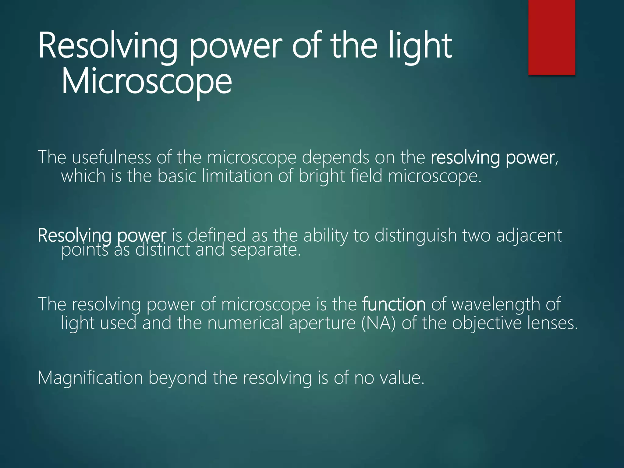
Magnification beyond the resolving power will result in larger images but less
distinct in their details and appearance.
Ocular Objective Magnification
10 X 10 X (low power) 100 X
10 X 45 X (high “dry”) 450 X
10 X 100 X (oil immersion) 1000 X](https://image.slidesharecdn.com/microscopesandmicroscopy-200501154720/75/Microscopes-and-microscopy-11-2048.jpg)