The light microscope uses visible light and a system of lenses to magnify small samples. Key developments included the first simple compound microscope in 1590 and Robert Hooke's compound microscope in 1665. Antonie van Leeuwenhoek developed the first simple microscope with 200-300x magnification in 1672.
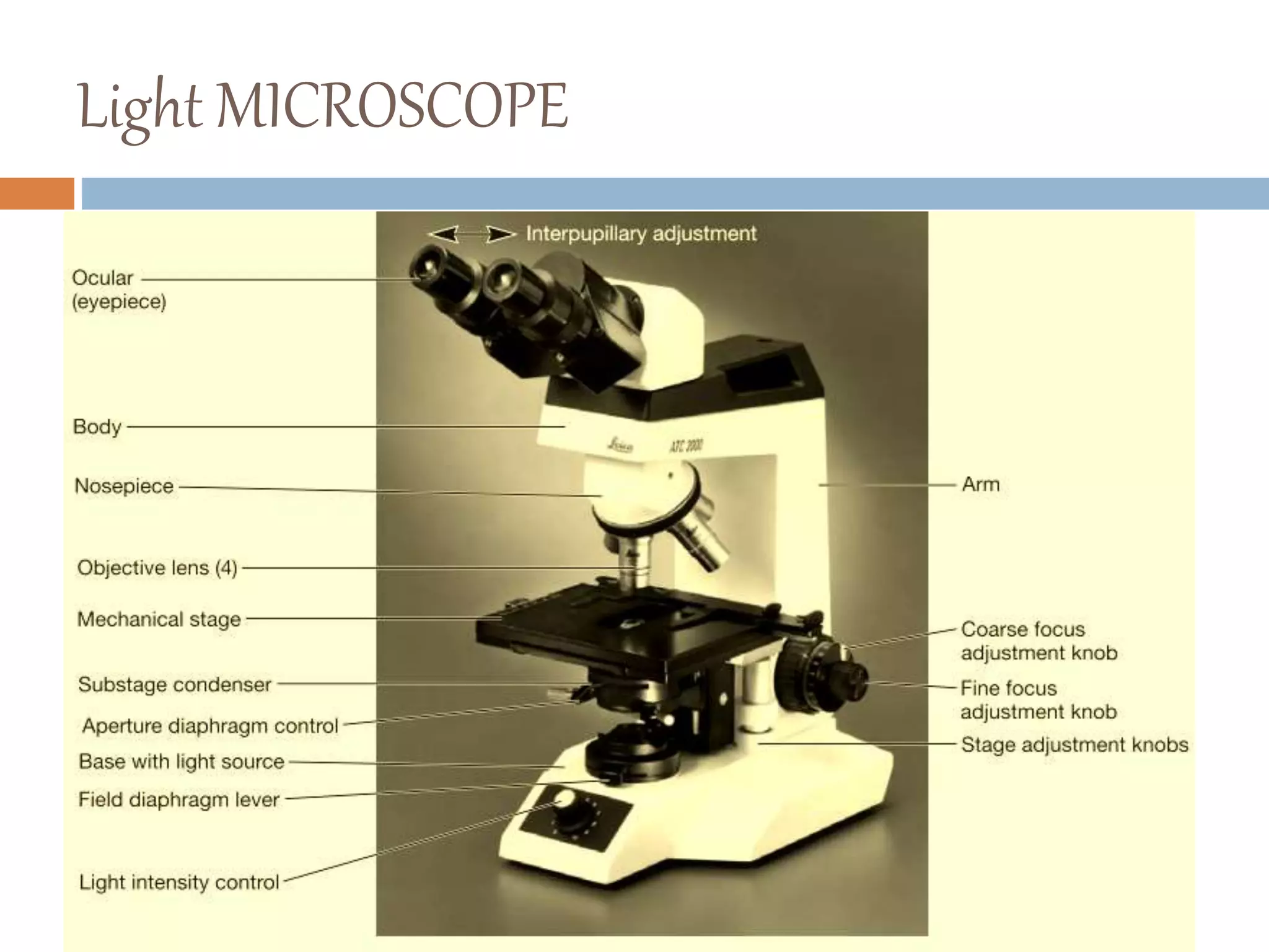
The main parts of a light microscope are the illuminator, condenser, stage, objective, nosepiece, iris diaphragm, base, focusing knobs, and ocular eyepiece. Light from the illuminator passes through the condenser, specimen, objective lens, and is magnified by the eyepiece for viewing. Resolution and magnification can be improved by using higher powered objectives, oil imm
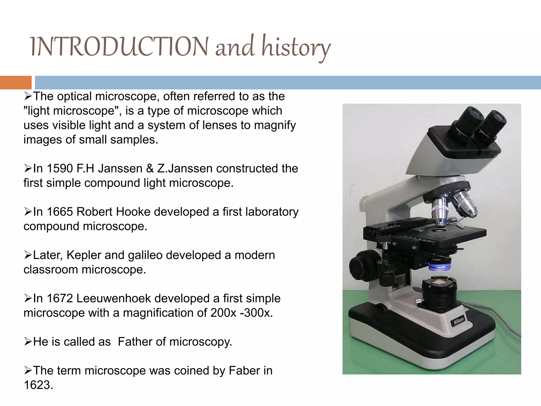
INTRODUCTION and history
Theoptical microscope, often referred to as the
"light microscope", is a type of microscope which
uses visible light and a system of lenses to magnify
images of small samples.
In 1590 F.H Janssen & Z.Janssen constructed the
first simple compound light microscope.
In 1665 Robert Hooke developed a first laboratory
compound microscope.
Later, Kepler and galileo developed a modern
classroom microscope.
In 1672 Leeuwenhoek developed a first simple
microscope with a magnification of 200x -300x.
He is called as Father of microscopy.
The term microscope was coined by Faber in
1623.
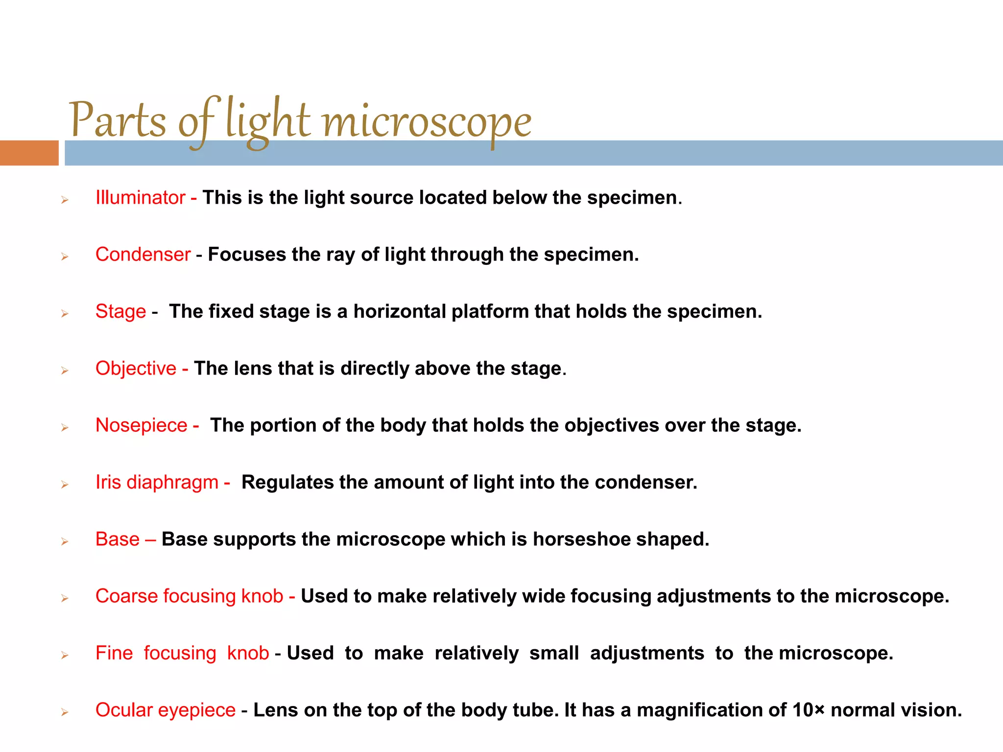
Parts of lightmicroscope
Illuminator - This is the light source located below the specimen.
Condenser - Focuses the ray of light through the specimen.
Stage - The fixed stage is a horizontal platform that holds the specimen.
Objective - The lens that is directly above the stage.
Nosepiece - The portion of the body that holds the objectives over the stage.
Iris diaphragm - Regulates the amount of light into the condenser.
Base – Base supports the microscope which is horseshoe shaped.
Coarse focusing knob - Used to make relatively wide focusing adjustments to the microscope.
Fine focusing knob - Used to make relatively small adjustments to the microscope.
Ocular eyepiece - Lens on the top of the body tube. It has a magnification of 10× normal vision.
5.
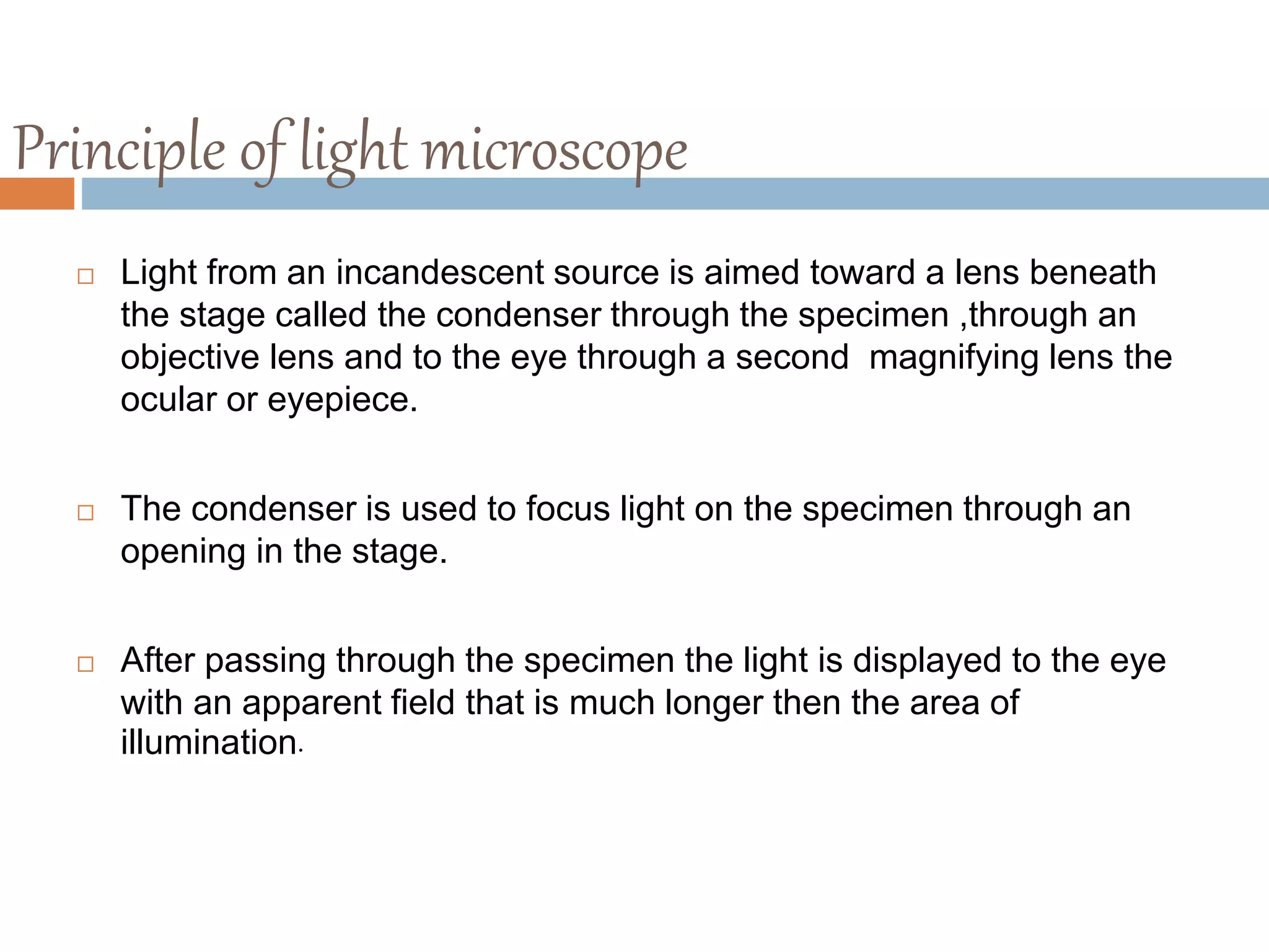
Principle of lightmicroscope
Light from an incandescent source is aimed toward a lens beneath
the stage called the condenser through the specimen ,through an
objective lens and to the eye through a second magnifying lens the
ocular or eyepiece.
The condenser is used to focus light on the specimen through an
opening in the stage.
After passing through the specimen the light is displayed to the eye
with an apparent field that is much longer then the area of
illumination.
6.
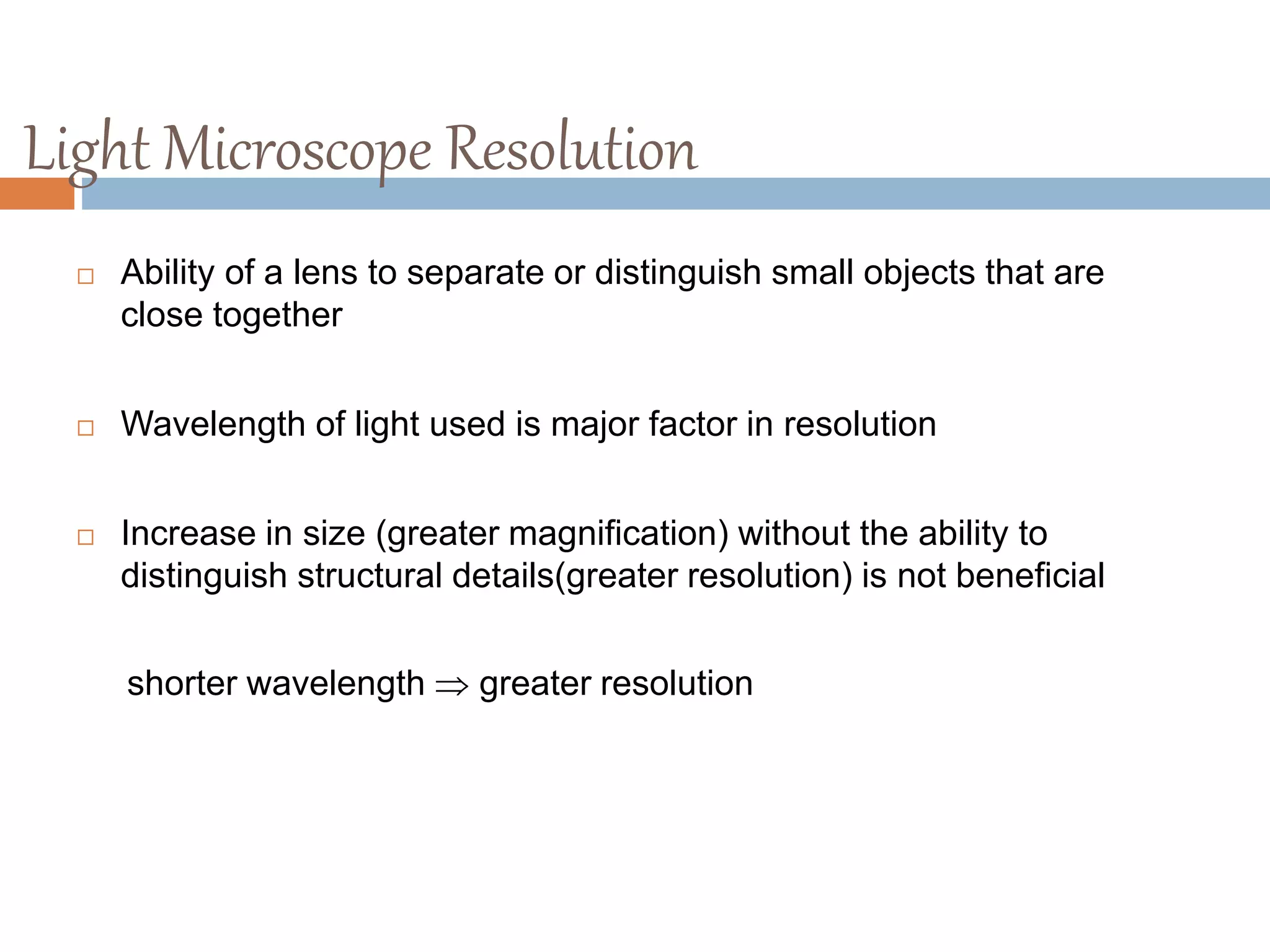
Light Microscope Resolution
Ability of a lens to separate or distinguish small objects that are
close together
Wavelength of light used is major factor in resolution
Increase in size (greater magnification) without the ability to
distinguish structural details(greater resolution) is not beneficial
shorter wavelength greater resolution
7.
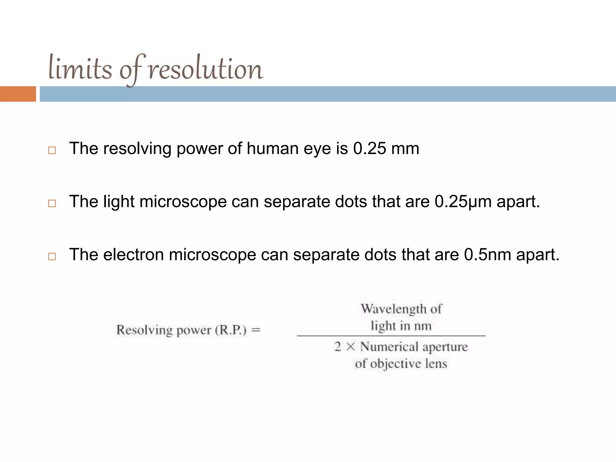
limits of resolution
The resolving power of human eye is 0.25 mm
The light microscope can separate dots that are 0.25µm apart.
The electron microscope can separate dots that are 0.5nm apart.
8.
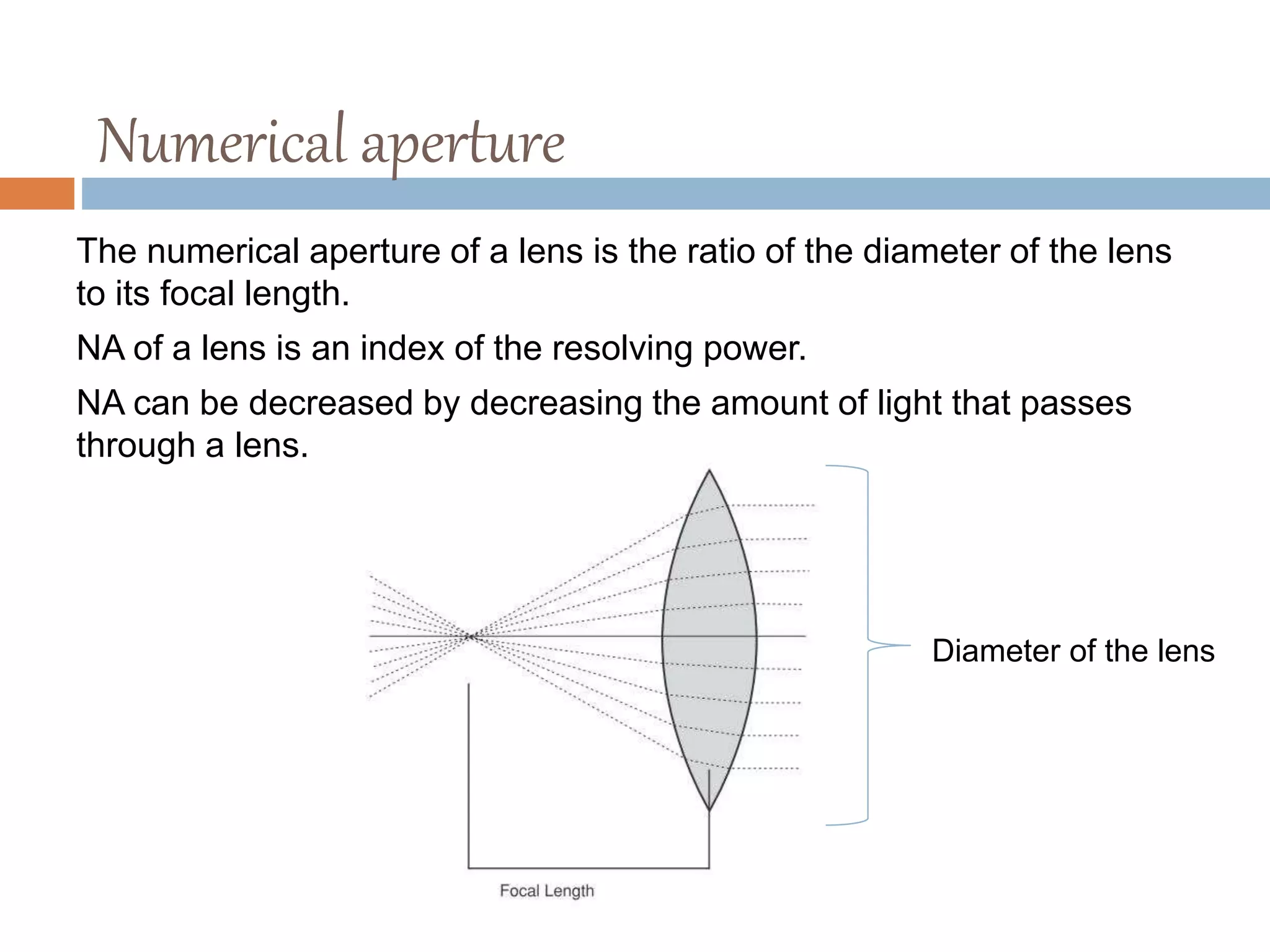
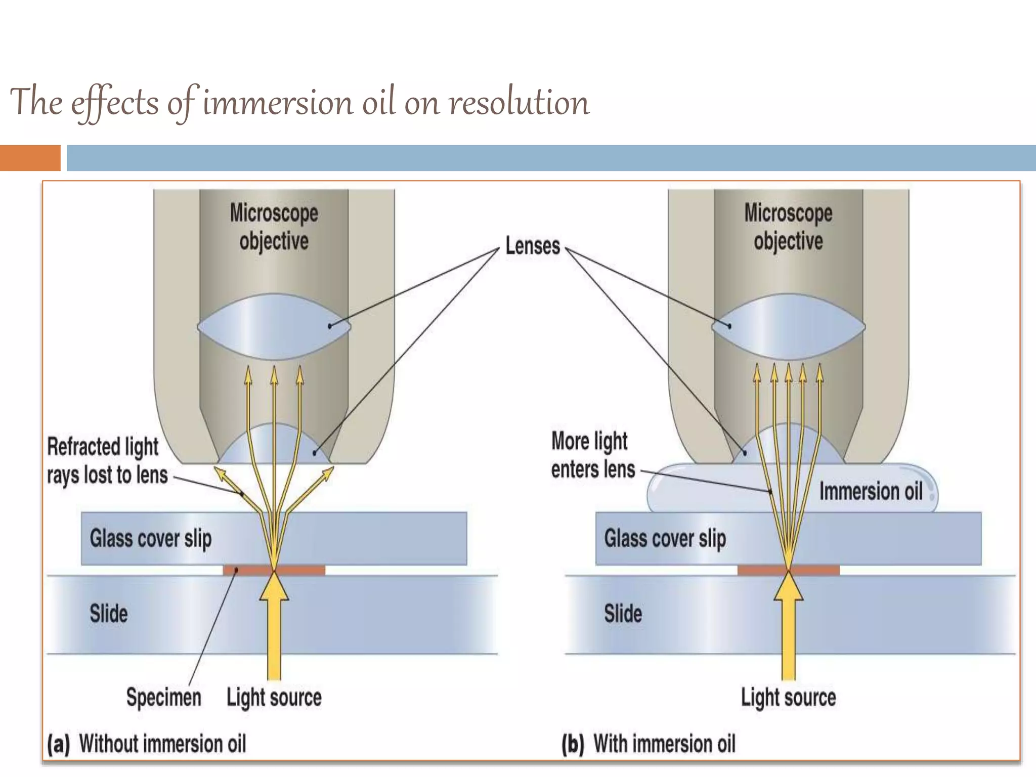
Numerical aperture
The numericalaperture of a lens is the ratio of the diameter of the lens
to its focal length.
NA of a lens is an index of the resolving power.
NA can be decreased by decreasing the amount of light that passes
through a lens.
Diameter of the lens
9.
magnification of lightmicroscope
It is the ratio of the size of an object seen under microscope to the
actual size observed with unaided eye.
The total magnification of microscope is calculated by multiplying the
magnifying power of the objective lens by that of eye piece
10.
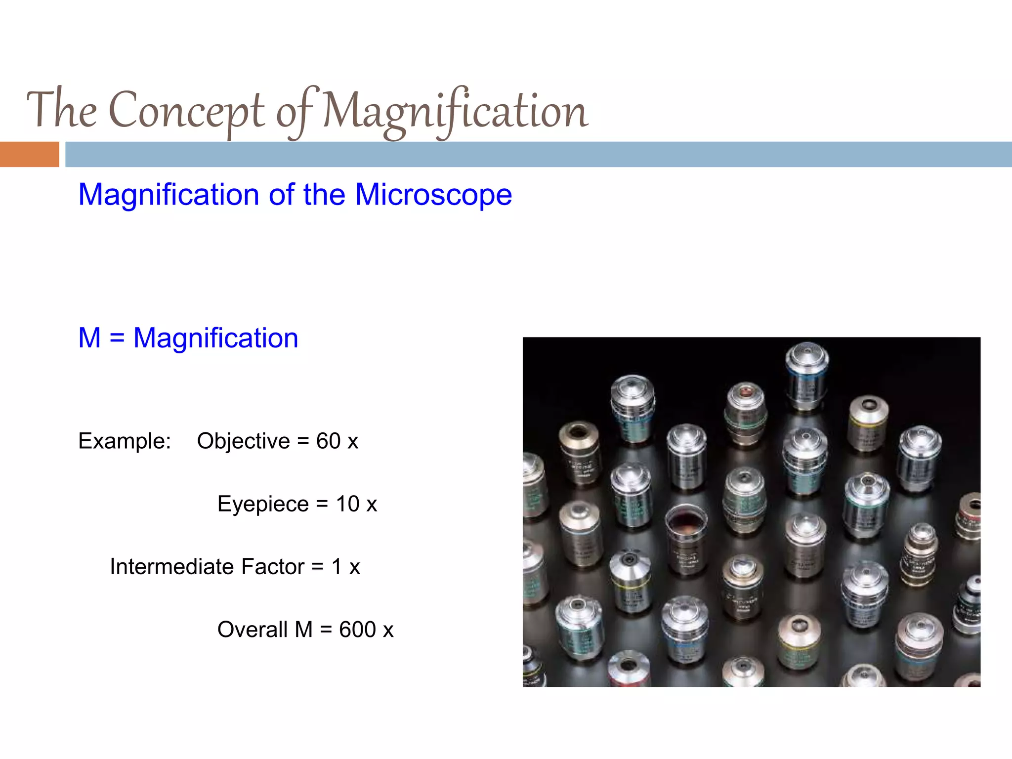
The Concept ofMagnification
Magnification of the Microscope
M Microscope = M Objective X M Eyepiece X M Intermediate Factor
M = Magnification
Example: Objective = 60 x
Eyepiece = 10 x
Intermediate Factor = 1 x
Overall M = 600 x
11.
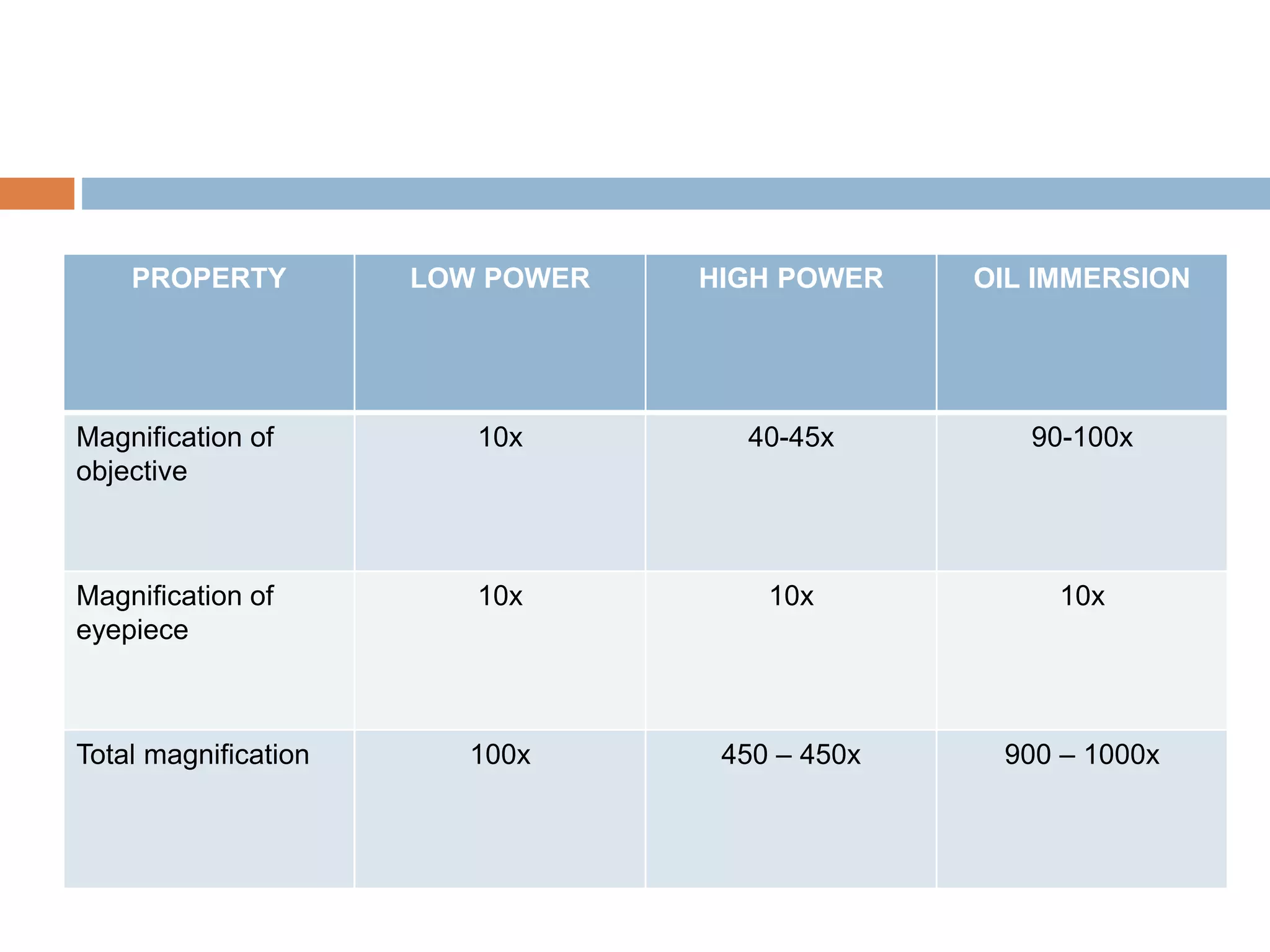
PROPERTY LOW POWERHIGH POWER OIL IMMERSION
Magnification of
objective
10x 40-45x 90-100x
Magnification of
eyepiece
10x 10x 10x
Total magnification 100x 450 – 450x 900 – 1000x
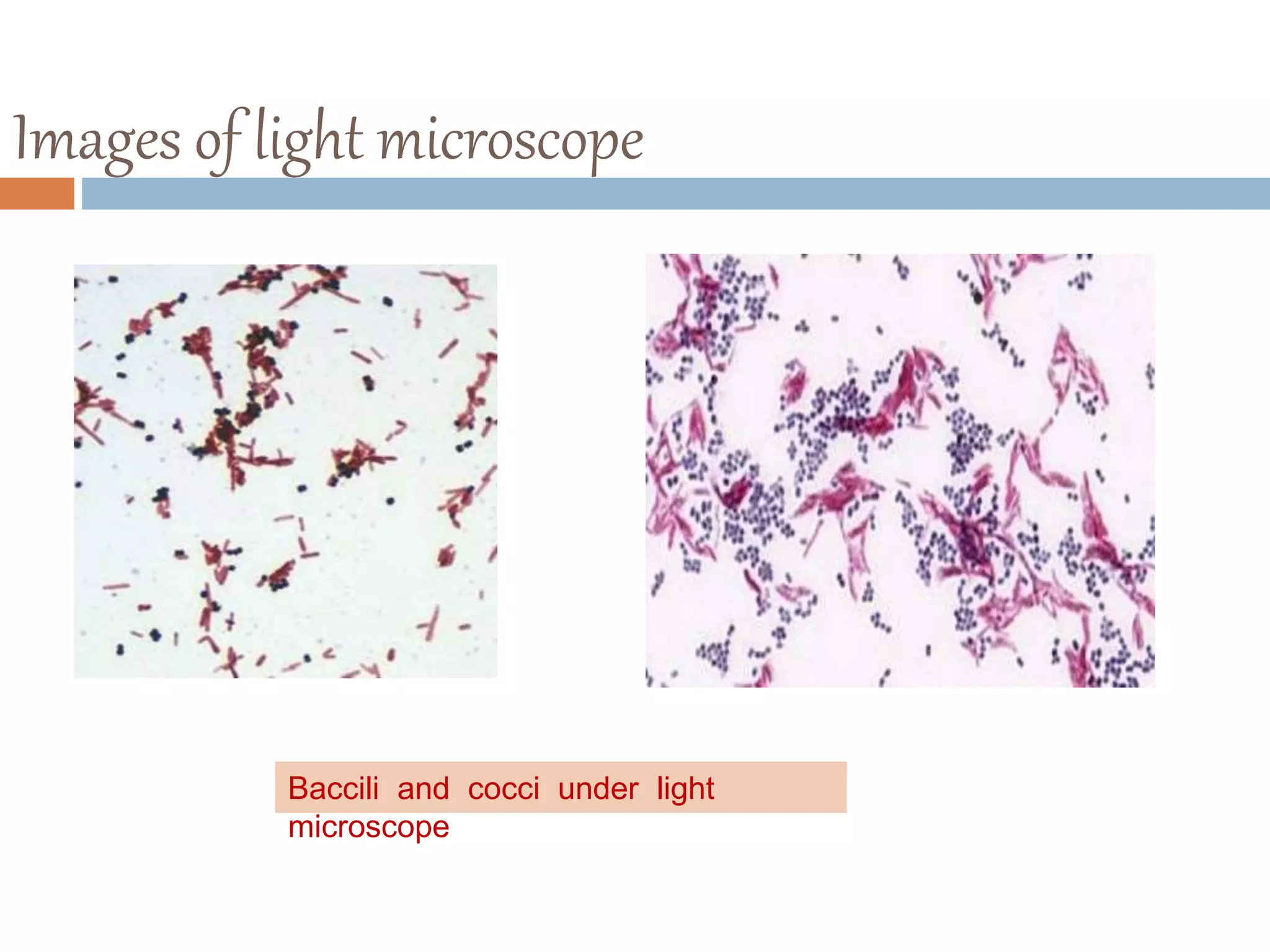
Images of lightmicroscope
Baccili and cocci under light
microscope
14.
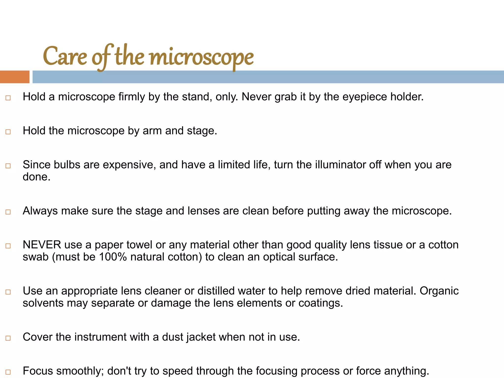
Care of themicroscope
Hold a microscope firmly by the stand, only. Never grab it by the eyepiece holder.
Hold the microscope by arm and stage.
Since bulbs are expensive, and have a limited life, turn the illuminator off when you are
done.
Always make sure the stage and lenses are clean before putting away the microscope.
NEVER use a paper towel or any material other than good quality lens tissue or a cotton
swab (must be 100% natural cotton) to clean an optical surface.
Use an appropriate lens cleaner or distilled water to help remove dried material. Organic
solvents may separate or damage the lens elements or coatings.
Cover the instrument with a dust jacket when not in use.
Focus smoothly; don't try to speed through the focusing process or force anything.