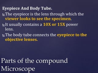
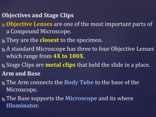
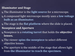
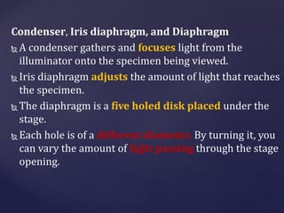
Download to read offline
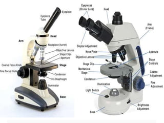
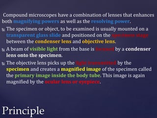


A compound microscope has more than one lens which allows for higher magnification and resolving power compared to a simple microscope. It consists of objective lenses close to the specimen and ocular lenses used by the observer. Light from the illuminator is focused on the specimen by the condenser lens, and the objective lens creates a primary magnified image inside the body tube which is further magnified by the ocular lens for viewing. Compound microscopes are used in various applications like pathology, forensics, biology and more due to their ability to reveal microscopic details.


![谷歌留痕技术 [ 𝙩𝙤𝙥 𝟮𝟯𝟯. 𝙘 𝙤𝙢 ]](https://cdn.slidesharecdn.com/ss_thumbnails/top233-260130174328-3833018c-thumbnail.jpg?width=640&height=640&fit=bounds)


