A microscopeis an instrument that
enables one to observe objects too small
to be seen clearly by the naked eye.
from the Greek: mikrós, "small" and
skopeîn, "to look" or "see")
3.
Compound Dissection or
Stereoscope
ConfocalMicr
oscope
Scanning Elec
tron Microsco
pe (SEM)
Transmission
Electron Micr
oscope (TEM)
Description Compound
microscopes are
light illuminated.
The image seen
with this type of
microscope is
two
dimensional.
This
microscope is
the most
commonly used.
You can view
individual cells,
even living ones.
It has high
magnification.
A dissection
microscope is
light illuminated.
The image that
appears is three
dimensional. It
is used for
dissection to
get a better
look at the
larger specimen.
You cannot see
individual cells
because it has a
low
magnification
This
microscope
uses a laser
light.This light is
used because of
the wavelength.
Laser light scan
across the
specimen with
the aid of
scanning
mirrors.Then
image is then
placed on a
digital computer
screen for
analyzing.
SEM use
electron
illumination.The
image is seen in
3-D. It has high
magnification
and high
resolution.The
specimen is
coated in gold
and the
electrons
bounce off to
give you and
exterior view of
the specimen.
TEM is electron
illuminated.This
gives a 2-D
view.Thin slices
of specimen are
obtained.The
electron beams
pass through
this. It has high
magnification
and high
resolution.
Source of
Radiation
visible light visible light laser light electrons electrons
Nature of
Lenses
glass glass glass lenses with
dichromatic
mirrors
one
electrostatic
lens with a few
electromagnetic
lenses
one
electrostatic
lens and a few
electromagnetic
lenses
4.
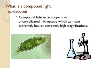
What is acompound light
microscope?
Compound light microscope is an
uncomplicated microscope which can have
extremely low or extremely high magnifications.
5.
As lightpasses through the object, the lens
nearest the object, called the objective lens,
produces an enlarged image of the object in
the primary image angle.The lens that you
look into, the eyepiece, acts as a magnifier
and produces an enlarged image of the
image produced by the objective lens.
A compound light microscope is limited to
about 2000X magnification
6.
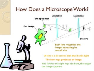
How Does aMicroscopeWork?
the eye
the image
the specimen
Each lens magnifies the
image, increasing its
overall size
A lens is a bi-convex disk that bends light
The farther the light rays are bent, the larger
the image appears
The bent rays produces an image
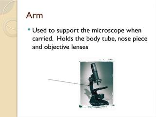
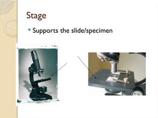
7.
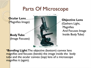
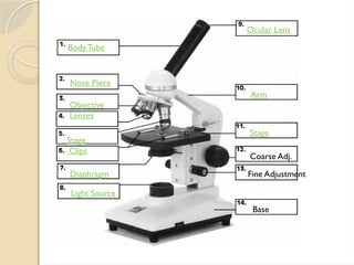
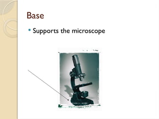
Parts Of Microscope
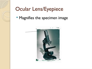
OcularLens
(Magnifies Image)
Objective Lens
(Gathers Light,
Magnifies
And Focuses Image
Inside Body Tube)
BodyTube
(Image Focuses)
•Bending Light:The objective (bottom) convex lens
magnifies and focuses (bends) the image inside the body
tube and the ocular convex (top) lens of a microscope
magnifies it (again).
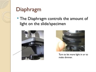
Diaphragm
The Diaphragmcontrols the amount of
light on the slide/specimen
Turn to let more light in or to
make dimmer.
12.
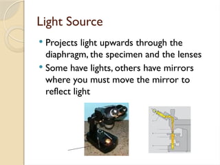
Light Source
Projectslight upwards through the
diaphragm, the specimen and the lenses
Some have lights, others have mirrors
where you must move the mirror to
reflect light
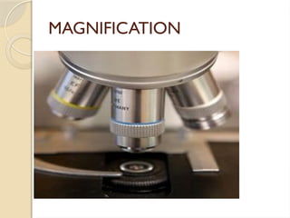
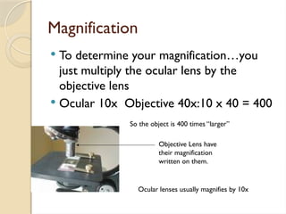
Magnification
To determineyour magnification…you
just multiply the ocular lens by the
objective lens
Ocular 10x Objective 40x:10 x 40 = 400
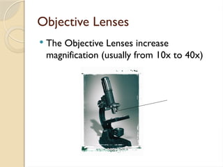
Objective Lens have
their magnification
written on them.
Ocular lenses usually magnifies by 10x
So the object is 400 times “larger”
21.
Using a Microscope
Start on the lowest magnification
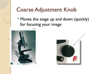
Don’t use the coarse adjustment knob on
high magnification…you’ll break the
slide!!!
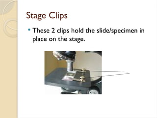
Place slide on stage and lock clips
Adjust light source (if it’s a mirror…don’t
stand in front of it!)
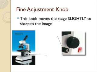
Use fine adjustment to focus
22.
APPLICATIONS:
A CompoundMicroscope is nowadays used
in several fields of sciences like the
Microbiology, Botany, Geology,
Genetics
Forensic experts and scientists can also
find out the country from which the drug
has come by viewing its particles under a
Compound Microscope as the shape of the
crystals can give a reference as to which
country the opium was grown in.
23.
Botanists canutilize a compound microscope
to examine plant parts to identify organisms that
exist on the surface of a plant.This can be for the
purpose of identifying fungi or other diseases
that may be present on the plant.
Microscopes also help in looking at the minute
details of the human cells and determine the
presence or absence of minerals, identify the
presence of metals, thus solve crimes and
discover medicines