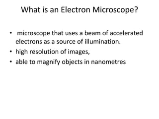
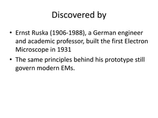
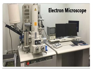
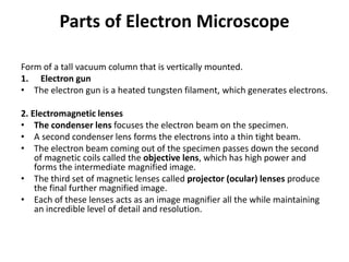
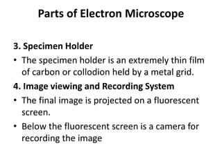
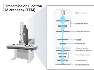
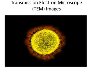
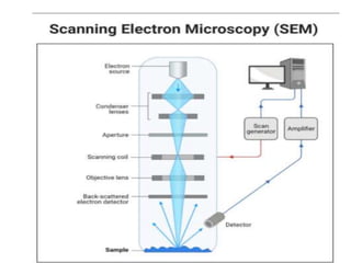
SlideShare a Scribd company logo
Electron microscope
Dr. Rasika Deshmukh
What is an Electron Microscope?
• microscope that uses a beam of accelerated
electrons as a source of illumination.
• high resolution of images,
• able to magnify objects in nanometres
Discovered by
• Ernst Ruska (1906-1988), a German engineer
and academic professor, built the first Electron
Microscope in 1931
• The same principles behind his prototype still
govern modern EMs.
Parts of Electron Microscope
Form of a tall vacuum column that is vertically mounted.
1. Electron gun
• The electron gun is a heated tungsten filament, which generates electrons.
2. Electromagnetic lenses
• The condenser lens focuses the electron beam on the specimen.
• A second condenser lens forms the electrons into a thin tight beam.
• The electron beam coming out of the specimen passes down the second
of magnetic coils called the objective lens, which has high power and
forms the intermediate magnified image.
• The third set of magnetic lenses called projector (ocular) lenses produce
the final further magnified image.
• Each of these lenses acts as an image magnifier all the while maintaining
an incredible level of detail and resolution.
Parts of Electron Microscope
3. Specimen Holder
• The specimen holder is an extremely thin film
of carbon or collodion held by a metal grid.
4. Image viewing and Recording System
• The final image is projected on a fluorescent
screen.
• Below the fluorescent screen is a camera for
recording the image
Working Principle of Electron
microscope
• The electron gun generates electrons.
• Two sets of condenser lenses focus the electron beam
on the specimen and then into a thin tight beam.
• To move electrons down the column, an accelerating
voltage (mostly between 100 kV-1000 kV) is applied
between the tungsten filament and anode.
• The specimen to be examined is made extremely thin,
at least 200 times thinner than those used in the
optical microscope. Ultra-thin sections of 20-100 nm
are cut which is already placed on the specimen holder.
Working Principle of Electron
microscope
• The electronic beam passes through the specimen and
electrons are scattered depending upon the thickness or
refractive index of different parts of the specimen.
• The denser regions in the specimen scatter more electrons
and therefore appear darker in the image since fewer
electrons strike that area of the screen. In contrast,
transparent regions are brighter.
• The electron beam coming out of the specimen passes to
the objective lens, which has high power and forms the
intermediate magnified image.
• The ocular lenses then produce the final further magnified
image.
Applications of Electron microscope:
• Investigate the ultrastructure of a wide range of
biological and inorganic specimens
• Used for quality control and failure analysis.
• Modern electron microscopes produce
electron micrographs using specialized digital
cameras and frame grabbers to capture the
images.
• Study of microorganisms like bacteria, virus, and
other pathogens have made the treatment of
diseases very effective.
Advantages of Electron microscope
• Very high magnification
• Incredibly high resolution
• Material rarely distorted by preparation
• It is possible to investigate a greater depth of
field
• Diverse applications
Limitations of Electron microscope
• The live specimen cannot be observed.
• The object should be ultra-thin
• The specimen is dried and cut into ultra-thin sections
before observation.
• As the EM works in a vacuum, the specimen should be
completely dry.
• Expensive to build and maintain
• Requiring researcher training
• Image artifacts resulting from specimen preparation.
• This type of microscope is large, cumbersome extremely
sensitive to vibration and external magnetic fields.
Types of electron microscope
• Transmission Electron Microscope (TEM)
• Scanning Electron Microscope (SEM)
Transmission Electron Microscope
(TEM)
• powerful electron microscope that uses a
beam of electrons to focus on a specimen
producing a highly magnified and detailed
image of the specimen.
• The magnification power is over 2 million
times, producing the image of the specimen
which enables easy characterization of the
image in its morphological features,
compositions
Principle of Transmission Electron
Microscope (TEM)
• similar to the light microscope.
• light microscopes use light rays to focus and
produce an image while the TEM uses a beam
of electrons to focus on the specimen, to
produce an image.
• Electrons have a shorter wavelength in
comparison to light which has a long
wavelength.
• Better resolution
Applications of Transmission Electron
Microscope (TEM)
• To visualize and study cell structures of bacteria,
viruses, and fungi
• To view bacteria flagella and plasmids
• To view the shapes and sizes of microbial cell
organelles
• To study and differentiate between plant and animal
cells.
• Its also used in nanotechnology to study nanoparticles
such as ZnO nanoparticles
• It is used to detect and identify fractures, damaged
microparticles which further enable repair mechanisms
of the particles.
Advantages of Transmission Electron
Microscope (TEM)
• It has a very powerful magnification of about 2 million
times that of the Light microscope.
• It can be used for a variety of applications ranging from
basic Biology to Nanotechnology, to education and
industrial uses.
• It can be used to acquire vast information on compounds
and their structures.
• It produces very efficient, high-quality images with high
clarity.
• It can produce permanent images.
• It is easy to train and use the Transmission Electron
Microscope
Limitations of Transmission Electron
Microscope (TEM)
• Generally, the TEMs are very expensive to purchase
• They are very big to handle.
• The preparation of specimens to be viewed under the TEM is very
tedious.
• The use of chemical fixations, dehydrators, and embedments can
cause the dangers of artifacts.
• They are laborious to maintain.
• It requires a constant inflow of voltage to operate.
• They are extremely sensitive to vibrations and electro-magnetic
movements hence they are used in isolated areas, where they are
not exposed.
• It produces monochromatic images, unless they use a fluorescent
screen at the end of visualization.
Transmission Electron Microscope
(TEM) Images
Scanning Electron Microscope (SEM)
• type of electron microscope that scans
surfaces of microorganisms that uses a beam
of electrons moving at low energy to focus
and scan specimens.
Principle of Scanning Electron
Microscope (SEM)
• applying kinetic energy to produce signals on the
interaction of the electrons.
• These electrons are secondary backscattered and
diffracted backscattered electrons which are used to
view crystallized elements and photons.
• Secondary and backscattered electrons are used to
produce an image.
• The secondary electrons are emitted from the
specimen play the primary role of detecting the
morphology and topography of the specimen while the
backscattered electrons show contrast in the
composition of the elements of the specimen.
Applications of the Scanning Electron
Microscope (SEM)
• It is used in a variety of fields including Industrial uses,
nanoscience studies, Biomedical studies, Microbiology
• Used for spot chemical analysis in energy-Dispersive X-
ray Spectroscopy.
• Used in the analysis of cosmetic components which are
very tiny in size.
• Used to study the filament structures of
microorganisms.
• Used to study the topography of elements used in
industries.
Advantages of the Scanning Electron
Microscope (SEM)
• They are easy to operate and have user-
friendly interfaces.
• They are used in a variety of industrial
applications to analyze surfaces of solid
objects.
• Some modern SEMs are able to generate
digital data that can be portable.
• It is easy to acquire data from the SEM, within
a short period of time of about 5 minutes.
Limitations
• They are very expensive to purchase
• They are bulky to carry
• They must be used in rooms that are free of
vibrations and free of electromagnetic
elements
• They must be maintained with a consistent
voltage
• They should be maintained with access to
cooling systems
Thank you

More Related Content

What's hot

Various types of microscopes and microscopy
Various types of microscopes and microscopy   Various types of microscopes and microscopy
Various types of microscopes and microscopy
Dr.Dinesh Jain
 
Principles and application of light, phase constrast and fluorescence microscope
Principles and application of light, phase constrast and fluorescence microscopePrinciples and application of light, phase constrast and fluorescence microscope
Principles and application of light, phase constrast and fluorescence microscope
MaitriThakor
 
Types of Microscope
Types of Microscope Types of Microscope
Types of Microscope
AmalMeraj
 
5. Microsocope ELECTRON MICROSCOPE (TEM & SEM ) - Basics
5. Microsocope   ELECTRON MICROSCOPE (TEM & SEM ) - Basics5. Microsocope   ELECTRON MICROSCOPE (TEM & SEM ) - Basics
5. Microsocope ELECTRON MICROSCOPE (TEM & SEM ) - Basics
Nethravathi Siri
 
Microscopy
MicroscopyMicroscopy
PHASE MICROSCOPY
PHASE MICROSCOPYPHASE MICROSCOPY
PHASE MICROSCOPY
Rajpal Choudhary
 
Electron microscope ppt
Electron microscope pptElectron microscope ppt
Electron microscope ppt
NithyaNandapal
 
Light microscope
Light microscopeLight microscope
Light microscope
Rasika Deshmukh
 
Light microscope
Light microscopeLight microscope
Light microscope
gohil sanjay bhagvanji
 
Bright field microscope
Bright field microscopeBright field microscope
Bright field microscope
Sivasangari Shanmugam
 
Light microscope
Light microscopeLight microscope
Light microscope
Sivasangari Shanmugam
 
Electron microscope
Electron microscopeElectron microscope
Electron microscope
ISHRAK MAHADI
 
Microscopy
MicroscopyMicroscopy
Microscopy
Pulipati Sowjanya
 
DARK FIELD MICROSCOPY
DARK FIELD MICROSCOPYDARK FIELD MICROSCOPY
DARK FIELD MICROSCOPY
Sivasangari Shanmugam
 
Transmission electron
Transmission        electronTransmission        electron
Transmission electron
Takeen Khurshid
 
Microscopy and Types of Microscopes
Microscopy and  Types of MicroscopesMicroscopy and  Types of Microscopes
Microscopy and Types of Microscopes
NCRIMS, Meerut
 
Fluorescence microscope
Fluorescence microscopeFluorescence microscope
Fluorescence microscope
gohil sanjay bhagvanji
 
Principles and application of light microscopy
Principles and application of light microscopyPrinciples and application of light microscopy
Principles and application of light microscopy
gohil sanjay bhagvanji
 
Electron microscope
Electron microscopeElectron microscope
Electron microscope
sanjaysingh1256
 

What's hot (20)

Various types of microscopes and microscopy
Various types of microscopes and microscopy   Various types of microscopes and microscopy
Various types of microscopes and microscopy
 
Principles and application of light, phase constrast and fluorescence microscope
Principles and application of light, phase constrast and fluorescence microscopePrinciples and application of light, phase constrast and fluorescence microscope
Principles and application of light, phase constrast and fluorescence microscope
 
Types of Microscope
Types of Microscope Types of Microscope
Types of Microscope
 
5. Microsocope ELECTRON MICROSCOPE (TEM & SEM ) - Basics
5. Microsocope   ELECTRON MICROSCOPE (TEM & SEM ) - Basics5. Microsocope   ELECTRON MICROSCOPE (TEM & SEM ) - Basics
5. Microsocope ELECTRON MICROSCOPE (TEM & SEM ) - Basics
 
Microscopy
MicroscopyMicroscopy
Microscopy
 
PHASE MICROSCOPY
PHASE MICROSCOPYPHASE MICROSCOPY
PHASE MICROSCOPY
 
Nomarski dic
Nomarski dicNomarski dic
Nomarski dic
 
Electron microscope ppt
Electron microscope pptElectron microscope ppt
Electron microscope ppt
 
Light microscope
Light microscopeLight microscope
Light microscope
 
Light microscope
Light microscopeLight microscope
Light microscope
 
Bright field microscope
Bright field microscopeBright field microscope
Bright field microscope
 
Light microscope
Light microscopeLight microscope
Light microscope
 
Electron microscope
Electron microscopeElectron microscope
Electron microscope
 
Microscopy
MicroscopyMicroscopy
Microscopy
 
DARK FIELD MICROSCOPY
DARK FIELD MICROSCOPYDARK FIELD MICROSCOPY
DARK FIELD MICROSCOPY
 
Transmission electron
Transmission        electronTransmission        electron
Transmission electron
 
Microscopy and Types of Microscopes
Microscopy and  Types of MicroscopesMicroscopy and  Types of Microscopes
Microscopy and Types of Microscopes
 
Fluorescence microscope
Fluorescence microscopeFluorescence microscope
Fluorescence microscope
 
Principles and application of light microscopy
Principles and application of light microscopyPrinciples and application of light microscopy
Principles and application of light microscopy
 
Electron microscope
Electron microscopeElectron microscope
Electron microscope
 

Similar to Electron microscope

Electron microscope
Electron microscopeElectron microscope
Electron microscope
SuganyaPaulraj
 
SEM TEM NOTES.pptx
SEM TEM NOTES.pptxSEM TEM NOTES.pptx
SEM TEM NOTES.pptx
SivananditaLoganatha2
 
Confocal and electron microscopy.pptx
Confocal and electron microscopy.pptxConfocal and electron microscopy.pptx
Confocal and electron microscopy.pptx
Sathish SV
 
Scanning and transmission electroscopy.pptx
Scanning and transmission electroscopy.pptxScanning and transmission electroscopy.pptx
Scanning and transmission electroscopy.pptx
Nirmal P J
 
electron microscope by Priyanka patel..pptx
electron microscope by Priyanka patel..pptxelectron microscope by Priyanka patel..pptx
electron microscope by Priyanka patel..pptx
MAMTESHBANJARE
 
ect 292 nano electronics
ect 292 nano electronicsect 292 nano electronics
ect 292 nano electronics
RenjithThomas13
 
Scanning Electron Microscopy
Scanning Electron MicroscopyScanning Electron Microscopy
Scanning Electron Microscopy
MalikNumairZeeshan
 
ELECTRON MICROSCOPE TYPES: SEM & TEM NOTES PPT
ELECTRON MICROSCOPE TYPES: SEM & TEM  NOTES PPTELECTRON MICROSCOPE TYPES: SEM & TEM  NOTES PPT
ELECTRON MICROSCOPE TYPES: SEM & TEM NOTES PPT
SruthyPB3
 
Scanning Electron Microscope
Scanning Electron MicroscopeScanning Electron Microscope
Scanning Electron Microscope
Marian L
 
Scanning electron microscope
Scanning electron microscopeScanning electron microscope
Scanning electron microscope
SuganyaPaulraj
 
Microscope
MicroscopeMicroscope
Microscope
shrishcg
 
Transmission electron microscope
Transmission electron microscopeTransmission electron microscope
Transmission electron microscope
soniaangeline
 
Tem
TemTem
Transmission Electron Microscopy
Transmission Electron MicroscopyTransmission Electron Microscopy
Transmission Electron Microscopy
BENJAMINFRANKLINSELV
 
Electron microscopy (EM)
Electron microscopy (EM) Electron microscopy (EM)
Electron microscopy (EM)
Sivasangari Shanmugam
 
Tem or transmitted electron microscopy
Tem or transmitted electron microscopyTem or transmitted electron microscopy
Tem or transmitted electron microscopy
vijayakumars66
 
Presenatation topic SEM, M.Rashad.pptx
Presenatation topic SEM,  M.Rashad.pptxPresenatation topic SEM,  M.Rashad.pptx
Presenatation topic SEM, M.Rashad.pptx
Chasim6
 
Microscopic Analysis of Ceramic Materials.pptx
Microscopic Analysis of Ceramic Materials.pptxMicroscopic Analysis of Ceramic Materials.pptx
Microscopic Analysis of Ceramic Materials.pptx
purnimasatapathy1234
 
NDT.pptx
NDT.pptxNDT.pptx

Similar to Electron microscope (20)

Electron microscope
Electron microscopeElectron microscope
Electron microscope
 
SEM TEM NOTES.pptx
SEM TEM NOTES.pptxSEM TEM NOTES.pptx
SEM TEM NOTES.pptx
 
Confocal and electron microscopy.pptx
Confocal and electron microscopy.pptxConfocal and electron microscopy.pptx
Confocal and electron microscopy.pptx
 
Scanning and transmission electroscopy.pptx
Scanning and transmission electroscopy.pptxScanning and transmission electroscopy.pptx
Scanning and transmission electroscopy.pptx
 
electron microscope by Priyanka patel..pptx
electron microscope by Priyanka patel..pptxelectron microscope by Priyanka patel..pptx
electron microscope by Priyanka patel..pptx
 
Sem n tem
Sem n temSem n tem
Sem n tem
 
ect 292 nano electronics
ect 292 nano electronicsect 292 nano electronics
ect 292 nano electronics
 
Scanning Electron Microscopy
Scanning Electron MicroscopyScanning Electron Microscopy
Scanning Electron Microscopy
 
ELECTRON MICROSCOPE TYPES: SEM & TEM NOTES PPT
ELECTRON MICROSCOPE TYPES: SEM & TEM  NOTES PPTELECTRON MICROSCOPE TYPES: SEM & TEM  NOTES PPT
ELECTRON MICROSCOPE TYPES: SEM & TEM NOTES PPT
 
Scanning Electron Microscope
Scanning Electron MicroscopeScanning Electron Microscope
Scanning Electron Microscope
 
Scanning electron microscope
Scanning electron microscopeScanning electron microscope
Scanning electron microscope
 
Microscope
MicroscopeMicroscope
Microscope
 
Transmission electron microscope
Transmission electron microscopeTransmission electron microscope
Transmission electron microscope
 
Tem
TemTem
Tem
 
Transmission Electron Microscopy
Transmission Electron MicroscopyTransmission Electron Microscopy
Transmission Electron Microscopy
 
Electron microscopy (EM)
Electron microscopy (EM) Electron microscopy (EM)
Electron microscopy (EM)
 
Tem or transmitted electron microscopy
Tem or transmitted electron microscopyTem or transmitted electron microscopy
Tem or transmitted electron microscopy
 
Presenatation topic SEM, M.Rashad.pptx
Presenatation topic SEM,  M.Rashad.pptxPresenatation topic SEM,  M.Rashad.pptx
Presenatation topic SEM, M.Rashad.pptx
 
Microscopic Analysis of Ceramic Materials.pptx
Microscopic Analysis of Ceramic Materials.pptxMicroscopic Analysis of Ceramic Materials.pptx
Microscopic Analysis of Ceramic Materials.pptx
 
NDT.pptx
NDT.pptxNDT.pptx
NDT.pptx
 

More from Rasika Deshmukh

Malaria
MalariaMalaria
HHV 8.pptx
HHV 8.pptxHHV 8.pptx
HHV 8.pptx
Rasika Deshmukh
 
Amp C (1).pptx
Amp C (1).pptxAmp C (1).pptx
Amp C (1).pptx
Rasika Deshmukh
 
atypical pneumonia.pptx
atypical pneumonia.pptxatypical pneumonia.pptx
atypical pneumonia.pptx
Rasika Deshmukh
 
malaria.pptx
malaria.pptxmalaria.pptx
malaria.pptx
Rasika Deshmukh
 
systems for processing blood cultures.pptx
systems for processing blood cultures.pptxsystems for processing blood cultures.pptx
systems for processing blood cultures.pptx
Rasika Deshmukh
 
Mechanism of immunoevasion in parasites 2018 06-17
Mechanism of immunoevasion in parasites  2018 06-17Mechanism of immunoevasion in parasites  2018 06-17
Mechanism of immunoevasion in parasites 2018 06-17
Rasika Deshmukh
 

More from Rasika Deshmukh (7)

Malaria
MalariaMalaria
Malaria
 
HHV 8.pptx
HHV 8.pptxHHV 8.pptx
HHV 8.pptx
 
Amp C (1).pptx
Amp C (1).pptxAmp C (1).pptx
Amp C (1).pptx
 
atypical pneumonia.pptx
atypical pneumonia.pptxatypical pneumonia.pptx
atypical pneumonia.pptx
 
malaria.pptx
malaria.pptxmalaria.pptx
malaria.pptx
 
systems for processing blood cultures.pptx
systems for processing blood cultures.pptxsystems for processing blood cultures.pptx
systems for processing blood cultures.pptx
 
Mechanism of immunoevasion in parasites 2018 06-17
Mechanism of immunoevasion in parasites  2018 06-17Mechanism of immunoevasion in parasites  2018 06-17
Mechanism of immunoevasion in parasites 2018 06-17
 

Recently uploaded

Report Back from SGO 2024: What’s the Latest in Cervical Cancer?
Report Back from SGO 2024: What’s the Latest in Cervical Cancer?Report Back from SGO 2024: What’s the Latest in Cervical Cancer?
Report Back from SGO 2024: What’s the Latest in Cervical Cancer?
bkling
 
HOT NEW PRODUCT! BIG SALES FAST SHIPPING NOW FROM CHINA!! EU KU DB BK substit...
HOT NEW PRODUCT! BIG SALES FAST SHIPPING NOW FROM CHINA!! EU KU DB BK substit...HOT NEW PRODUCT! BIG SALES FAST SHIPPING NOW FROM CHINA!! EU KU DB BK substit...
HOT NEW PRODUCT! BIG SALES FAST SHIPPING NOW FROM CHINA!! EU KU DB BK substit...
GL Anaacs
 
How to Give Better Lectures: Some Tips for Doctors
How to Give Better Lectures: Some Tips for DoctorsHow to Give Better Lectures: Some Tips for Doctors
How to Give Better Lectures: Some Tips for Doctors
LanceCatedral
 
BRACHYTHERAPY OVERVIEW AND APPLICATORS
BRACHYTHERAPY OVERVIEW  AND  APPLICATORSBRACHYTHERAPY OVERVIEW  AND  APPLICATORS
BRACHYTHERAPY OVERVIEW AND APPLICATORS
Krishan Murari
 
BENIGN PROSTATIC HYPERPLASIA.BPH. BPHpdf
BENIGN PROSTATIC HYPERPLASIA.BPH. BPHpdfBENIGN PROSTATIC HYPERPLASIA.BPH. BPHpdf
BENIGN PROSTATIC HYPERPLASIA.BPH. BPHpdf
DR SETH JOTHAM
 
KDIGO 2024 guidelines for diabetologists
KDIGO 2024 guidelines for diabetologistsKDIGO 2024 guidelines for diabetologists
KDIGO 2024 guidelines for diabetologists
د.محمود نجيب
 
Tom Selleck Health: A Comprehensive Look at the Iconic Actor’s Wellness Journey
Tom Selleck Health: A Comprehensive Look at the Iconic Actor’s Wellness JourneyTom Selleck Health: A Comprehensive Look at the Iconic Actor’s Wellness Journey
Tom Selleck Health: A Comprehensive Look at the Iconic Actor’s Wellness Journey
greendigital
 
Hemodialysis: Chapter 3, Dialysis Water Unit - Dr.Gawad
Hemodialysis: Chapter 3, Dialysis Water Unit - Dr.GawadHemodialysis: Chapter 3, Dialysis Water Unit - Dr.Gawad
Hemodialysis: Chapter 3, Dialysis Water Unit - Dr.Gawad
NephroTube - Dr.Gawad
 
Triangles of Neck and Clinical Correlation by Dr. RIG.pptx
Triangles of Neck and Clinical Correlation by Dr. RIG.pptxTriangles of Neck and Clinical Correlation by Dr. RIG.pptx
Triangles of Neck and Clinical Correlation by Dr. RIG.pptx
Dr. Rabia Inam Gandapore
 
The hemodynamic and autonomic determinants of elevated blood pressure in obes...
The hemodynamic and autonomic determinants of elevated blood pressure in obes...The hemodynamic and autonomic determinants of elevated blood pressure in obes...
The hemodynamic and autonomic determinants of elevated blood pressure in obes...
Catherine Liao
 
The Normal Electrocardiogram - Part I of II
The Normal Electrocardiogram - Part I of IIThe Normal Electrocardiogram - Part I of II
The Normal Electrocardiogram - Part I of II
MedicoseAcademics
 
MANAGEMENT OF ATRIOVENTRICULAR CONDUCTION BLOCK.pdf
MANAGEMENT OF ATRIOVENTRICULAR CONDUCTION BLOCK.pdfMANAGEMENT OF ATRIOVENTRICULAR CONDUCTION BLOCK.pdf
MANAGEMENT OF ATRIOVENTRICULAR CONDUCTION BLOCK.pdf
Jim Jacob Roy
 
Antiulcer drugs Advance Pharmacology .pptx
Antiulcer drugs Advance Pharmacology .pptxAntiulcer drugs Advance Pharmacology .pptx
Antiulcer drugs Advance Pharmacology .pptx
Rohit chaurpagar
 
Surgical Site Infections, pathophysiology, and prevention.pptx
Surgical Site Infections, pathophysiology, and prevention.pptxSurgical Site Infections, pathophysiology, and prevention.pptx
Surgical Site Infections, pathophysiology, and prevention.pptx
jval Landero
 
New Directions in Targeted Therapeutic Approaches for Older Adults With Mantl...
New Directions in Targeted Therapeutic Approaches for Older Adults With Mantl...New Directions in Targeted Therapeutic Approaches for Older Adults With Mantl...
New Directions in Targeted Therapeutic Approaches for Older Adults With Mantl...
i3 Health
 
Evaluation of antidepressant activity of clitoris ternatea in animals
Evaluation of antidepressant activity of clitoris ternatea in animalsEvaluation of antidepressant activity of clitoris ternatea in animals
Evaluation of antidepressant activity of clitoris ternatea in animals
Shweta
 
Lung Cancer: Artificial Intelligence, Synergetics, Complex System Analysis, S...
Lung Cancer: Artificial Intelligence, Synergetics, Complex System Analysis, S...Lung Cancer: Artificial Intelligence, Synergetics, Complex System Analysis, S...
Lung Cancer: Artificial Intelligence, Synergetics, Complex System Analysis, S...
Oleg Kshivets
 
Charaka Samhita Sutra sthana Chapter 15 Upakalpaniyaadhyaya
Charaka Samhita Sutra sthana Chapter 15 UpakalpaniyaadhyayaCharaka Samhita Sutra sthana Chapter 15 Upakalpaniyaadhyaya
Charaka Samhita Sutra sthana Chapter 15 Upakalpaniyaadhyaya
Dr KHALID B.M
 
THOA 2.ppt Human Organ Transplantation Act
THOA 2.ppt Human Organ Transplantation ActTHOA 2.ppt Human Organ Transplantation Act
THOA 2.ppt Human Organ Transplantation Act
DrSathishMS1
 
Prix Galien International 2024 Forum Program
Prix Galien International 2024 Forum ProgramPrix Galien International 2024 Forum Program
Prix Galien International 2024 Forum Program
Levi Shapiro
 

Recently uploaded (20)

Report Back from SGO 2024: What’s the Latest in Cervical Cancer?
Report Back from SGO 2024: What’s the Latest in Cervical Cancer?Report Back from SGO 2024: What’s the Latest in Cervical Cancer?
Report Back from SGO 2024: What’s the Latest in Cervical Cancer?
 
HOT NEW PRODUCT! BIG SALES FAST SHIPPING NOW FROM CHINA!! EU KU DB BK substit...
HOT NEW PRODUCT! BIG SALES FAST SHIPPING NOW FROM CHINA!! EU KU DB BK substit...HOT NEW PRODUCT! BIG SALES FAST SHIPPING NOW FROM CHINA!! EU KU DB BK substit...
HOT NEW PRODUCT! BIG SALES FAST SHIPPING NOW FROM CHINA!! EU KU DB BK substit...
 
How to Give Better Lectures: Some Tips for Doctors
How to Give Better Lectures: Some Tips for DoctorsHow to Give Better Lectures: Some Tips for Doctors
How to Give Better Lectures: Some Tips for Doctors
 
BRACHYTHERAPY OVERVIEW AND APPLICATORS
BRACHYTHERAPY OVERVIEW  AND  APPLICATORSBRACHYTHERAPY OVERVIEW  AND  APPLICATORS
BRACHYTHERAPY OVERVIEW AND APPLICATORS
 
BENIGN PROSTATIC HYPERPLASIA.BPH. BPHpdf
BENIGN PROSTATIC HYPERPLASIA.BPH. BPHpdfBENIGN PROSTATIC HYPERPLASIA.BPH. BPHpdf
BENIGN PROSTATIC HYPERPLASIA.BPH. BPHpdf
 
KDIGO 2024 guidelines for diabetologists
KDIGO 2024 guidelines for diabetologistsKDIGO 2024 guidelines for diabetologists
KDIGO 2024 guidelines for diabetologists
 
Tom Selleck Health: A Comprehensive Look at the Iconic Actor’s Wellness Journey
Tom Selleck Health: A Comprehensive Look at the Iconic Actor’s Wellness JourneyTom Selleck Health: A Comprehensive Look at the Iconic Actor’s Wellness Journey
Tom Selleck Health: A Comprehensive Look at the Iconic Actor’s Wellness Journey
 
Hemodialysis: Chapter 3, Dialysis Water Unit - Dr.Gawad
Hemodialysis: Chapter 3, Dialysis Water Unit - Dr.GawadHemodialysis: Chapter 3, Dialysis Water Unit - Dr.Gawad
Hemodialysis: Chapter 3, Dialysis Water Unit - Dr.Gawad
 
Triangles of Neck and Clinical Correlation by Dr. RIG.pptx
Triangles of Neck and Clinical Correlation by Dr. RIG.pptxTriangles of Neck and Clinical Correlation by Dr. RIG.pptx
Triangles of Neck and Clinical Correlation by Dr. RIG.pptx
 
The hemodynamic and autonomic determinants of elevated blood pressure in obes...
The hemodynamic and autonomic determinants of elevated blood pressure in obes...The hemodynamic and autonomic determinants of elevated blood pressure in obes...
The hemodynamic and autonomic determinants of elevated blood pressure in obes...
 
The Normal Electrocardiogram - Part I of II
The Normal Electrocardiogram - Part I of IIThe Normal Electrocardiogram - Part I of II
The Normal Electrocardiogram - Part I of II
 
MANAGEMENT OF ATRIOVENTRICULAR CONDUCTION BLOCK.pdf
MANAGEMENT OF ATRIOVENTRICULAR CONDUCTION BLOCK.pdfMANAGEMENT OF ATRIOVENTRICULAR CONDUCTION BLOCK.pdf
MANAGEMENT OF ATRIOVENTRICULAR CONDUCTION BLOCK.pdf
 
Antiulcer drugs Advance Pharmacology .pptx
Antiulcer drugs Advance Pharmacology .pptxAntiulcer drugs Advance Pharmacology .pptx
Antiulcer drugs Advance Pharmacology .pptx
 
Surgical Site Infections, pathophysiology, and prevention.pptx
Surgical Site Infections, pathophysiology, and prevention.pptxSurgical Site Infections, pathophysiology, and prevention.pptx
Surgical Site Infections, pathophysiology, and prevention.pptx
 
New Directions in Targeted Therapeutic Approaches for Older Adults With Mantl...
New Directions in Targeted Therapeutic Approaches for Older Adults With Mantl...New Directions in Targeted Therapeutic Approaches for Older Adults With Mantl...
New Directions in Targeted Therapeutic Approaches for Older Adults With Mantl...
 
Evaluation of antidepressant activity of clitoris ternatea in animals
Evaluation of antidepressant activity of clitoris ternatea in animalsEvaluation of antidepressant activity of clitoris ternatea in animals
Evaluation of antidepressant activity of clitoris ternatea in animals
 
Lung Cancer: Artificial Intelligence, Synergetics, Complex System Analysis, S...
Lung Cancer: Artificial Intelligence, Synergetics, Complex System Analysis, S...Lung Cancer: Artificial Intelligence, Synergetics, Complex System Analysis, S...
Lung Cancer: Artificial Intelligence, Synergetics, Complex System Analysis, S...
 
Charaka Samhita Sutra sthana Chapter 15 Upakalpaniyaadhyaya
Charaka Samhita Sutra sthana Chapter 15 UpakalpaniyaadhyayaCharaka Samhita Sutra sthana Chapter 15 Upakalpaniyaadhyaya
Charaka Samhita Sutra sthana Chapter 15 Upakalpaniyaadhyaya
 
THOA 2.ppt Human Organ Transplantation Act
THOA 2.ppt Human Organ Transplantation ActTHOA 2.ppt Human Organ Transplantation Act
THOA 2.ppt Human Organ Transplantation Act
 
Prix Galien International 2024 Forum Program
Prix Galien International 2024 Forum ProgramPrix Galien International 2024 Forum Program
Prix Galien International 2024 Forum Program
 

Electron microscope

  • 2. What is an Electron Microscope? • microscope that uses a beam of accelerated electrons as a source of illumination. • high resolution of images, • able to magnify objects in nanometres
  • 3. Discovered by • Ernst Ruska (1906-1988), a German engineer and academic professor, built the first Electron Microscope in 1931 • The same principles behind his prototype still govern modern EMs.
  • 4.
  • 5. Parts of Electron Microscope Form of a tall vacuum column that is vertically mounted. 1. Electron gun • The electron gun is a heated tungsten filament, which generates electrons. 2. Electromagnetic lenses • The condenser lens focuses the electron beam on the specimen. • A second condenser lens forms the electrons into a thin tight beam. • The electron beam coming out of the specimen passes down the second of magnetic coils called the objective lens, which has high power and forms the intermediate magnified image. • The third set of magnetic lenses called projector (ocular) lenses produce the final further magnified image. • Each of these lenses acts as an image magnifier all the while maintaining an incredible level of detail and resolution.
  • 6. Parts of Electron Microscope 3. Specimen Holder • The specimen holder is an extremely thin film of carbon or collodion held by a metal grid. 4. Image viewing and Recording System • The final image is projected on a fluorescent screen. • Below the fluorescent screen is a camera for recording the image
  • 7. Working Principle of Electron microscope • The electron gun generates electrons. • Two sets of condenser lenses focus the electron beam on the specimen and then into a thin tight beam. • To move electrons down the column, an accelerating voltage (mostly between 100 kV-1000 kV) is applied between the tungsten filament and anode. • The specimen to be examined is made extremely thin, at least 200 times thinner than those used in the optical microscope. Ultra-thin sections of 20-100 nm are cut which is already placed on the specimen holder.
  • 8. Working Principle of Electron microscope • The electronic beam passes through the specimen and electrons are scattered depending upon the thickness or refractive index of different parts of the specimen. • The denser regions in the specimen scatter more electrons and therefore appear darker in the image since fewer electrons strike that area of the screen. In contrast, transparent regions are brighter. • The electron beam coming out of the specimen passes to the objective lens, which has high power and forms the intermediate magnified image. • The ocular lenses then produce the final further magnified image.
  • 9. Applications of Electron microscope: • Investigate the ultrastructure of a wide range of biological and inorganic specimens • Used for quality control and failure analysis. • Modern electron microscopes produce electron micrographs using specialized digital cameras and frame grabbers to capture the images. • Study of microorganisms like bacteria, virus, and other pathogens have made the treatment of diseases very effective.
  • 10. Advantages of Electron microscope • Very high magnification • Incredibly high resolution • Material rarely distorted by preparation • It is possible to investigate a greater depth of field • Diverse applications
  • 11. Limitations of Electron microscope • The live specimen cannot be observed. • The object should be ultra-thin • The specimen is dried and cut into ultra-thin sections before observation. • As the EM works in a vacuum, the specimen should be completely dry. • Expensive to build and maintain • Requiring researcher training • Image artifacts resulting from specimen preparation. • This type of microscope is large, cumbersome extremely sensitive to vibration and external magnetic fields.
  • 12. Types of electron microscope • Transmission Electron Microscope (TEM) • Scanning Electron Microscope (SEM)
  • 13. Transmission Electron Microscope (TEM) • powerful electron microscope that uses a beam of electrons to focus on a specimen producing a highly magnified and detailed image of the specimen. • The magnification power is over 2 million times, producing the image of the specimen which enables easy characterization of the image in its morphological features, compositions
  • 14. Principle of Transmission Electron Microscope (TEM) • similar to the light microscope. • light microscopes use light rays to focus and produce an image while the TEM uses a beam of electrons to focus on the specimen, to produce an image. • Electrons have a shorter wavelength in comparison to light which has a long wavelength. • Better resolution
  • 15.
  • 16. Applications of Transmission Electron Microscope (TEM) • To visualize and study cell structures of bacteria, viruses, and fungi • To view bacteria flagella and plasmids • To view the shapes and sizes of microbial cell organelles • To study and differentiate between plant and animal cells. • Its also used in nanotechnology to study nanoparticles such as ZnO nanoparticles • It is used to detect and identify fractures, damaged microparticles which further enable repair mechanisms of the particles.
  • 17. Advantages of Transmission Electron Microscope (TEM) • It has a very powerful magnification of about 2 million times that of the Light microscope. • It can be used for a variety of applications ranging from basic Biology to Nanotechnology, to education and industrial uses. • It can be used to acquire vast information on compounds and their structures. • It produces very efficient, high-quality images with high clarity. • It can produce permanent images. • It is easy to train and use the Transmission Electron Microscope
  • 18. Limitations of Transmission Electron Microscope (TEM) • Generally, the TEMs are very expensive to purchase • They are very big to handle. • The preparation of specimens to be viewed under the TEM is very tedious. • The use of chemical fixations, dehydrators, and embedments can cause the dangers of artifacts. • They are laborious to maintain. • It requires a constant inflow of voltage to operate. • They are extremely sensitive to vibrations and electro-magnetic movements hence they are used in isolated areas, where they are not exposed. • It produces monochromatic images, unless they use a fluorescent screen at the end of visualization.
  • 20. Scanning Electron Microscope (SEM) • type of electron microscope that scans surfaces of microorganisms that uses a beam of electrons moving at low energy to focus and scan specimens.
  • 21. Principle of Scanning Electron Microscope (SEM) • applying kinetic energy to produce signals on the interaction of the electrons. • These electrons are secondary backscattered and diffracted backscattered electrons which are used to view crystallized elements and photons. • Secondary and backscattered electrons are used to produce an image. • The secondary electrons are emitted from the specimen play the primary role of detecting the morphology and topography of the specimen while the backscattered electrons show contrast in the composition of the elements of the specimen.
  • 22.
  • 23.
  • 24. Applications of the Scanning Electron Microscope (SEM) • It is used in a variety of fields including Industrial uses, nanoscience studies, Biomedical studies, Microbiology • Used for spot chemical analysis in energy-Dispersive X- ray Spectroscopy. • Used in the analysis of cosmetic components which are very tiny in size. • Used to study the filament structures of microorganisms. • Used to study the topography of elements used in industries.
  • 25. Advantages of the Scanning Electron Microscope (SEM) • They are easy to operate and have user- friendly interfaces. • They are used in a variety of industrial applications to analyze surfaces of solid objects. • Some modern SEMs are able to generate digital data that can be portable. • It is easy to acquire data from the SEM, within a short period of time of about 5 minutes.
  • 26. Limitations • They are very expensive to purchase • They are bulky to carry • They must be used in rooms that are free of vibrations and free of electromagnetic elements • They must be maintained with a consistent voltage • They should be maintained with access to cooling systems
  • 27.
  • 28.
  • 29.
  • 30.
  • 31.
  • 32.

Editor's Notes

  1. As the penetration power of the electron beam is very low,