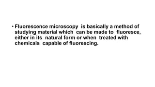
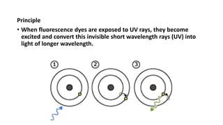
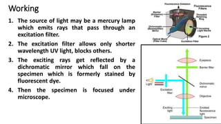
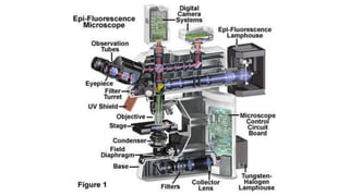
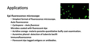
Fluorescence Microscope
Mr. Leyon Selvin Raj
Assistant Professor
Vivek Institute of Laboratory Medicine, Nagercoil
• Fluorescence microscopy is basically a method of
studying material which can be made to fluoresce,
either in its natural form or when treated with
chemicals capable of fluorescing.
Principle
• When fluorescence dyes are exposed to UV rays, they become
excited and convert this invisible short wavelength rays (UV) into
light of longer wavelength.
Working
1. The source of light may be a mercury lamp
which emits rays that pass through an
excitation filter.
2. The excitation filter allows only shorter
wavelength UV light, blocks others.
3. The exciting rays get reflected by a
dichromatic mirror which fall on the
specimen which is formerly stained by
fluorescent dye.
4. Then the specimen is focused under
microscope.
5. The fluorescent dye absorbs the exciting
rays and emits fluorescent rays of
higher wavelength.
6. A barrier filter positioned after the
objective lens removes the UV light,
which could damage the viewer’s eyes.
And also the blue/violet light which
would reduce the contrast.
Applications
Epi-fluorescence microscope
• Simplest format of fluorescence microscope.
Auto fluorescence
• Cyclospora – Auto fluoresce
Microbes coated with fluorescent dye
• Acridine orange- malaria parasite-quantitative buffy coat examination.
• Auramine phenol- detection of tubercle bacilli
Immunofluorescence
• Florescent dye tagged antigens or antibodies.
THANK YOU

Fluorescence microscope

  • 1.
    Fluorescence Microscope Mr. LeyonSelvin Raj Assistant Professor Vivek Institute of Laboratory Medicine, Nagercoil
  • 2.
    • Fluorescence microscopyis basically a method of studying material which can be made to fluoresce, either in its natural form or when treated with chemicals capable of fluorescing.
  • 3.
    Principle • When fluorescencedyes are exposed to UV rays, they become excited and convert this invisible short wavelength rays (UV) into light of longer wavelength.
  • 4.
    Working 1. The sourceof light may be a mercury lamp which emits rays that pass through an excitation filter. 2. The excitation filter allows only shorter wavelength UV light, blocks others. 3. The exciting rays get reflected by a dichromatic mirror which fall on the specimen which is formerly stained by fluorescent dye. 4. Then the specimen is focused under microscope.
  • 5.
    5. The fluorescentdye absorbs the exciting rays and emits fluorescent rays of higher wavelength. 6. A barrier filter positioned after the objective lens removes the UV light, which could damage the viewer’s eyes. And also the blue/violet light which would reduce the contrast.
  • 7.
    Applications Epi-fluorescence microscope • Simplestformat of fluorescence microscope. Auto fluorescence • Cyclospora – Auto fluoresce Microbes coated with fluorescent dye • Acridine orange- malaria parasite-quantitative buffy coat examination. • Auramine phenol- detection of tubercle bacilli Immunofluorescence • Florescent dye tagged antigens or antibodies.
  • 8.