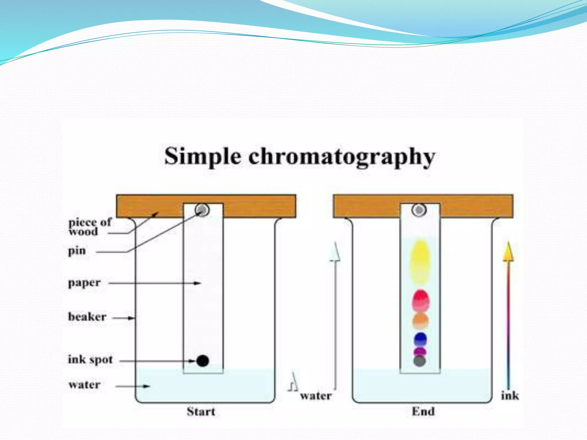
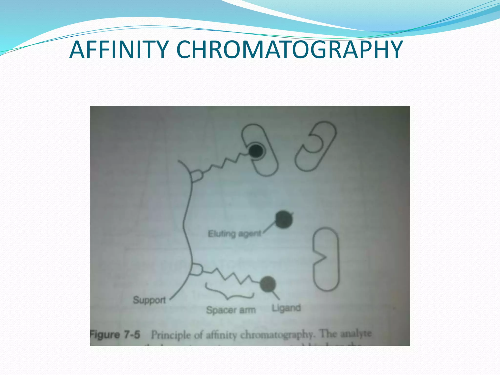
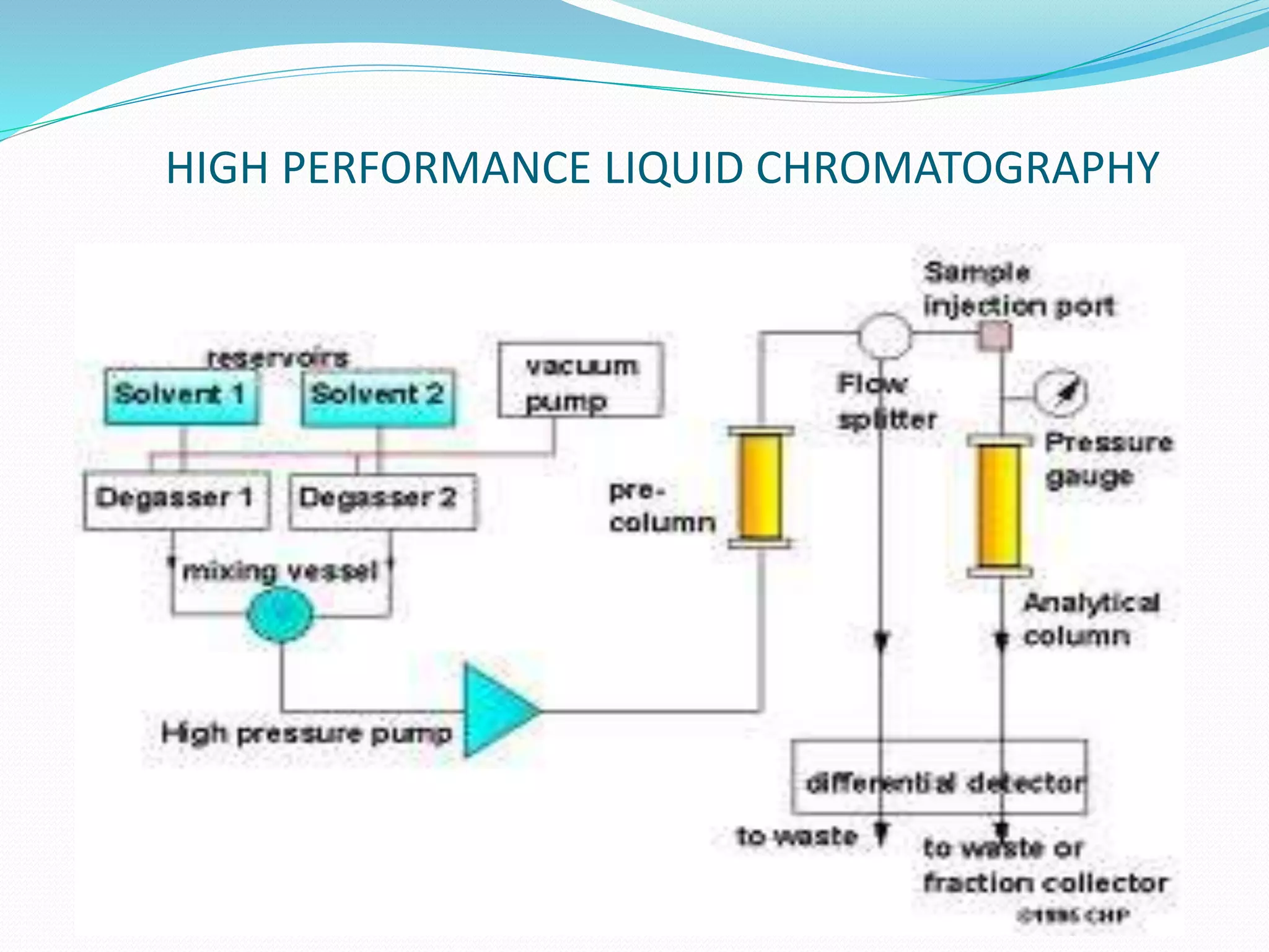
Chromatography is a technique used to separate mixtures based on how their components interact with both a mobile and stationary phase. It was first developed in 1900 by Russian scientist Mikhail Tsvet to separate plant pigments. There are several types of chromatography that differ based on the phases used, including paper chromatography, thin layer chromatography, gas chromatography, ion exchange chromatography, gel filtration chromatography, and affinity chromatography. High performance liquid chromatography is a modern technique that uses small particle sizes and high pressure to improve separation efficiency.
Sepration of molecules on the basis of applied Electric Field
Categorized into 1) Zone Electrophoresis 2) Moving Boundary Electrophoresis
We can seprate macromolecules (DNA , RNA, PROTEINS )on the basis of their charge, size shape & molecular weight
Sepration of molecules on the basis of applied Electric Field
Categorized into 1) Zone Electrophoresis 2) Moving Boundary Electrophoresis
We can seprate macromolecules (DNA , RNA, PROTEINS )on the basis of their charge, size shape & molecular weight
Spectroscopy techniques, it's principle, types and applications NizadSultana
Spectroscopy and it's applications as well as it's types like Infrared spectroscopy and ultraviolet spectroscopy and principle of spectroscopy why we use spectroscopy.
ION EXCHANGE CHROMATOGRAPHY
ByM.Vharshini
B.Sc. Bio Medical Science
Sri Ramachandra University
ION EXCHANGE CHROMATOGRAPHY
Ion-exchange chromatography is a process that allows the separation of ions and polar molecules based on their affinity to the ion exchanger.
It can be used for almost any kind of charged molecule including large proteins, small nucleotides and amino acids.
Cations or Anions can be separated using this method.
PRINCIPLE
It is based on the reversible electrostatic interaction of ions with the separation matrix (i.e.)
The separation occurs by reversible exchange of ions between the ions present in the solution and those present in the ion exchange resin.
CLASSIFICATION OF RESINS
According to the chemical nature they classified as-
1. Strong cation exchange resin
2. Weak cation exchange resin
3. Strong anion exchange resin
4. Weak anion exchange resin
According to the Source they can -
Natural resins : Cation - Zeolytes, Clay
Anion - Dolomite
Synthetic resins: Inorganic & Organic resins
◘Organic resins are polymeric resin matrix.
The resin composed of –
Polystyrene (sites for exchangeable functional groups)
Divinyl benzene(Cross linking agent)-offers stability.
Ion exchange resin should have following requirements
»It must be chemically stable.
»It should be insoluble in common solvents.
» It should have a sufficient degree of cross linking.
»The swollen resin must be denser than water.
»It must contain sufficient no. of ion exchange groups.
Physical properties of ion exchange resins
Cross linking:
It affects swelling & strength & solubility
Swelling:
When resin swells, polymer chain spreads apart
Polar solvents → swelling
Non-polar solvents → contraction
Swelling also affected electrolyte concentration.
Particle size and porosity
Increase in surface area & decrease in particle size will increase the rate of ion exchange.
Regeneration
Cation exchange resin are regenerated by treatment with acid, then washing with water.
Anion exchange resin are regenerated by treatment with NaOH, then washing with water until neutral.
EXPERIMENTAL SETUP OF ION EXCHANGE CHROMATOGRAPHY
Metrohm 850 Ion chromatography system
Instrumentation of ion exchange chromatography
PRACTICAL REQUIREMENTS
1.Column
» glass, stainless steel or polymers
2.Packing the column
» Wet packing method:
A slurry is prepared of the eluent with the stationary phase powder and then carefully poured into the column. Care must be taken to avoid air bubbles.
3.Application of the sample
After packing, sample is added to the top of the stationary phase, use syringe or pipette.
This layer is usually topped with a small layer of sand or with cotton or glass wool to protect the shape of the organic layer from the velocity of newly added eluent.
4.Mobile phase
Acids, alkalis, buffers…
6.Stationary phase
The ionic
Electrophoresis is the movement of charged particles through an electrode when subjected to an electric Field
Cations move towards cathode
Anions move towards anode
By this technique solutes are separated by their different rates of travel through an electric field.
Commonly used in biological analysis, particularly in the separations of proteins, peptides and nucleic acids
HPLC Principle,Instrumentation and ApplicationAlakesh Pradhan
HPLC Chromatography and its principle
Liquid chromatography
High Performance Liquid Chromatography ( HPLC )
The components of the high performance liquid chromatograph (HPLC).
The separation process.
The chromatogram
• Chromatography is a method of separation in which the components to be separated are distributed between two phases, one of these is called a stationary phase and the other is a mobile phase which moves on stationary phase in a definite direction
Chromatography is based on the principle where molecules in mixture applied onto the surface or into the solid, and fluid stationary phase (stable phase) is separating from each other while moving with the aid of a mobile phase.
The factors effective on this separation process include molecular characteristics related to adsorption (liquid-solid), partition (liquid-solid), and affinity or differences among their molecular weights
Because of these differences, some components of the mixture stay longer in the stationary phase, and they move slowly in the chromatography system, while others pass rapidly into mobile phase, and leave the system faster.
Slide 1: Title Slide
Extrachromosomal Inheritance
Slide 2: Introduction to Extrachromosomal Inheritance
Definition: Extrachromosomal inheritance refers to the transmission of genetic material that is not found within the nucleus.
Key Components: Involves genes located in mitochondria, chloroplasts, and plasmids.
Slide 3: Mitochondrial Inheritance
Mitochondria: Organelles responsible for energy production.
Mitochondrial DNA (mtDNA): Circular DNA molecule found in mitochondria.
Inheritance Pattern: Maternally inherited, meaning it is passed from mothers to all their offspring.
Diseases: Examples include Leber’s hereditary optic neuropathy (LHON) and mitochondrial myopathy.
Slide 4: Chloroplast Inheritance
Chloroplasts: Organelles responsible for photosynthesis in plants.
Chloroplast DNA (cpDNA): Circular DNA molecule found in chloroplasts.
Inheritance Pattern: Often maternally inherited in most plants, but can vary in some species.
Examples: Variegation in plants, where leaf color patterns are determined by chloroplast DNA.
Slide 5: Plasmid Inheritance
Plasmids: Small, circular DNA molecules found in bacteria and some eukaryotes.
Features: Can carry antibiotic resistance genes and can be transferred between cells through processes like conjugation.
Significance: Important in biotechnology for gene cloning and genetic engineering.
Slide 6: Mechanisms of Extrachromosomal Inheritance
Non-Mendelian Patterns: Do not follow Mendel’s laws of inheritance.
Cytoplasmic Segregation: During cell division, organelles like mitochondria and chloroplasts are randomly distributed to daughter cells.
Heteroplasmy: Presence of more than one type of organellar genome within a cell, leading to variation in expression.
Slide 7: Examples of Extrachromosomal Inheritance
Four O’clock Plant (Mirabilis jalapa): Shows variegated leaves due to different cpDNA in leaf cells.
Petite Mutants in Yeast: Result from mutations in mitochondrial DNA affecting respiration.
Slide 8: Importance of Extrachromosomal Inheritance
Evolution: Provides insight into the evolution of eukaryotic cells.
Medicine: Understanding mitochondrial inheritance helps in diagnosing and treating mitochondrial diseases.
Agriculture: Chloroplast inheritance can be used in plant breeding and genetic modification.
Slide 9: Recent Research and Advances
Gene Editing: Techniques like CRISPR-Cas9 are being used to edit mitochondrial and chloroplast DNA.
Therapies: Development of mitochondrial replacement therapy (MRT) for preventing mitochondrial diseases.
Slide 10: Conclusion
Summary: Extrachromosomal inheritance involves the transmission of genetic material outside the nucleus and plays a crucial role in genetics, medicine, and biotechnology.
Future Directions: Continued research and technological advancements hold promise for new treatments and applications.
Slide 11: Questions and Discussion
Invite Audience: Open the floor for any questions or further discussion on the topic.
Introduction:
RNA interference (RNAi) or Post-Transcriptional Gene Silencing (PTGS) is an important biological process for modulating eukaryotic gene expression.
It is highly conserved process of posttranscriptional gene silencing by which double stranded RNA (dsRNA) causes sequence-specific degradation of mRNA sequences.
dsRNA-induced gene silencing (RNAi) is reported in a wide range of eukaryotes ranging from worms, insects, mammals and plants.
This process mediates resistance to both endogenous parasitic and exogenous pathogenic nucleic acids, and regulates the expression of protein-coding genes.
What are small ncRNAs?
micro RNA (miRNA)
short interfering RNA (siRNA)
Properties of small non-coding RNA:
Involved in silencing mRNA transcripts.
Called “small” because they are usually only about 21-24 nucleotides long.
Synthesized by first cutting up longer precursor sequences (like the 61nt one that Lee discovered).
Silence an mRNA by base pairing with some sequence on the mRNA.
Discovery of siRNA?
The first small RNA:
In 1993 Rosalind Lee (Victor Ambros lab) was studying a non- coding gene in C. elegans, lin-4, that was involved in silencing of another gene, lin-14, at the appropriate time in the
development of the worm C. elegans.
Two small transcripts of lin-4 (22nt and 61nt) were found to be complementary to a sequence in the 3' UTR of lin-14.
Because lin-4 encoded no protein, she deduced that it must be these transcripts that are causing the silencing by RNA-RNA interactions.
Types of RNAi ( non coding RNA)
MiRNA
Length (23-25 nt)
Trans acting
Binds with target MRNA in mismatch
Translation inhibition
Si RNA
Length 21 nt.
Cis acting
Bind with target Mrna in perfect complementary sequence
Piwi-RNA
Length ; 25 to 36 nt.
Expressed in Germ Cells
Regulates trnasposomes activity
MECHANISM OF RNAI:
First the double-stranded RNA teams up with a protein complex named Dicer, which cuts the long RNA into short pieces.
Then another protein complex called RISC (RNA-induced silencing complex) discards one of the two RNA strands.
The RISC-docked, single-stranded RNA then pairs with the homologous mRNA and destroys it.
THE RISC COMPLEX:
RISC is large(>500kD) RNA multi- protein Binding complex which triggers MRNA degradation in response to MRNA
Unwinding of double stranded Si RNA by ATP independent Helicase
Active component of RISC is Ago proteins( ENDONUCLEASE) which cleave target MRNA.
DICER: endonuclease (RNase Family III)
Argonaute: Central Component of the RNA-Induced Silencing Complex (RISC)
One strand of the dsRNA produced by Dicer is retained in the RISC complex in association with Argonaute
ARGONAUTE PROTEIN :
1.PAZ(PIWI/Argonaute/ Zwille)- Recognition of target MRNA
2.PIWI (p-element induced wimpy Testis)- breaks Phosphodiester bond of mRNA.)RNAse H activity.
MiRNA:
The Double-stranded RNAs are naturally produced in eukaryotic cells during development, and they have a key role in regulating gene expression .
(May 29th, 2024) Advancements in Intravital Microscopy- Insights for Preclini...Scintica Instrumentation
Intravital microscopy (IVM) is a powerful tool utilized to study cellular behavior over time and space in vivo. Much of our understanding of cell biology has been accomplished using various in vitro and ex vivo methods; however, these studies do not necessarily reflect the natural dynamics of biological processes. Unlike traditional cell culture or fixed tissue imaging, IVM allows for the ultra-fast high-resolution imaging of cellular processes over time and space and were studied in its natural environment. Real-time visualization of biological processes in the context of an intact organism helps maintain physiological relevance and provide insights into the progression of disease, response to treatments or developmental processes.
In this webinar we give an overview of advanced applications of the IVM system in preclinical research. IVIM technology is a provider of all-in-one intravital microscopy systems and solutions optimized for in vivo imaging of live animal models at sub-micron resolution. The system’s unique features and user-friendly software enables researchers to probe fast dynamic biological processes such as immune cell tracking, cell-cell interaction as well as vascularization and tumor metastasis with exceptional detail. This webinar will also give an overview of IVM being utilized in drug development, offering a view into the intricate interaction between drugs/nanoparticles and tissues in vivo and allows for the evaluation of therapeutic intervention in a variety of tissues and organs. This interdisciplinary collaboration continues to drive the advancements of novel therapeutic strategies.
THE IMPORTANCE OF MARTIAN ATMOSPHERE SAMPLE RETURN.Sérgio Sacani
The return of a sample of near-surface atmosphere from Mars would facilitate answers to several first-order science questions surrounding the formation and evolution of the planet. One of the important aspects of terrestrial planet formation in general is the role that primary atmospheres played in influencing the chemistry and structure of the planets and their antecedents. Studies of the martian atmosphere can be used to investigate the role of a primary atmosphere in its history. Atmosphere samples would also inform our understanding of the near-surface chemistry of the planet, and ultimately the prospects for life. High-precision isotopic analyses of constituent gases are needed to address these questions, requiring that the analyses are made on returned samples rather than in situ.
The increased availability of biomedical data, particularly in the public domain, offers the opportunity to better understand human health and to develop effective therapeutics for a wide range of unmet medical needs. However, data scientists remain stymied by the fact that data remain hard to find and to productively reuse because data and their metadata i) are wholly inaccessible, ii) are in non-standard or incompatible representations, iii) do not conform to community standards, and iv) have unclear or highly restricted terms and conditions that preclude legitimate reuse. These limitations require a rethink on data can be made machine and AI-ready - the key motivation behind the FAIR Guiding Principles. Concurrently, while recent efforts have explored the use of deep learning to fuse disparate data into predictive models for a wide range of biomedical applications, these models often fail even when the correct answer is already known, and fail to explain individual predictions in terms that data scientists can appreciate. These limitations suggest that new methods to produce practical artificial intelligence are still needed.
In this talk, I will discuss our work in (1) building an integrative knowledge infrastructure to prepare FAIR and "AI-ready" data and services along with (2) neurosymbolic AI methods to improve the quality of predictions and to generate plausible explanations. Attention is given to standards, platforms, and methods to wrangle knowledge into simple, but effective semantic and latent representations, and to make these available into standards-compliant and discoverable interfaces that can be used in model building, validation, and explanation. Our work, and those of others in the field, creates a baseline for building trustworthy and easy to deploy AI models in biomedicine.
Bio
Dr. Michel Dumontier is the Distinguished Professor of Data Science at Maastricht University, founder and executive director of the Institute of Data Science, and co-founder of the FAIR (Findable, Accessible, Interoperable and Reusable) data principles. His research explores socio-technological approaches for responsible discovery science, which includes collaborative multi-modal knowledge graphs, privacy-preserving distributed data mining, and AI methods for drug discovery and personalized medicine. His work is supported through the Dutch National Research Agenda, the Netherlands Organisation for Scientific Research, Horizon Europe, the European Open Science Cloud, the US National Institutes of Health, and a Marie-Curie Innovative Training Network. He is the editor-in-chief for the journal Data Science and is internationally recognized for his contributions in bioinformatics, biomedical informatics, and semantic technologies including ontologies and linked data.
Multi-source connectivity as the driver of solar wind variability in the heli...Sérgio Sacani
The ambient solar wind that flls the heliosphere originates from multiple
sources in the solar corona and is highly structured. It is often described
as high-speed, relatively homogeneous, plasma streams from coronal
holes and slow-speed, highly variable, streams whose source regions are
under debate. A key goal of ESA/NASA’s Solar Orbiter mission is to identify
solar wind sources and understand what drives the complexity seen in the
heliosphere. By combining magnetic feld modelling and spectroscopic
techniques with high-resolution observations and measurements, we show
that the solar wind variability detected in situ by Solar Orbiter in March
2022 is driven by spatio-temporal changes in the magnetic connectivity to
multiple sources in the solar atmosphere. The magnetic feld footpoints
connected to the spacecraft moved from the boundaries of a coronal hole
to one active region (12961) and then across to another region (12957). This
is refected in the in situ measurements, which show the transition from fast
to highly Alfvénic then to slow solar wind that is disrupted by the arrival of
a coronal mass ejection. Our results describe solar wind variability at 0.5 au
but are applicable to near-Earth observatories.
A brief information about the SCOP protein database used in bioinformatics.
The Structural Classification of Proteins (SCOP) database is a comprehensive and authoritative resource for the structural and evolutionary relationships of proteins. It provides a detailed and curated classification of protein structures, grouping them into families, superfamilies, and folds based on their structural and sequence similarities.
2. Chromatography
Chromatography (from Greek chroma "color
and graphein "to write") is the collective term for a set
of laboratory techniques for the separation of
mixtures. The mixture is dissolved in a fluid called
the mobile phase, which carries it through a structure
holding another material called the stationary
phase. The various constituents of the mixture travel at
different speeds, causing them to separate. The
separation is based on differential partitioning
between the mobile and stationary phases.
3. Chromatography, literally "color writing", was first employed
by Russian scientist Mikhail Tsvet in 1900. He continued to
work with chromatography in the first decade of the 20th
century, primarily for the separation of plant pigments such
as chlorophyll, carotenes, and xanthophylls. Since these
components have different colors (green, orange, and
yellow,respectively) they gave the technique its name.
4. PRINCIPLES
Chromatography usually consists of mobile phase and
stationary phase.The mobile phase refers to the
mixture of substances to be separated dissolved in a
liquid or a gas.The stationary phase is a porous solid
matrix through which the sample contained in the
mobile phase percolates.The interaction between the
mobile phase and the stationary phase results in the
separation of the compound from the mixture.
5. APPLICATIONS OF CHROMATOGRAPHY
The chromatographic technique is used for the
separation of amino acids,proteins & carbohydrates.
It is also used for the analysis of
drugs,hormones,vitamins
Helpful for the qualitative & quantitative analysis of
complex mixtures.
The technique is also useful for the determination of
molecular weight of proteins.
6. Types of Chromatography
There are following types of Chromatography
Paper Chromatography
Thin Layer Chromatography(TLC)
Gel Chromatography
Column Chromatography
Ion Exchange Chromatography
Gel Filtration Chromatography
Gas Liquid Chromatography
Affinity Chromatography
7. Paper chromatography
Paper chromatography is a technique that involves
placing a small dot or line of sample solution onto a
strip ofchromatography paper. The paper is placed in a
jar containing a shallow layer of solvent and sealed. As
the solvent rises through the paper, it meets the
sample mixture, which starts to travel up the paper
with the solvent.
8.
9.
10.
Rf= Distance travelled by the substance
Distance travelled by the solvent front
The Rf value helps for the identification of unknown.
11. Sometimes,it is rather difficult to separate a
complex mixture of substances by a single run with
one solvent system. In such a case, a second run is
carried out by a different solvent system, in a
direction perpendicular to the first run. This is
referred to as two dimensional chromatography.
12.
13. Thin layer chromatography
Thin layer chromatography (TLC) is a widely employed
laboratory technique and is similar to paper
chromatography. However, instead of using a
stationary phase of paper, it involves a stationary phase
of a thin layer of adsorbent like silica gel, alumina,
or cellulose . Compared to paper, it has the advantage
of faster runs, better separations, and the choice
between different adsorbents.
15. GAS-LIQUID CHROMATOGRAPHY
Gas chromatography (GC), also sometimes known as Gas-Liquid chromatography, (GLC),
is a separation technique in which the mobile phase is a gas.It is the method of choice for
the separation of volatile substances or the volatile derivatives of certain non-volatile
substances.
Stationary phase is an inert solid material impregnated with a non-volatile liquid.
In gas chromatography, a sample is rapidly heated and vaporized at the injection port.
The sample is transported through the column by a mobile phase consisiting of an inert
gas. Sample components are separated based on their boiling points and relative affinity
for the stationary phase, which is most often a viscous liquid (wax) within the column.
The higher a component's affinity for the stationary phase, the slower it comes off the
column. The components are then detected and represented as peaks on a
chromatogram.
16. The mixture of volatile material is injected into the
column along with the mobile phase.
The separation of the volatile mixture is based on the
partition of the components between the mobile
phase(gas) and stationary phase (liq.), hence the name
GAS-LIQUID CHROMATOGRAPHY.
17.
18. It is well suited for use in
the petrochemical,environmental monitoring and
industrial chemical fields.
Sensitive, rapid and reliable.
20. ADSORPTION COLUMN
CHROMATOGRAPHY
Column chromatography is a separation technique in
which the stationary bed is within a tube.Adsorbents
are packed into a column in a glass tube.This serves as
the stationary phase,leaving an open unrestricted path
for the mobile phase in the middle of the tube.
Adsorbents such as silica gel, alumina, charcoal
powder & calcium hydroxyapatite are used.
21. The sample mixture in a solvent is loaded on this
column.The individual compounds get differentially
adsorbed on to the adsorbent.
The elution is carried out by the buffer system which is
the mobile phase.
The individual compounds come out of the column at
different rates which may be collected separately &
identified.
23. ION EXCHANGE CHROMATOGRAPHY
Ion exchange chromatography (usually referred to as
ion chromatography) uses an ion exchange
mechanism to separate molecules on the basis of their
electrical charges.Ion exchange chromatography uses a
charged stationary phase to separate charged
compounds including anions, cations, amino
acids, peptides, and proteins.
24. Cation exchangers & anion exchangers are used as ion
exchange resins.
In conventional methods the stationary phase is
an ion exchange resin that carries charged functional
groups that interact with oppositely charged groups of
the compound to retain.
26. GEL FILTRATION CHROMATOGRAPHY
Size-exclusion chromatography (SEC) is also known
as gel permeation chromatography (GPC) or gel
filtration chromatography and separates molecules
according to their size, shape & molecular weight.
It is also referred to as molecular sieving or molecular
exclusion chromatography.
27. Smaller molecules are able to enter the pores of the
media and, therefore, molecules are trapped and
removed from the flow of the mobile phase. However,
molecules that are larger than the average pore size of
the packing are excluded and thus suffer essentially no
retention; such species are the first to be eluted. This is
how the molecules are separated.
28. It is generally a low-resolution chromatography
technique and thus it is often reserved for the final,
"polishing" step of a purification. It is also useful for
determining the tertiary structure and quaternary
structure of purified proteins, especially since it can be
carried out under native solution conditions.
30. AFFINITY CHROMATOGRAPHY
Affinity chromatographyis based on selective non-
covalent interaction between an analyte and specific
molecules, referred to as ligands.
The immobilized ligands act as molecular fish-hooks
& selectively pick up desired proteins while the
remaining protein pass through the column.
33. HIGH PERFORMANCE LIQUID
CHROMATOGRAPHY (HPLC )
Liquid chromatography (LC) is a separation technique in
which the mobile phase is a liquid. Liquid chromatography
can be carried out either in a column or a plane. Present
day liquid chromatography that generally utilizes very
small packing particles and a relatively high pressure is
referred to as high performance liquid
chromatography (HPLC).
Since the chromatographic techniques are slow & time
consuming,hence the separation can be greatly improved
by using high pressure in the range of 5000-10000
psi(pounds per square inch),hence this technique is also
referred to as high pressure liquid chromatography.
34. In HPLC the sample is forced by a liquid at high
pressure (the mobile phase) through a column that is
packed with a stationary phase composed of
irregularly or spherically shaped particles.
The interaction between the mobile and the stationary
phase leads to the separation of the mixture.