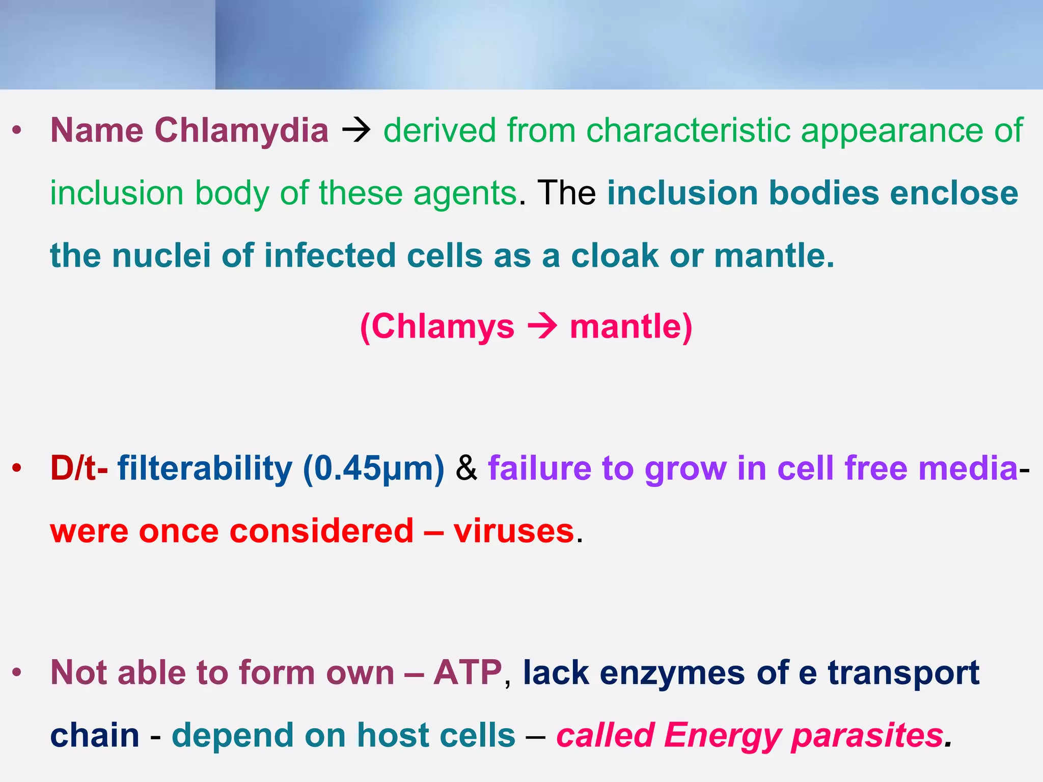
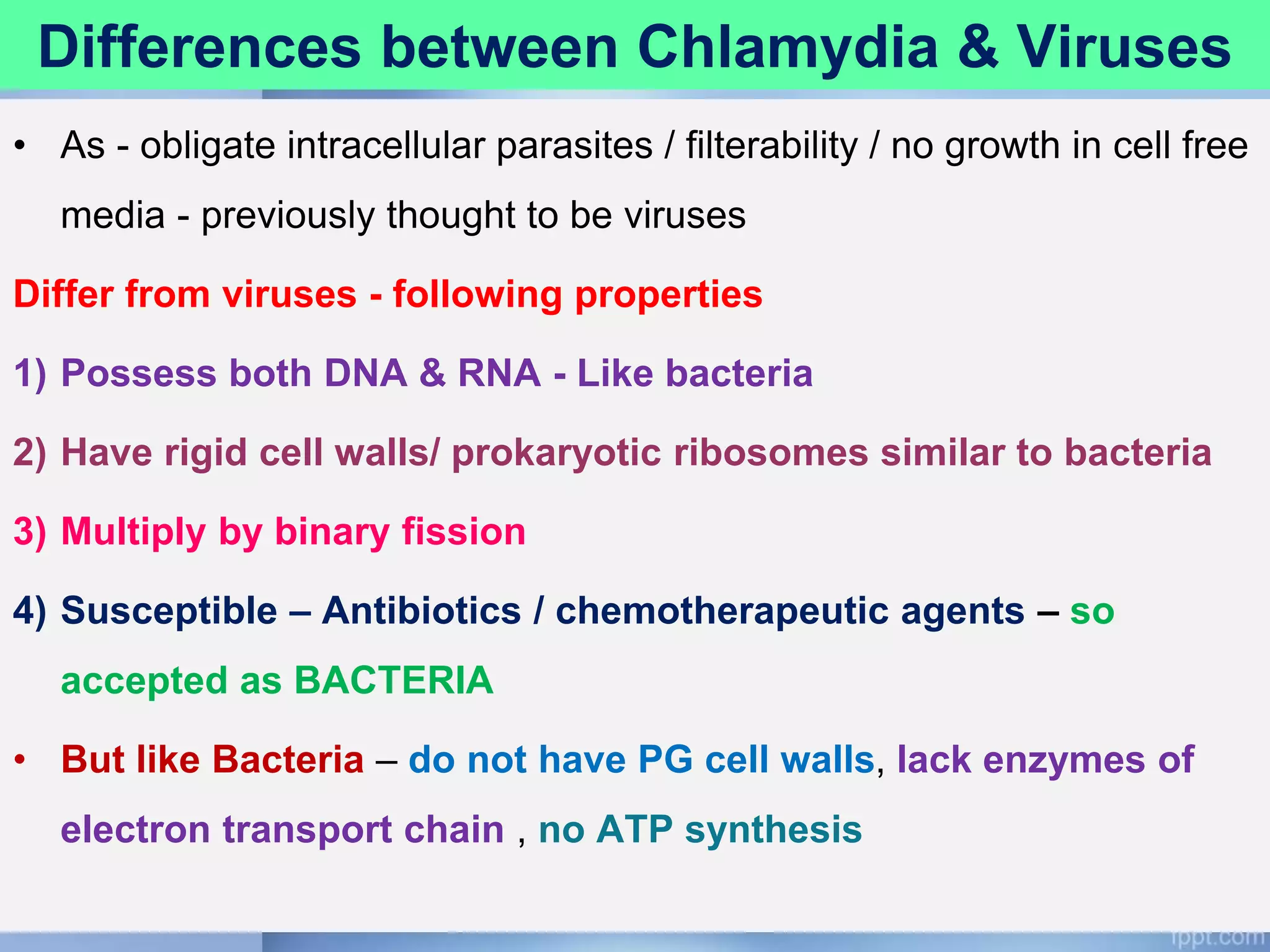
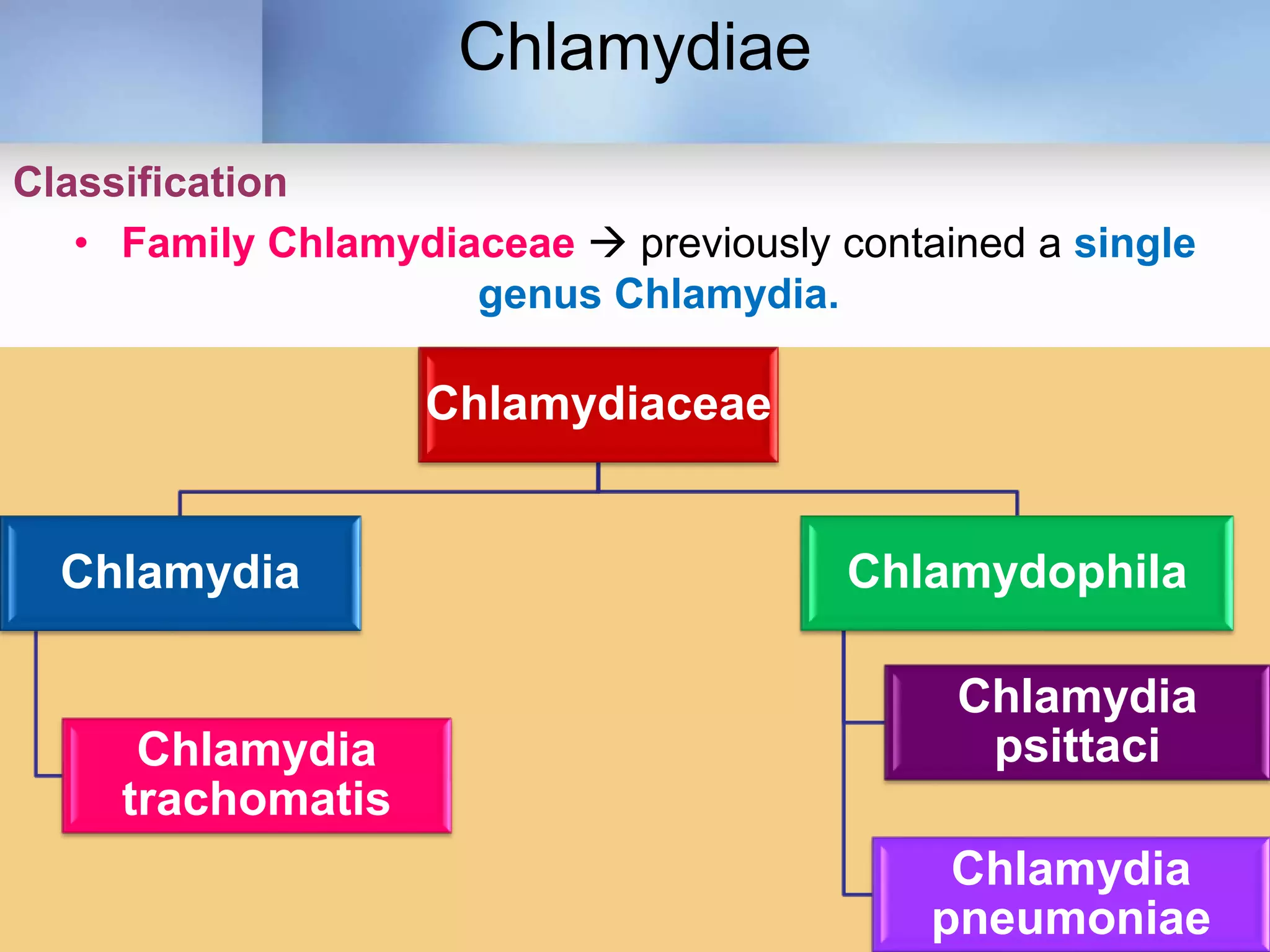
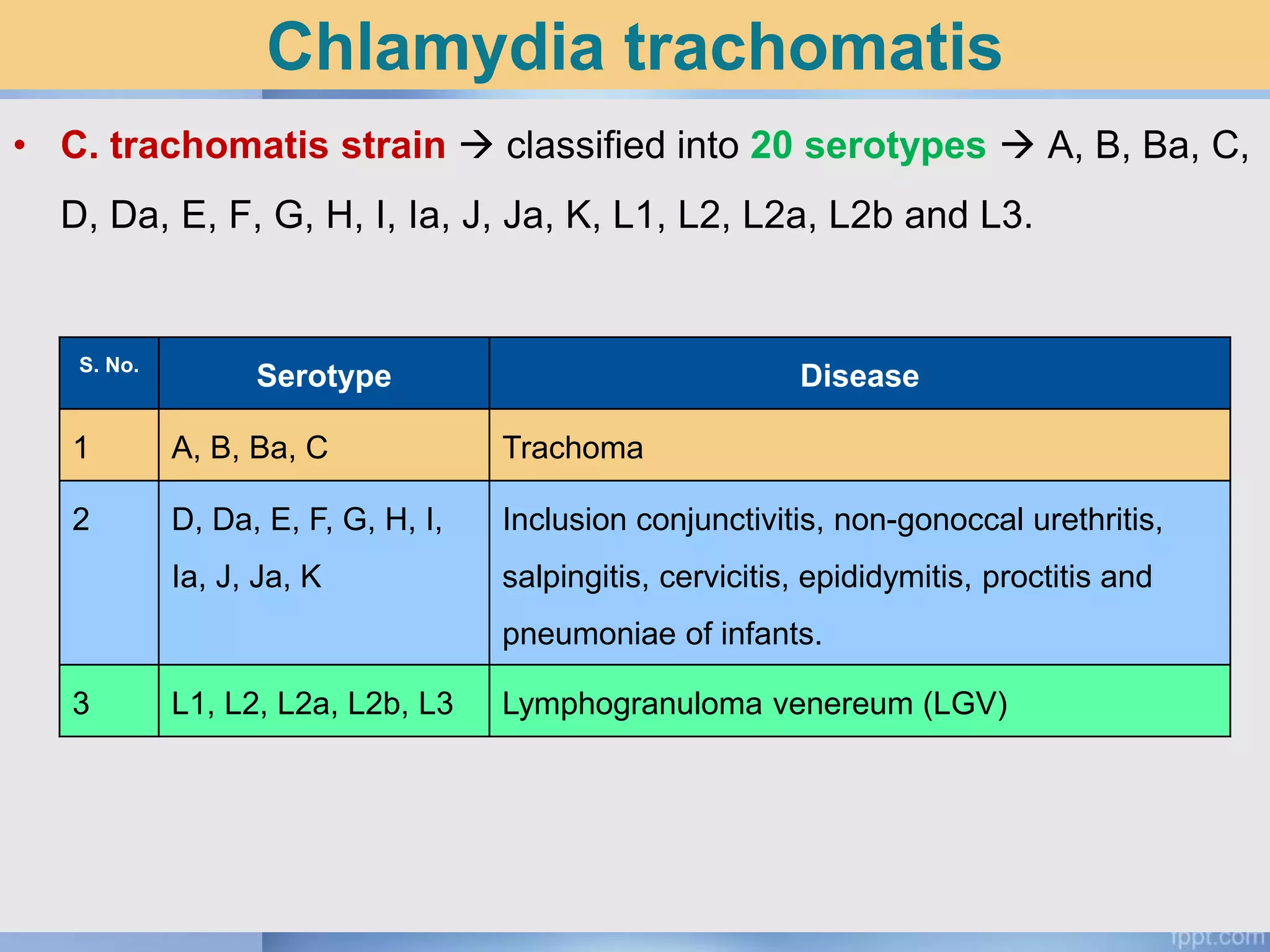
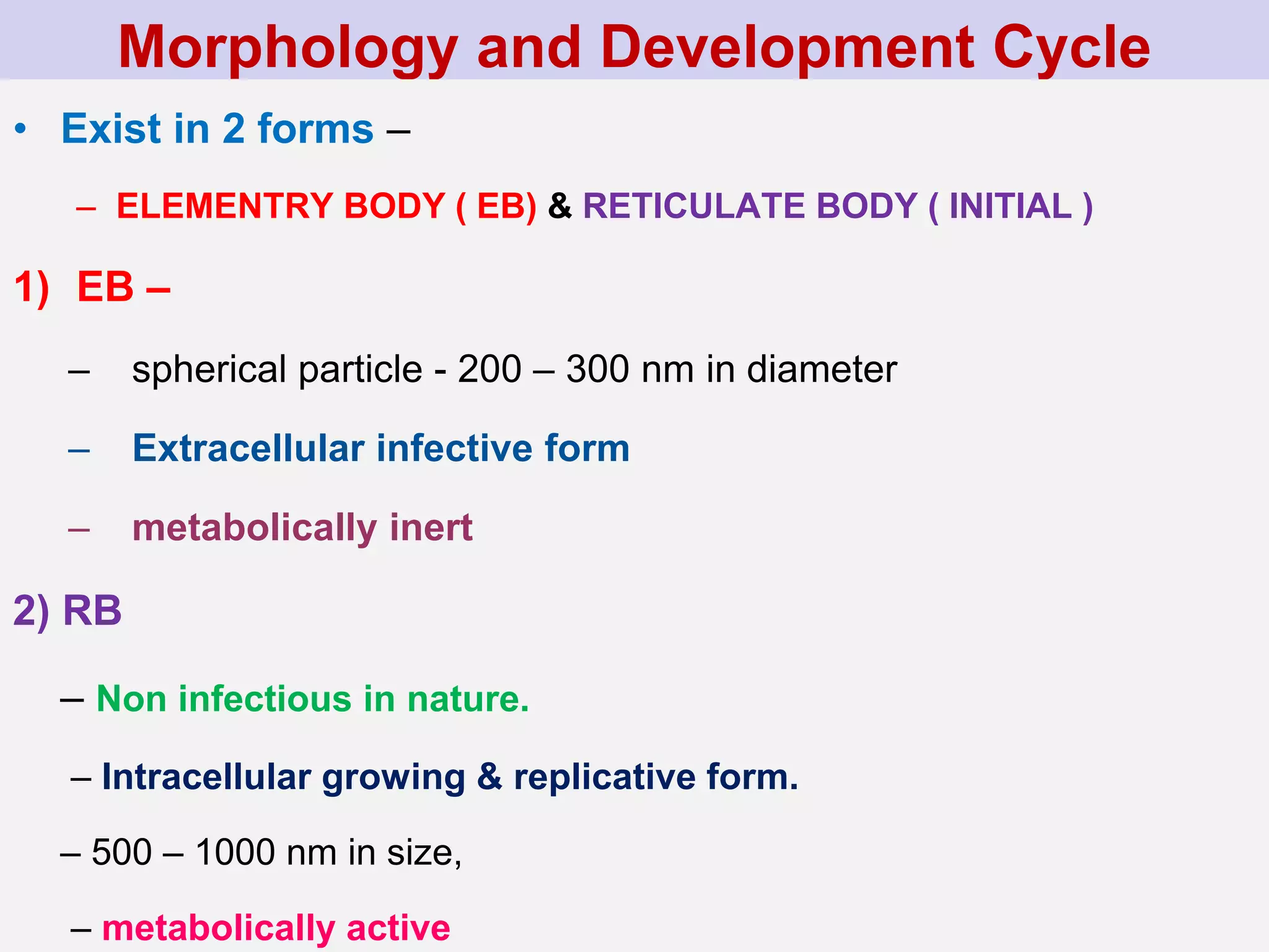
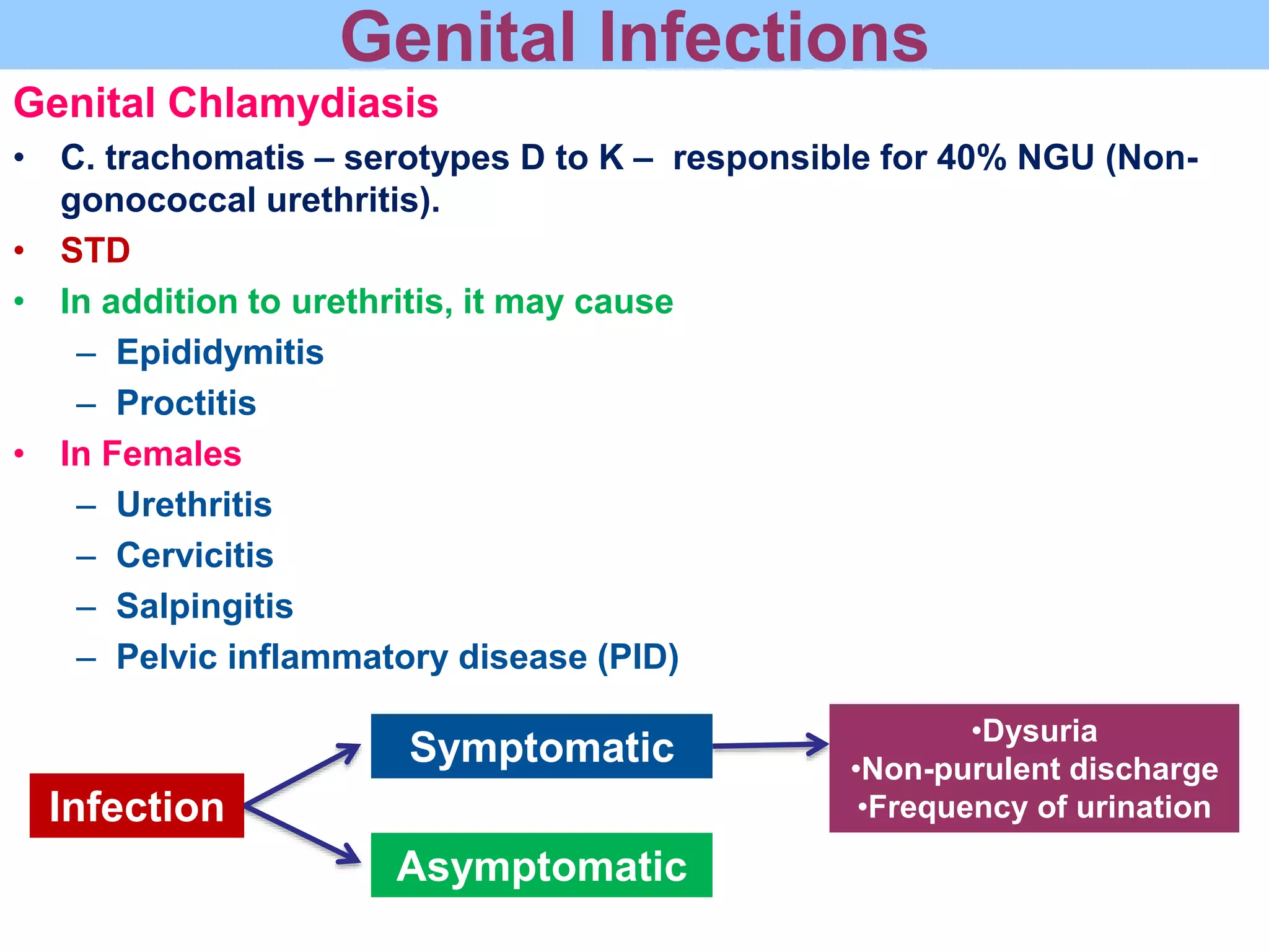
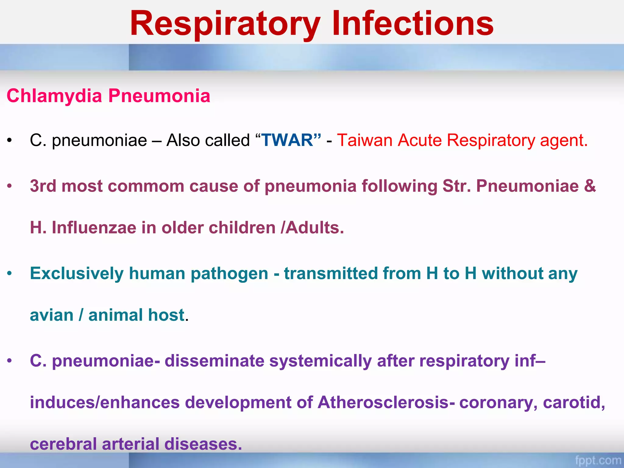
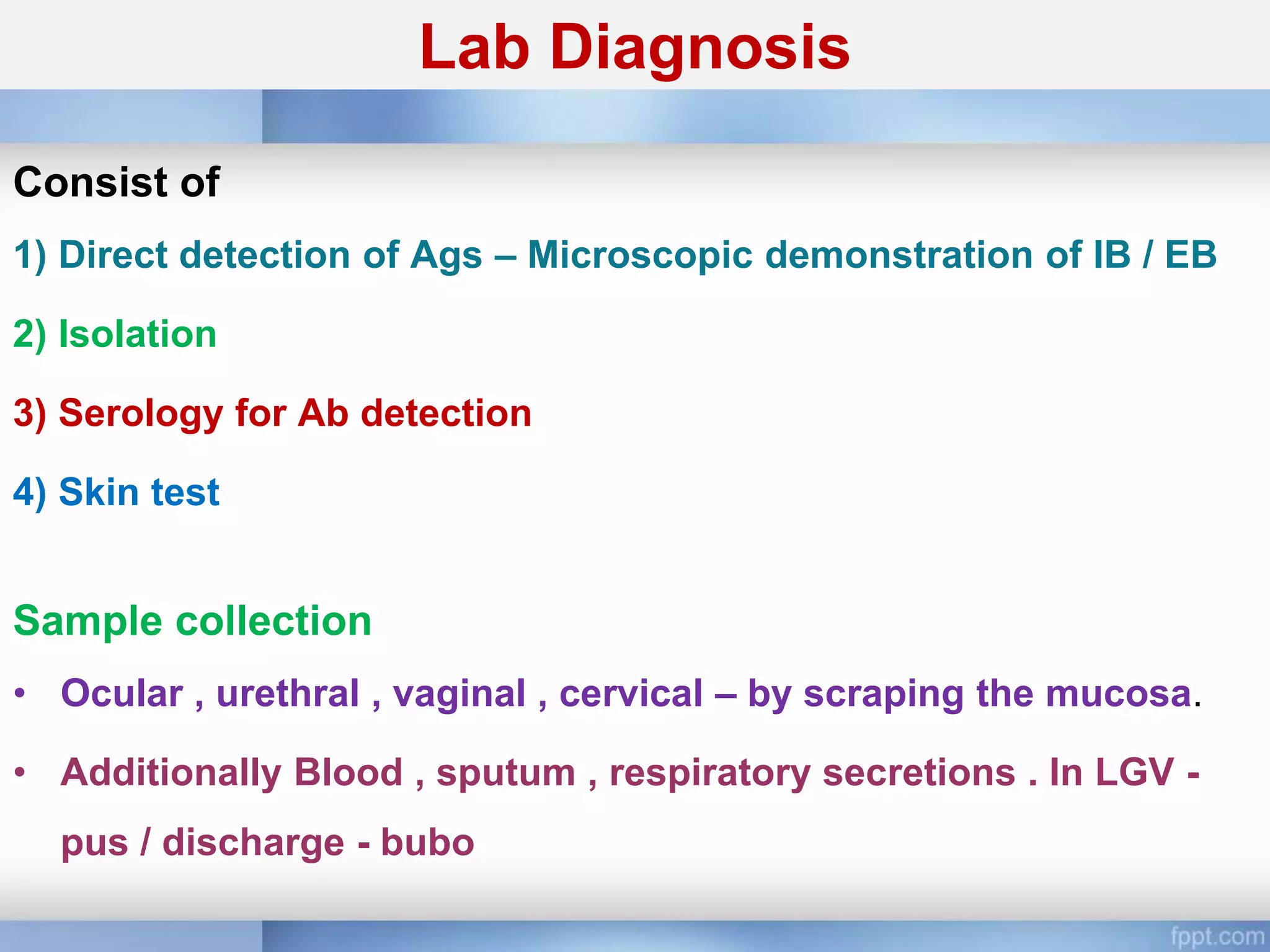
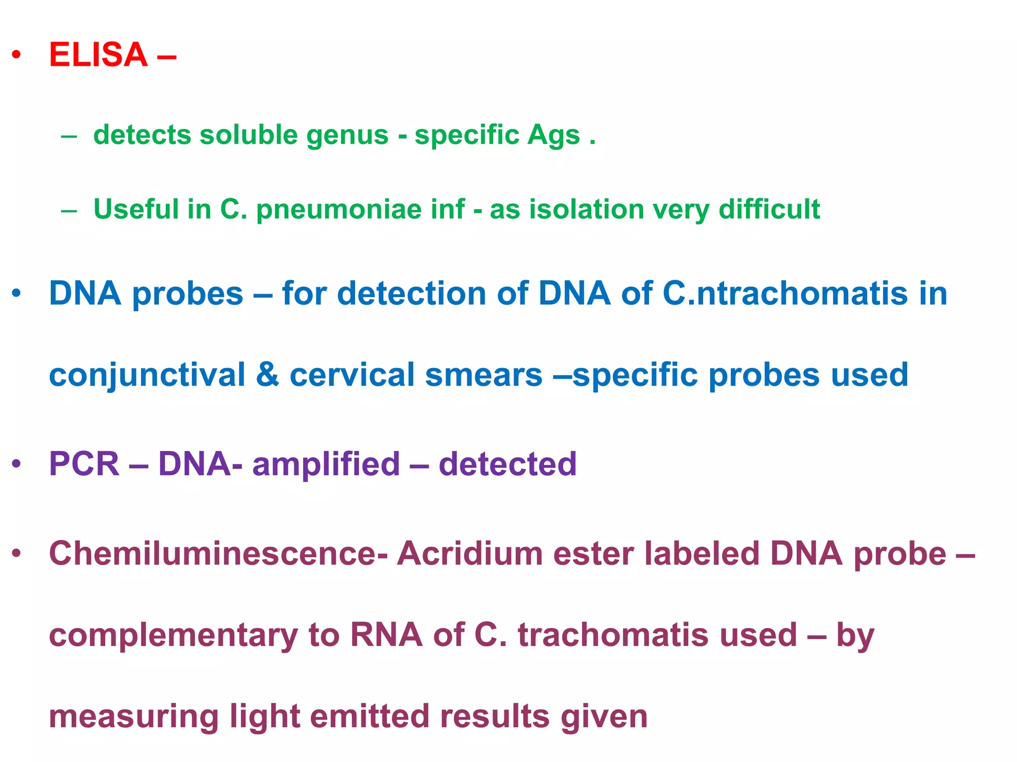
Chlamydia
By Dr. Rakesh Prasad Sah
Associate Professor, Microbiology
Introduction
• Obligate intracellular parasites
– Small
– Gram-negative
– Non-motile
• Causes
– Trachoma
– Lymphogranuloma venerum (LGV)
– Psittacosis in man
– Were called  “psittacosis-lymphogranuloma-trachoma (PLT)” or
“TRIC (trachoma-inclusion conjunctivitis) organisms”.
• Name Chlamydia  derived from characteristic appearance of
inclusion body of these agents. The inclusion bodies enclose
the nuclei of infected cells as a cloak or mantle.
(Chlamys  mantle)
• D/t- filterability (0.45μm) & failure to grow in cell free media-
were once considered – viruses.
• Not able to form own – ATP, lack enzymes of e transport
chain - depend on host cells – called Energy parasites.
Differences between Chlamydia & Viruses
• As - obligate intracellular parasites / filterability / no growth in cell free
media - previously thought to be viruses
Differ from viruses - following properties
1) Possess both DNA & RNA - Like bacteria
2) Have rigid cell walls/ prokaryotic ribosomes similar to bacteria
3) Multiply by binary fission
4) Susceptible – Antibiotics / chemotherapeutic agents – so
accepted as BACTERIA
• But like Bacteria – do not have PG cell walls, lack enzymes of
electron transport chain , no ATP synthesis
Chlamydiae
Classification
• Family Chlamydiaceae  previously contained a single
genus Chlamydia.
Chlamydiaceae
Chlamydia
Chlamydia
trachomatis
Chlamydophila
Chlamydia
psittaci
Chlamydia
pneumoniae
Chlamydia trachomatis
• C. trachomatis strain  classified into 20 serotypes  A, B, Ba, C,
D, Da, E, F, G, H, I, Ia, J, Ja, K, L1, L2, L2a, L2b and L3.
S. No.
Serotype Disease
1 A, B, Ba, C Trachoma
2 D, Da, E, F, G, H, I,
Ia, J, Ja, K
Inclusion conjunctivitis, non-gonoccal urethritis,
salpingitis, cervicitis, epididymitis, proctitis and
pneumoniae of infants.
3 L1, L2, L2a, L2b, L3 Lymphogranuloma venereum (LGV)
Chlamydophila pneumoniae
• Formerly  “TWAR strain”
• Causes
– Community acquired atypical pneumoniae
– atherosclerosis
S.No Species Serotype Disease
1 C. Psittaci Many
serotypes
Psittacosis (atypical interstitial
pneumonia)
2 C. Trachomatis A, B, Ba, C Trachoma
3 C. Trachomatis D, Da, E, F, G,
H, I, Ia, J, Ja, K
•Inclusion conjunctivitis
•Genital chlamydiasis
•Infant pneumonia
4 C. Trachomatis L1, L2, L2a,
L2b, L3
Lymphogranuloma venereum
5 C. Pneumoniae One serotype •Community acquired atypical
pneumoniae
•Associated with atherosclerosis
Morphology and Development Cycle
• Exist in 2 forms –
– ELEMENTRY BODY ( EB) & RETICULATE BODY ( INITIAL )
1) EB –
– spherical particle - 200 – 300 nm in diameter
– Extracellular infective form
– metabolically inert
2) RB
– Non infectious in nature.
– Intracellular growing & replicative form.
– 500 – 1000 nm in size,
– metabolically active
• Chlamydia – EB/ IB – large enough to be seen – L. microscope
• Chlamydia is GM –ve , other better stains
– Castaneda
– Machiavello
– Giemsa
• C. trachomatis stained by – Lugol’s iodine – glycogen matrix
Host Cell
Phagocytosis
Elementary
body
Release of Elementary Bodies Reticulate
body
Cycle of development
Attachment- to surface of
susceptible Epithelial cell
Enters the host cell by phagocytosis/
endocytosis –
Once internalized remain in
phagosome –Prevent
phagosome Lysosome fusion
Enlarges / under go reorganization -
Reticulate body
( RB ) - 500 – 1000 nm in size
Some RB become EB
Liberated from host cell by cell lysis
Divides rapidly by B. F. -- To
produce large no of RB
( newly formed infectious particles
within host cell - IB )
Mature IB -100 -500 RB .
Duration
is
24-48hrs
Ag Structure
1) Genus sp Ags
• Heat stable - common to all chlamydiae.
• LPS in nature.
• Similar to LPS of GNB.
• Present in all stages of the developmental cycle.
• Identified by CFT.
2) Species specific protein Ags
• at envelope surface-
• Help in classifying chlamydiae in to different species
3) Serotype specific Ags
• Helps in intraspecies typing
• Ex- C. trachomatis – 15 serotypes A- K & L1 – L3
• located on MOMP(Major outer membrane protein)- can be demonstrated
by – micro IF
RESISTANCE
• Heat labile – inactivated with in minutes at 56oc
• Susceptible to ethanol, ether , phenol & formalin
• Infectivity is maintained for several days at 4oc
• Can be preserved frozen at – 70oc or lyophilised.
CULTURE --Chlamydia can be isolated by
1. Animal inoculation – mice
2. Yolk sac inoculation – Embryonated egg
3. Tissue culture
Animal inoculation
Mice
ELEMENTARY bodies
Inoculated by intranasal, intraperitoneal , intracerebral
Die 10 days
smears - various tissues – lung, spleen, brain, peritoneal
exudate
Yolk Sac Inoculation
• OF 6-8day old chick embryos  Group sp. CF Ag / EB & IB
-Organisms detected in impression smears –stained by
Giemsa, Gimenez method  Tedious / insensitive
Embryonated Hen’s Egg
AMNIOTIC SAC..---------------------
YOLK SAC
AIR SAC
EYE
EGG SHELL
CHORIOALLONTOIC
MEMB------
------- ALLANTOIC CAVITY
Tissue Culture
• Preferred mode
• commonly used cell lines-
– McCoy cells pretreated by irradiation or with cycloheximide
– HeLa
– Monkey kidney cells .
• Organisms in tissue culture - detected by staining for inclusion
or elementary bodies
Pathogenesis
• Occurs in three forms
– Ocular infections
– Genital infections
– Respiratory infections
Trachoma
• Chronic keratoconjunctivitis  formation of conjunctival
follicles  enlarges/ hypertrophy  Necrosis of it  Scarring 
Blindness
• Trachoma- Greek trakhus – rough – roughness of conjunctiva in
the disease
• Is a chronic keratoconjunctivitis  major cause of blindness.
• Characterized by
– Follicles
– Paplliray hyperplasia
– Annus formation
– In the late stage  cicatrization.
• C. trachomatis serotypes A, B, Ba &C causative agents
• MOT
– by fingers, towels , clothing, flies.
• I.P.  3-10 days
• Children below – 9 yrs frequently affected Trachoma –
classified – four stages I- IV.
• Infectivity maximum – early stages
Inclusion Conjunctivitis
• Caused by C. trachomatis serotypes D – K.
• Natural habitat – genital tract
• Most prevalent in sexually active young people
• Spread from – genital secretions to the eye – hand inf. Contact.
• Also obtain from contaminated swimming pools  so formerly
“Swimming pool conjunctivitis”also known as “Paratrachoma”.
Ophthalmia neonatorum/Neonatal Conjunctivitis
• Neonatal form of inclusion conjunctivitis.
• Infant acquires infection – passage through infected birth
canal of mother.
• Infection becomes apparent between 5 – 12 days after
birth.
• 20- 50 % infants of infected mothers develop infection
Genital Infections
Genital Chlamydiasis
• C. trachomatis – serotypes D to K – responsible for 40% NGU (Non-
gonococcal urethritis).
• STD
• In addition to urethritis, it may cause
– Epididymitis
– Proctitis
• In Females
– Urethritis
– Cervicitis
– Salpingitis
– Pelvic inflammatory disease (PID)
Infection
Asymptomatic
Symptomatic •Dysuria
•Non-purulent discharge
•Frequency of urination
Lymphogranuloma Venereum
• STD – Caused by C.trachomatis serotypes L1 – L3.
• small painless papule / vesicle  external genitalia  ulcerate
 heal.
• Regional lymph nodes (m- inguinal & f- intrapelvic, pararectal)
enlarged , tender , adherent to the skin  break open with
formation of sinuses - discharging pus.
• Enlarged inguinal LN – called “Buboes”.
• Complication –
– Perianal abscesses
– fistula
• Late sequelae - rectal strictures, elephantiasis of vulva
• Metastatic complications- involvement of joints , eyes,
meninges.
Respiratory Infections
Chlamydia Pneumonia
• C. pneumoniae – Also called “TWAR” - Taiwan Acute Respiratory agent.
• 3rd most commom cause of pneumonia following Str. Pneumoniae &
H. Influenzae in older children /Adults.
• Exclusively human pathogen - transmitted from H to H without any
avian / animal host.
• C. pneumoniae- disseminate systemically after respiratory inf–
induces/enhances development of Atherosclerosis- coronary, carotid,
cerebral arterial diseases.
Psittacosis
• Is a disease of Parrots / other psittacine birds.
• Non psittacine birds- Ornithosis
• In birds – subclinical inf / carrier shed in droppings , nasal discharge.
• Human infection – inhalation of dried faeces / rarely bite.
• Disease - vary from a mild influenza like syndrome to severe
illness with
– Pneumonia
– Septicemia
– Meningoencephalitis.
Lab Diagnosis
Consist of
1) Direct detection of Ags – Microscopic demonstration of IB / EB
2) Isolation
3) Serology for Ab detection
4) Skin test
Sample collection
• Ocular , urethral , vaginal , cervical – by scraping the mucosa.
• Additionally Blood , sputum , respiratory secretions . In LGV -
pus / discharge - bubo
Direct Detection Of Ags
By light microscopy
• infection of conjunctiva, urethra & cx – by C. trachomatis -
diagnosed by demonstration of inclusion bodies – smear stained –
Giemsa / Castanaeda / Machiavello.
Direct Detection Of Ags
• I. B. – Trachoma / inclusion conjunctivitis TRIC –
Halberstaedter Prowazek or HP bodies where as E.B. C. psittacia –
called Levinthal Cole Lillie or LCL bodies.
Direct Detection Of Ags
• I.B. – C. trachomatis - contain glycogen matrix - stained with Iodine
solution, poor sensitivity - useful – rapid screening for chlamydial
Inclusions in cell cultures
HP BODY –conjunctival epithelial cell with large reniform I. B. ,
surrounding the nucleus
Immunofluorescence
• More sensitive & specific
• Rapid test . Smear stained - fluorescent labeled monoclonal Abs
– for detection of both – I. B. /extracellular E. B.
– more S than Iodine staining for detection of I.B.- in cell cultures
• Besides ocular infs- useful in examination of cervical & urethral
specimens  contain E. B. & few intact intracellular inclusions
• ELISA –
– detects soluble genus - specific Ags .
– Useful in C. pneumoniae inf - as isolation very difficult
• DNA probes – for detection of DNA of C.ntrachomatis in
conjunctival & cervical smears –specific probes used
• PCR – DNA- amplified – detected
• Chemiluminescence- Acridium ester labeled DNA probe –
complementary to RNA of C. trachomatis used – by
measuring light emitted results given
Isolation
• Isolated by inoculation in to
• Mice - no longer in use
• Yolk sac of chick embryo – Tedious / insensitive
• Tissue cultures – better method
Serology For Ab Detection
• BY CFT – in psittacosis & LGV – 4 fold rise significant little
value – in TRIC inf
• By Microimmunofluorescence – C. trachomatis & C.
pneumoniae.
• By ELISA- IgM detection diagnostic – in neonatal chlamydial
pneumonia.
Skin Test – Frei’s test – LGV Ag – nodule 7mm in dia
Chlamydia
Chlamydia

Chlamydia

  • 1.
    Chlamydia By Dr. RakeshPrasad Sah Associate Professor, Microbiology
  • 2.
    Introduction • Obligate intracellularparasites – Small – Gram-negative – Non-motile • Causes – Trachoma – Lymphogranuloma venerum (LGV) – Psittacosis in man – Were called  “psittacosis-lymphogranuloma-trachoma (PLT)” or “TRIC (trachoma-inclusion conjunctivitis) organisms”.
  • 3.
    • Name Chlamydia derived from characteristic appearance of inclusion body of these agents. The inclusion bodies enclose the nuclei of infected cells as a cloak or mantle. (Chlamys  mantle) • D/t- filterability (0.45μm) & failure to grow in cell free media- were once considered – viruses. • Not able to form own – ATP, lack enzymes of e transport chain - depend on host cells – called Energy parasites.
  • 4.
    Differences between Chlamydia& Viruses • As - obligate intracellular parasites / filterability / no growth in cell free media - previously thought to be viruses Differ from viruses - following properties 1) Possess both DNA & RNA - Like bacteria 2) Have rigid cell walls/ prokaryotic ribosomes similar to bacteria 3) Multiply by binary fission 4) Susceptible – Antibiotics / chemotherapeutic agents – so accepted as BACTERIA • But like Bacteria – do not have PG cell walls, lack enzymes of electron transport chain , no ATP synthesis
  • 5.
    Chlamydiae Classification • Family Chlamydiaceae previously contained a single genus Chlamydia. Chlamydiaceae Chlamydia Chlamydia trachomatis Chlamydophila Chlamydia psittaci Chlamydia pneumoniae
  • 6.
    Chlamydia trachomatis • C.trachomatis strain  classified into 20 serotypes  A, B, Ba, C, D, Da, E, F, G, H, I, Ia, J, Ja, K, L1, L2, L2a, L2b and L3. S. No. Serotype Disease 1 A, B, Ba, C Trachoma 2 D, Da, E, F, G, H, I, Ia, J, Ja, K Inclusion conjunctivitis, non-gonoccal urethritis, salpingitis, cervicitis, epididymitis, proctitis and pneumoniae of infants. 3 L1, L2, L2a, L2b, L3 Lymphogranuloma venereum (LGV)
  • 7.
    Chlamydophila pneumoniae • Formerly “TWAR strain” • Causes – Community acquired atypical pneumoniae – atherosclerosis S.No Species Serotype Disease 1 C. Psittaci Many serotypes Psittacosis (atypical interstitial pneumonia) 2 C. Trachomatis A, B, Ba, C Trachoma 3 C. Trachomatis D, Da, E, F, G, H, I, Ia, J, Ja, K •Inclusion conjunctivitis •Genital chlamydiasis •Infant pneumonia 4 C. Trachomatis L1, L2, L2a, L2b, L3 Lymphogranuloma venereum 5 C. Pneumoniae One serotype •Community acquired atypical pneumoniae •Associated with atherosclerosis
  • 8.
    Morphology and DevelopmentCycle • Exist in 2 forms – – ELEMENTRY BODY ( EB) & RETICULATE BODY ( INITIAL ) 1) EB – – spherical particle - 200 – 300 nm in diameter – Extracellular infective form – metabolically inert 2) RB – Non infectious in nature. – Intracellular growing & replicative form. – 500 – 1000 nm in size, – metabolically active
  • 9.
    • Chlamydia –EB/ IB – large enough to be seen – L. microscope • Chlamydia is GM –ve , other better stains – Castaneda – Machiavello – Giemsa • C. trachomatis stained by – Lugol’s iodine – glycogen matrix
  • 10.
    Host Cell Phagocytosis Elementary body Release ofElementary Bodies Reticulate body
  • 11.
    Cycle of development Attachment-to surface of susceptible Epithelial cell Enters the host cell by phagocytosis/ endocytosis – Once internalized remain in phagosome –Prevent phagosome Lysosome fusion Enlarges / under go reorganization - Reticulate body ( RB ) - 500 – 1000 nm in size Some RB become EB Liberated from host cell by cell lysis Divides rapidly by B. F. -- To produce large no of RB ( newly formed infectious particles within host cell - IB ) Mature IB -100 -500 RB . Duration is 24-48hrs
  • 14.
    Ag Structure 1) Genussp Ags • Heat stable - common to all chlamydiae. • LPS in nature. • Similar to LPS of GNB. • Present in all stages of the developmental cycle. • Identified by CFT. 2) Species specific protein Ags • at envelope surface- • Help in classifying chlamydiae in to different species 3) Serotype specific Ags • Helps in intraspecies typing • Ex- C. trachomatis – 15 serotypes A- K & L1 – L3 • located on MOMP(Major outer membrane protein)- can be demonstrated by – micro IF
  • 15.
    RESISTANCE • Heat labile– inactivated with in minutes at 56oc • Susceptible to ethanol, ether , phenol & formalin • Infectivity is maintained for several days at 4oc • Can be preserved frozen at – 70oc or lyophilised.
  • 16.
    CULTURE --Chlamydia canbe isolated by 1. Animal inoculation – mice 2. Yolk sac inoculation – Embryonated egg 3. Tissue culture
  • 17.
    Animal inoculation Mice ELEMENTARY bodies Inoculatedby intranasal, intraperitoneal , intracerebral Die 10 days smears - various tissues – lung, spleen, brain, peritoneal exudate
  • 19.
    Yolk Sac Inoculation •OF 6-8day old chick embryos  Group sp. CF Ag / EB & IB -Organisms detected in impression smears –stained by Giemsa, Gimenez method  Tedious / insensitive
  • 20.
    Embryonated Hen’s Egg AMNIOTICSAC..--------------------- YOLK SAC AIR SAC EYE EGG SHELL CHORIOALLONTOIC MEMB------ ------- ALLANTOIC CAVITY
  • 21.
    Tissue Culture • Preferredmode • commonly used cell lines- – McCoy cells pretreated by irradiation or with cycloheximide – HeLa – Monkey kidney cells . • Organisms in tissue culture - detected by staining for inclusion or elementary bodies
  • 22.
    Pathogenesis • Occurs inthree forms – Ocular infections – Genital infections – Respiratory infections
  • 23.
    Trachoma • Chronic keratoconjunctivitis formation of conjunctival follicles  enlarges/ hypertrophy  Necrosis of it  Scarring  Blindness • Trachoma- Greek trakhus – rough – roughness of conjunctiva in the disease • Is a chronic keratoconjunctivitis  major cause of blindness. • Characterized by – Follicles – Paplliray hyperplasia – Annus formation – In the late stage  cicatrization.
  • 24.
    • C. trachomatisserotypes A, B, Ba &C causative agents • MOT – by fingers, towels , clothing, flies. • I.P.  3-10 days • Children below – 9 yrs frequently affected Trachoma – classified – four stages I- IV. • Infectivity maximum – early stages
  • 25.
    Inclusion Conjunctivitis • Causedby C. trachomatis serotypes D – K. • Natural habitat – genital tract • Most prevalent in sexually active young people • Spread from – genital secretions to the eye – hand inf. Contact. • Also obtain from contaminated swimming pools  so formerly “Swimming pool conjunctivitis”also known as “Paratrachoma”.
  • 26.
    Ophthalmia neonatorum/Neonatal Conjunctivitis •Neonatal form of inclusion conjunctivitis. • Infant acquires infection – passage through infected birth canal of mother. • Infection becomes apparent between 5 – 12 days after birth. • 20- 50 % infants of infected mothers develop infection
  • 27.
    Genital Infections Genital Chlamydiasis •C. trachomatis – serotypes D to K – responsible for 40% NGU (Non- gonococcal urethritis). • STD • In addition to urethritis, it may cause – Epididymitis – Proctitis • In Females – Urethritis – Cervicitis – Salpingitis – Pelvic inflammatory disease (PID) Infection Asymptomatic Symptomatic •Dysuria •Non-purulent discharge •Frequency of urination
  • 28.
    Lymphogranuloma Venereum • STD– Caused by C.trachomatis serotypes L1 – L3. • small painless papule / vesicle  external genitalia  ulcerate  heal. • Regional lymph nodes (m- inguinal & f- intrapelvic, pararectal) enlarged , tender , adherent to the skin  break open with formation of sinuses - discharging pus. • Enlarged inguinal LN – called “Buboes”.
  • 30.
    • Complication – –Perianal abscesses – fistula • Late sequelae - rectal strictures, elephantiasis of vulva • Metastatic complications- involvement of joints , eyes, meninges.
  • 31.
    Respiratory Infections Chlamydia Pneumonia •C. pneumoniae – Also called “TWAR” - Taiwan Acute Respiratory agent. • 3rd most commom cause of pneumonia following Str. Pneumoniae & H. Influenzae in older children /Adults. • Exclusively human pathogen - transmitted from H to H without any avian / animal host. • C. pneumoniae- disseminate systemically after respiratory inf– induces/enhances development of Atherosclerosis- coronary, carotid, cerebral arterial diseases.
  • 32.
    Psittacosis • Is adisease of Parrots / other psittacine birds. • Non psittacine birds- Ornithosis • In birds – subclinical inf / carrier shed in droppings , nasal discharge. • Human infection – inhalation of dried faeces / rarely bite. • Disease - vary from a mild influenza like syndrome to severe illness with – Pneumonia – Septicemia – Meningoencephalitis.
  • 33.
    Lab Diagnosis Consist of 1)Direct detection of Ags – Microscopic demonstration of IB / EB 2) Isolation 3) Serology for Ab detection 4) Skin test Sample collection • Ocular , urethral , vaginal , cervical – by scraping the mucosa. • Additionally Blood , sputum , respiratory secretions . In LGV - pus / discharge - bubo
  • 34.
    Direct Detection OfAgs By light microscopy • infection of conjunctiva, urethra & cx – by C. trachomatis - diagnosed by demonstration of inclusion bodies – smear stained – Giemsa / Castanaeda / Machiavello.
  • 35.
    Direct Detection OfAgs • I. B. – Trachoma / inclusion conjunctivitis TRIC – Halberstaedter Prowazek or HP bodies where as E.B. C. psittacia – called Levinthal Cole Lillie or LCL bodies.
  • 36.
    Direct Detection OfAgs • I.B. – C. trachomatis - contain glycogen matrix - stained with Iodine solution, poor sensitivity - useful – rapid screening for chlamydial Inclusions in cell cultures
  • 37.
    HP BODY –conjunctivalepithelial cell with large reniform I. B. , surrounding the nucleus
  • 39.
    Immunofluorescence • More sensitive& specific • Rapid test . Smear stained - fluorescent labeled monoclonal Abs – for detection of both – I. B. /extracellular E. B. – more S than Iodine staining for detection of I.B.- in cell cultures • Besides ocular infs- useful in examination of cervical & urethral specimens  contain E. B. & few intact intracellular inclusions
  • 40.
    • ELISA – –detects soluble genus - specific Ags . – Useful in C. pneumoniae inf - as isolation very difficult • DNA probes – for detection of DNA of C.ntrachomatis in conjunctival & cervical smears –specific probes used • PCR – DNA- amplified – detected • Chemiluminescence- Acridium ester labeled DNA probe – complementary to RNA of C. trachomatis used – by measuring light emitted results given
  • 41.
    Isolation • Isolated byinoculation in to • Mice - no longer in use • Yolk sac of chick embryo – Tedious / insensitive • Tissue cultures – better method Serology For Ab Detection • BY CFT – in psittacosis & LGV – 4 fold rise significant little value – in TRIC inf • By Microimmunofluorescence – C. trachomatis & C. pneumoniae. • By ELISA- IgM detection diagnostic – in neonatal chlamydial pneumonia. Skin Test – Frei’s test – LGV Ag – nodule 7mm in dia