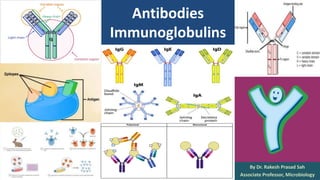
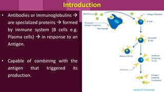
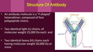
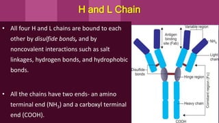
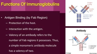
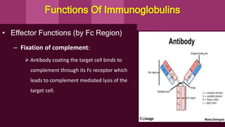
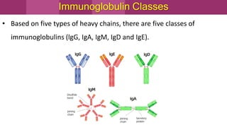
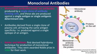
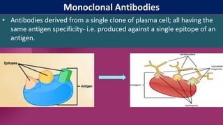
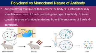
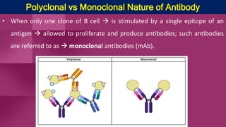
Antibodies
Immunoglobulins
By Dr. Rakesh Prasad Sah
Associate Professor, Microbiology
Introduction
• Antibodies or immunoglobulins 
are specialized proteins  formed
by immune system (B cells e.g.
Plasma cells)  in response to an
Antigen.
• Capable of combining with the
antigen that triggered its
production.
Introduction
• Both the terms, immunoglobulin (Ig) and antibody are used
interchangeably; representing the physiological &functional
properties of same molecule respectively.
• Immunoglobulin (Ig) constitutes 20-25 % of total serum proteins.
• There are five classes (or isotypes) of immunoglobulins recognised-
IgG, IgA, IgM, IgD and IgE.
Structure Of Antibody
• An antibody molecule is a ‘Y-shaped’
heterodimer; composed of four
polypeptide chains.
• Two identical light (L) chains, of
molecular weight 25,000 Da each and
• Two identical heavy (H) chains each
having molecular weight 50,000 Da or
more.
H and L Chain
• All four H and L chains are bound to each
other by disulfide bonds, and by
noncovalent interactions such as salt
linkages, hydrogen bonds, and hydrophobic
bonds.
• All the chains have two ends- an amino
terminal end (NH3) and a carboxyl terminal
end (COOH).
• There are five classes of heavy (H) chains and two classes of light (L)
chains.
• L chains are of two types- kappa (κ) and lambda (λ), named after
Korngold and Lapari who originally described them.
Immunoglobulin class Heavy chain type
IgG γ(gamma)
IgA α (alpha)
IgM µ(mu)
IgD δ(delta)
IgE ε(epsilon)
Variable and Constant Regions
• Each H and L chain comprises of two regions- variable and constant region.
• Variable Region: Represents the antigen binding site of the antibody.
Paratope:
• The site on the hypervariable regions
that make actual contact with the
epitope of an antigen is called as
paratope.
Constant Region
• Constitutes the remaining part of an
Ig molecule other than that of
variable region.
• A single antibody molecule has two
identical heavy chains and two
identical light chains; H2L2
Hinge Region
• Rich in proline and cysteine.
• Quite flexible, allowing the Ig molecule
to assume different positions, thus
helps the antibody in reaching towards
the antigen.
• Hinge region is sensitive to various
enzymatic digestions.
Enzymatic Digestion
• When an immunoglobulin molecule is subjected to enzymatic digestion, it
generates various fragments.
• Types:
 Papain digestion
 Pepsin digestion
 Mercaptoethanol reduction
Functions Of Immunoglobulins
• Antigen Binding (by Fab Region)
– Protection of the host.
– Interaction with the antigen.
– Valency of an antibody refers to the
number of Fab regions it possesses. Thus,
a simple monomeric antibody molecule
has a valency of two.
• Effector Functions (by Fc Region)
– Fixation of complement:
 Antibody coating the target cell binds to
complement through its Fc receptor which
leads to complement mediated lysis of the
target cell.
Functions Of Immunoglobulins
• Binding to various cell types
 Phagocytic cells, lymphocytes, platelets, mast cells, NK cell, eosinophils and
basophils bear Fc receptors (FcR) that bind to Fc region of immunoglobulins.
 Binding can activate the cells to perform some biological functions.
 Some immunoglobulins (e.g. IgG) also bind to receptors on placental
trophoblasts, which results in transfer of the immunoglobulin across the
placenta.
Functions Of Immunoglobulins
Immunoglobulin Classes
• Based on five types of heavy chains, there are five classes of
immunoglobulins (lgG, IgA, IgM, IgD and IgE).
IgG
• IgG has four subclasses- IgG1, IgG2, IgG3 and IgG4; all differ from
each other in the amino acid sequences.
Functions
• IgG can cross placenta - hence provide immunity to the fetus and new born.
 Among subclasses, IgG2 has the poorest ability to cross placenta.
• Complement fixing: Complement fixing ability of subclasses varies - IgG3>
IgG1> IgG2. IgG4 does not fix complements.
• Phagocytosis
Functions of IgG
• Mediates precipitation and neutralization reactions.
• IgG plays a major role in neutralization of toxins as it can easily
diffuse into extravascular space.
• IgG is raised after long time following infection and represents
chronic or past infection (recovery).
Monoclonal Antibodies
• Monoclonal antibodies (mAb) are antibodies
produced by a single clone of antibody
forming cells, and these abs are produced
against a single antigen or single antigenic
determinant site.
• Antibodies derived from a single clone of
plasma cell; all having the same antigen
specificity- i.e. produced against a single
epitope of an antigen.
• Kohler and Milstein first devised hybridoma
technique for production of monoclonal
antibodies. They were awarded Noble prize in
1984 for their work.
Monoclonal Antibodies
• Antibodies derived from a single clone of plasma cell; all having the
same antigen specificity- i.e. produced against a single epitope of an
antigen.
Polyclonal vs Monoclonal Nature of Antibody
• Antigen having multiple epitopes enters the body  each epitope may
stimulate one clone of B cells producing one type of antibody  Serum
contains mixture of antibodies derived from different clones of B cells 
polyclonal.
Polyclonal vs Monoclonal Nature of Antibody
• When only one clone of B cell  is stimulated by a single epitope of an
antigen  allowed to proliferate and produce antibodies; such antibodies
are referred to as  monoclonal antibodies (mAb).
Technique
• Myeloma cells – are malignant cells with capacity to multiply
indefinitely .
• Splenic cells – these are T cells with capacity to form antibodies
against given antigen for limited period.
Hybridoma cells have capacity to produce the required specific
antibodies for a long time.
Preparation of Monoclonal Antibodies
Uses of monoclonal antibodies
• Used in serological reactions which require high degree of specificity eg.- ELISA,
Radio immuno assay, Immunofluorescence, Immunochromatography
• Monoclonal antibodies are widely used for diagnosis of bacterial, viral, parasitic
diseases (e.g. HIV infection, Hepatitis B infection, Auto disease etc).
• Tests for tissue transplantation- tissue typing.
• Tests for vaccines and their industrial production.
Abnormal Immunoglobulins
Abnormal proteins, having structure similar to Ig, but no antibody activity.
Bence Jones proteins - Found in urine of patients with multiplemyeloma &
waldenstroms macroglobulinemia
Property: While heating urine samples,BJ proteins coagulate at 50°C&
redissolve at70°C. These are the light chains of Ig -Either K or L chains.
Property
1. Multiple myeloma- malignant condition affection plasma cells which
produce IgG, IgA, IgD, IgE.
2. Waldestroms macroglobulinemia- malignant condition IgM
producing plasma cells. Thus M proteins & BJ proteins are produced.
3. Heavy chain disease- overproduction of Fc parts of Heavy chains.
4. Eryoglobulinemia- When serum is cooled–Formation of gel
IgG/IgM.
When serum is warmed– gel re-dissolves.
Found in myelomas, macroglobulinemias,
autoimmune diseases
Thank
You…

Antibodies.pptxa.pptx

  • 1.
    Antibodies Immunoglobulins By Dr. RakeshPrasad Sah Associate Professor, Microbiology
  • 2.
    Introduction • Antibodies orimmunoglobulins  are specialized proteins  formed by immune system (B cells e.g. Plasma cells)  in response to an Antigen. • Capable of combining with the antigen that triggered its production.
  • 3.
    Introduction • Both theterms, immunoglobulin (Ig) and antibody are used interchangeably; representing the physiological &functional properties of same molecule respectively. • Immunoglobulin (Ig) constitutes 20-25 % of total serum proteins. • There are five classes (or isotypes) of immunoglobulins recognised- IgG, IgA, IgM, IgD and IgE.
  • 4.
    Structure Of Antibody •An antibody molecule is a ‘Y-shaped’ heterodimer; composed of four polypeptide chains. • Two identical light (L) chains, of molecular weight 25,000 Da each and • Two identical heavy (H) chains each having molecular weight 50,000 Da or more.
  • 5.
    H and LChain • All four H and L chains are bound to each other by disulfide bonds, and by noncovalent interactions such as salt linkages, hydrogen bonds, and hydrophobic bonds. • All the chains have two ends- an amino terminal end (NH3) and a carboxyl terminal end (COOH).
  • 6.
    • There arefive classes of heavy (H) chains and two classes of light (L) chains. • L chains are of two types- kappa (κ) and lambda (λ), named after Korngold and Lapari who originally described them. Immunoglobulin class Heavy chain type IgG γ(gamma) IgA α (alpha) IgM µ(mu) IgD δ(delta) IgE ε(epsilon)
  • 7.
    Variable and ConstantRegions • Each H and L chain comprises of two regions- variable and constant region. • Variable Region: Represents the antigen binding site of the antibody. Paratope: • The site on the hypervariable regions that make actual contact with the epitope of an antigen is called as paratope.
  • 8.
    Constant Region • Constitutesthe remaining part of an Ig molecule other than that of variable region. • A single antibody molecule has two identical heavy chains and two identical light chains; H2L2
  • 9.
    Hinge Region • Richin proline and cysteine. • Quite flexible, allowing the Ig molecule to assume different positions, thus helps the antibody in reaching towards the antigen. • Hinge region is sensitive to various enzymatic digestions.
  • 10.
    Enzymatic Digestion • Whenan immunoglobulin molecule is subjected to enzymatic digestion, it generates various fragments. • Types:  Papain digestion  Pepsin digestion  Mercaptoethanol reduction
  • 11.
    Functions Of Immunoglobulins •Antigen Binding (by Fab Region) – Protection of the host. – Interaction with the antigen. – Valency of an antibody refers to the number of Fab regions it possesses. Thus, a simple monomeric antibody molecule has a valency of two.
  • 12.
    • Effector Functions(by Fc Region) – Fixation of complement:  Antibody coating the target cell binds to complement through its Fc receptor which leads to complement mediated lysis of the target cell. Functions Of Immunoglobulins
  • 13.
    • Binding tovarious cell types  Phagocytic cells, lymphocytes, platelets, mast cells, NK cell, eosinophils and basophils bear Fc receptors (FcR) that bind to Fc region of immunoglobulins.  Binding can activate the cells to perform some biological functions.  Some immunoglobulins (e.g. IgG) also bind to receptors on placental trophoblasts, which results in transfer of the immunoglobulin across the placenta. Functions Of Immunoglobulins
  • 14.
    Immunoglobulin Classes • Basedon five types of heavy chains, there are five classes of immunoglobulins (lgG, IgA, IgM, IgD and IgE).
  • 16.
    IgG • IgG hasfour subclasses- IgG1, IgG2, IgG3 and IgG4; all differ from each other in the amino acid sequences. Functions • IgG can cross placenta - hence provide immunity to the fetus and new born.  Among subclasses, IgG2 has the poorest ability to cross placenta. • Complement fixing: Complement fixing ability of subclasses varies - IgG3> IgG1> IgG2. IgG4 does not fix complements. • Phagocytosis
  • 17.
    Functions of IgG •Mediates precipitation and neutralization reactions. • IgG plays a major role in neutralization of toxins as it can easily diffuse into extravascular space. • IgG is raised after long time following infection and represents chronic or past infection (recovery).
  • 18.
    Monoclonal Antibodies • Monoclonalantibodies (mAb) are antibodies produced by a single clone of antibody forming cells, and these abs are produced against a single antigen or single antigenic determinant site. • Antibodies derived from a single clone of plasma cell; all having the same antigen specificity- i.e. produced against a single epitope of an antigen. • Kohler and Milstein first devised hybridoma technique for production of monoclonal antibodies. They were awarded Noble prize in 1984 for their work.
  • 19.
    Monoclonal Antibodies • Antibodiesderived from a single clone of plasma cell; all having the same antigen specificity- i.e. produced against a single epitope of an antigen.
  • 20.
    Polyclonal vs MonoclonalNature of Antibody • Antigen having multiple epitopes enters the body  each epitope may stimulate one clone of B cells producing one type of antibody  Serum contains mixture of antibodies derived from different clones of B cells  polyclonal.
  • 22.
    Polyclonal vs MonoclonalNature of Antibody • When only one clone of B cell  is stimulated by a single epitope of an antigen  allowed to proliferate and produce antibodies; such antibodies are referred to as  monoclonal antibodies (mAb).
  • 23.
    Technique • Myeloma cells– are malignant cells with capacity to multiply indefinitely . • Splenic cells – these are T cells with capacity to form antibodies against given antigen for limited period. Hybridoma cells have capacity to produce the required specific antibodies for a long time.
  • 24.
  • 28.
    Uses of monoclonalantibodies • Used in serological reactions which require high degree of specificity eg.- ELISA, Radio immuno assay, Immunofluorescence, Immunochromatography • Monoclonal antibodies are widely used for diagnosis of bacterial, viral, parasitic diseases (e.g. HIV infection, Hepatitis B infection, Auto disease etc). • Tests for tissue transplantation- tissue typing. • Tests for vaccines and their industrial production.
  • 29.
    Abnormal Immunoglobulins Abnormal proteins,having structure similar to Ig, but no antibody activity. Bence Jones proteins - Found in urine of patients with multiplemyeloma & waldenstroms macroglobulinemia Property: While heating urine samples,BJ proteins coagulate at 50°C& redissolve at70°C. These are the light chains of Ig -Either K or L chains.
  • 30.
    Property 1. Multiple myeloma-malignant condition affection plasma cells which produce IgG, IgA, IgD, IgE. 2. Waldestroms macroglobulinemia- malignant condition IgM producing plasma cells. Thus M proteins & BJ proteins are produced. 3. Heavy chain disease- overproduction of Fc parts of Heavy chains.
  • 31.
    4. Eryoglobulinemia- Whenserum is cooled–Formation of gel IgG/IgM. When serum is warmed– gel re-dissolves. Found in myelomas, macroglobulinemias, autoimmune diseases
  • 32.