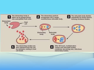
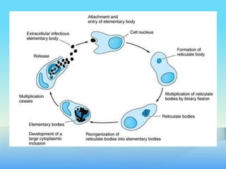
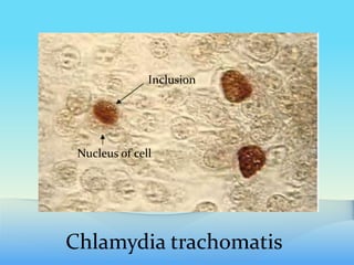
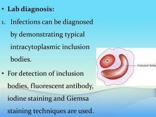
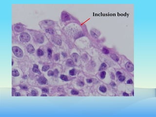
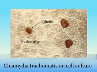
CHLAMYDIA
General features:
• Chlamydiae are obligate intracellular organisms
• Depend on the host cell for energy (ATP).
• Therefore can grow only inside host cells.
• Chlamydiae are small, round-to-ovoid organisms
that vary in size during different stages of their
life cycle
• There are three important human pathogens:
1. Chlamydia trachomatis
2. Chlamydophila psittaci
3. Chlamydophila pneumoniae.
• C. pneumoniae and C. psittaci are different
molecularly from C. trachomatis.
• Therefore have been placed into a new genus
called Chlamydophila.
• Thus Chlamydophila pneumoniae and
Chlamydophila psittaci.
• But still known as Chlamydia pneumoniae and
Chlamydia psittaci
Structure:
• They have a rigid cell wall but do not have a
typical peptidoglycan layer.
• C. trachomatis is the most common cause of
STDs.
• Chlamydia pneumoniae causes atypical
pneumonia.
• C. psittaci causes psittacosis ( mainly disease of
birds)
Pathogenesis:
• Chlamydiae have a unique life cycle, with distinct
infectious and reproductive forms.
• The infectious form is called elementary body.
• It is a tiny, condensed structure that is
extracellular and initiates an infection.
• The elementary body enters by phagocytosis into
susceptible host cells.
• The elementary body is present in phagosome
inside the host cell.
• Inside the cell, the elementary body prevents
fusion of the phagosome and lysosome.
• The particle reorganizes over the next 8 hours
into a larger, noninfectious reticulate body.
• The phagosome now becomes a larger structure.
• This vacoule/larger phagosome is called
inclusion body.
• Reticulate body divides repeatedly by binary
fission.
• After 48 hours, multiplication ceases, and
reticulate bodies condense to become new
infectious elementary bodies.
• This whole process causes host cell death and
release of elementary bodies.
• C. psittaci and C. pneumoniae each have 1
immunotype, whereas C. trachomatis has at least 15.
• Laboratory identification
1. Chlamydiae are not seen by Gram stain
• Can be visualized under light microscope by
Giemsa stain.
2. Direct immunofluorescence is also a common and
useful procedure
Chlamydia trachomatis
Inclusion
Nucleus of cell
CHLAMYDIA TRACHOMATIS
• C. trachomatis has 15 serotypes, which are related
to different diseases.
• C. trachomatis infects only humans.
• Mainly causes NGU (Non Gonococcal Urethritis)
and Trachoma.
• Usually transmitted by close personal contact.
• Pathogenesis & Clinical Findings:
• C. trachomatis exists in more than 15
immunotypes (A–L).
A. Trachoma:
• Types A, B, and C cause trachoma, a chronic
keratoconjunctivitis.
• Endemic in Africa and Asia.
• Previously known as Egyptian ophthalmia.
• Primarily affects the superficial epithelium of
conjunctiva and cornea simultaneously.
• The word 'trachoma' ('rough' )comes from the
Greek word because surface appearance of the
conjunctiva in chronic trachoma is rough or
granular.
• It is still one of the leading causes of preventable
blindness in the world.
• Because of persistent /repeated infection over
several years leads to scarring causing permanent
opacities of the cornea and distortion of eyelids.
• Trachoma can be further divided into
1. Blinding trachoma
2. Non blinding trachoma
3. Paratrachoma
1. Blinding trachoma.
• It is caused by serotypes A, B, Ba and C of
Chlamydia trachomatis.
• It is associated with secondary bacterial infection.
• It is transmitted from eye to eye by transfer of
ocular discharge.
• There is mucopurulent conjunctivtis
2. Non-blinding trachoma.
• It is also caused by serotypes A, B, Ba, and C; but is
not associated with secondary bacterial infections.
• It is a mild form of disease with limited
transmission due to improved hygiene.
• Symptoms are minimal and include mild foreign
body sensation in the eyes.
3. Paratrachoma.
• It is caused by serotypes D to K and L1,L2 and L3
as well.
• It spreads from genitals to eye.
• It manifests as either
a. Adult inclusion conjunctivitis or
b. Chlamydial ophthalmia neonatorum.
• Trachoma is a chronic infection.
Follicular
conujnctivitis
Intense
follicular
inflammation
Tarsal conjunctival scarring
Blinding trachoma with entropion and
trichiasis
B. Genital infections:
• Types D–K cause genital tract infections.
• In men, it causes NGU, which may progress to
epididymitis or prostatitis.
• In women it causes cervicitis which may progress to
salpingitis and pelvic inflammatory disease (PID).
• Repeated episodes of salpingitis or PID can result in
infertility or ectopic pregnancy.
a. Neonatal conjunctivitis (chlamydial
ophthalmia neonatorum) :
• In infants born to genitally infected women.
b. Inclusion conjunctivitis:
• Conjunctivitis also occurs in adults due to transfer of
organisms from the genitals to the eye.
• Patients with genital infections have a high chance
of developing Reiter's syndrome (urethritis,
arthritis and uveitis)
• C. trachomatis directed antibodies cross react.
c. Lymphogranuloma venereum:
• C. trachomatis serotypes L1, L2, and L3 cause LGV
• More invasive STD.
• It is uncommon in the United States but endemic
in Asia, Africa, and South America.
• Formation of transient papules on genitalia,
followed in 1 to 2 months by painful swelling of
inguinal and perirectal lymph nodes.
• The affected lymph nodes suppurate and then
chronic inflammation and fibrosis leads to
blockage of regional lymphatic drainage.
• Lab diagnosis:
1. Infections can be diagnosed
by demonstrating typical
intracytoplasmic inclusion
bodies.
• For detection of inclusion
bodies, fluorescent antibody,
iodine staining and Giemsa
staining techniques are used.
Inclusion body
2. Chlamydial antigens can also be detected in
exudates or urine by ELISA.
• PCR based test using the patient's urine can also
be used to diagnose chlamydial STD.
3. Cell culture:
• In this case suspected specimen is added to
special cells, which are then cultured in
laboratory.
• The infected cells are stained with iodine to
detect the glycogen-positive inclusions.
• The presence of chlamydial inclusions can be
demonstrated after 2 to 7 days of incubation.
Chlamydia trachomatis on cell culture
Inclusion
Nucleus of cell
• Treatment:
• All chlamydiae are susceptible to tetracyclines,
(doxycyline), and macrolides (erythromycin and
azithromycin).
• The drug of choice for STDs is azithromycin.
• The drug of choice for chlamydial ophthalmia
neonatorum is erythromycin.
• Because of co-infection with gonococci, any
patient with a diagnosis of genital infections
should also be treated for Gonorrhoea and vice
versa.
• Prevention:
• There is no vaccine against any chlamydial
disease.
• So other preventive measures should be used.

CHLAMYDIA by Dr munir HMC PICO.slideshare

  • 1.
  • 2.
    General features: • Chlamydiaeare obligate intracellular organisms • Depend on the host cell for energy (ATP). • Therefore can grow only inside host cells. • Chlamydiae are small, round-to-ovoid organisms that vary in size during different stages of their life cycle
  • 3.
    • There arethree important human pathogens: 1. Chlamydia trachomatis 2. Chlamydophila psittaci 3. Chlamydophila pneumoniae. • C. pneumoniae and C. psittaci are different molecularly from C. trachomatis.
  • 4.
    • Therefore havebeen placed into a new genus called Chlamydophila. • Thus Chlamydophila pneumoniae and Chlamydophila psittaci. • But still known as Chlamydia pneumoniae and Chlamydia psittaci
  • 5.
    Structure: • They havea rigid cell wall but do not have a typical peptidoglycan layer. • C. trachomatis is the most common cause of STDs. • Chlamydia pneumoniae causes atypical pneumonia. • C. psittaci causes psittacosis ( mainly disease of birds)
  • 6.
    Pathogenesis: • Chlamydiae havea unique life cycle, with distinct infectious and reproductive forms. • The infectious form is called elementary body. • It is a tiny, condensed structure that is extracellular and initiates an infection. • The elementary body enters by phagocytosis into susceptible host cells.
  • 7.
    • The elementarybody is present in phagosome inside the host cell. • Inside the cell, the elementary body prevents fusion of the phagosome and lysosome. • The particle reorganizes over the next 8 hours into a larger, noninfectious reticulate body. • The phagosome now becomes a larger structure.
  • 9.
    • This vacoule/largerphagosome is called inclusion body. • Reticulate body divides repeatedly by binary fission. • After 48 hours, multiplication ceases, and reticulate bodies condense to become new infectious elementary bodies. • This whole process causes host cell death and release of elementary bodies.
  • 12.
    • C. psittaciand C. pneumoniae each have 1 immunotype, whereas C. trachomatis has at least 15. • Laboratory identification 1. Chlamydiae are not seen by Gram stain • Can be visualized under light microscope by Giemsa stain. 2. Direct immunofluorescence is also a common and useful procedure
  • 13.
  • 14.
    CHLAMYDIA TRACHOMATIS • C.trachomatis has 15 serotypes, which are related to different diseases. • C. trachomatis infects only humans. • Mainly causes NGU (Non Gonococcal Urethritis) and Trachoma. • Usually transmitted by close personal contact.
  • 15.
    • Pathogenesis &Clinical Findings: • C. trachomatis exists in more than 15 immunotypes (A–L). A. Trachoma: • Types A, B, and C cause trachoma, a chronic keratoconjunctivitis. • Endemic in Africa and Asia.
  • 16.
    • Previously knownas Egyptian ophthalmia. • Primarily affects the superficial epithelium of conjunctiva and cornea simultaneously. • The word 'trachoma' ('rough' )comes from the Greek word because surface appearance of the conjunctiva in chronic trachoma is rough or granular.
  • 17.
    • It isstill one of the leading causes of preventable blindness in the world. • Because of persistent /repeated infection over several years leads to scarring causing permanent opacities of the cornea and distortion of eyelids.
  • 18.
    • Trachoma canbe further divided into 1. Blinding trachoma 2. Non blinding trachoma 3. Paratrachoma 1. Blinding trachoma. • It is caused by serotypes A, B, Ba and C of Chlamydia trachomatis.
  • 19.
    • It isassociated with secondary bacterial infection. • It is transmitted from eye to eye by transfer of ocular discharge. • There is mucopurulent conjunctivtis 2. Non-blinding trachoma. • It is also caused by serotypes A, B, Ba, and C; but is not associated with secondary bacterial infections.
  • 20.
    • It isa mild form of disease with limited transmission due to improved hygiene. • Symptoms are minimal and include mild foreign body sensation in the eyes. 3. Paratrachoma. • It is caused by serotypes D to K and L1,L2 and L3 as well. • It spreads from genitals to eye.
  • 21.
    • It manifestsas either a. Adult inclusion conjunctivitis or b. Chlamydial ophthalmia neonatorum. • Trachoma is a chronic infection.
  • 22.
  • 23.
    Tarsal conjunctival scarring Blindingtrachoma with entropion and trichiasis
  • 24.
    B. Genital infections: •Types D–K cause genital tract infections. • In men, it causes NGU, which may progress to epididymitis or prostatitis. • In women it causes cervicitis which may progress to salpingitis and pelvic inflammatory disease (PID). • Repeated episodes of salpingitis or PID can result in infertility or ectopic pregnancy.
  • 25.
    a. Neonatal conjunctivitis(chlamydial ophthalmia neonatorum) : • In infants born to genitally infected women. b. Inclusion conjunctivitis: • Conjunctivitis also occurs in adults due to transfer of organisms from the genitals to the eye. • Patients with genital infections have a high chance
  • 26.
    of developing Reiter'ssyndrome (urethritis, arthritis and uveitis) • C. trachomatis directed antibodies cross react. c. Lymphogranuloma venereum: • C. trachomatis serotypes L1, L2, and L3 cause LGV • More invasive STD. • It is uncommon in the United States but endemic in Asia, Africa, and South America.
  • 27.
    • Formation oftransient papules on genitalia, followed in 1 to 2 months by painful swelling of inguinal and perirectal lymph nodes. • The affected lymph nodes suppurate and then chronic inflammation and fibrosis leads to blockage of regional lymphatic drainage.
  • 28.
    • Lab diagnosis: 1.Infections can be diagnosed by demonstrating typical intracytoplasmic inclusion bodies. • For detection of inclusion bodies, fluorescent antibody, iodine staining and Giemsa staining techniques are used.
  • 29.
  • 30.
    2. Chlamydial antigenscan also be detected in exudates or urine by ELISA. • PCR based test using the patient's urine can also be used to diagnose chlamydial STD. 3. Cell culture: • In this case suspected specimen is added to special cells, which are then cultured in laboratory.
  • 31.
    • The infectedcells are stained with iodine to detect the glycogen-positive inclusions. • The presence of chlamydial inclusions can be demonstrated after 2 to 7 days of incubation.
  • 32.
    Chlamydia trachomatis oncell culture Inclusion Nucleus of cell
  • 34.
    • Treatment: • Allchlamydiae are susceptible to tetracyclines, (doxycyline), and macrolides (erythromycin and azithromycin). • The drug of choice for STDs is azithromycin. • The drug of choice for chlamydial ophthalmia neonatorum is erythromycin.
  • 35.
    • Because ofco-infection with gonococci, any patient with a diagnosis of genital infections should also be treated for Gonorrhoea and vice versa. • Prevention: • There is no vaccine against any chlamydial disease. • So other preventive measures should be used.