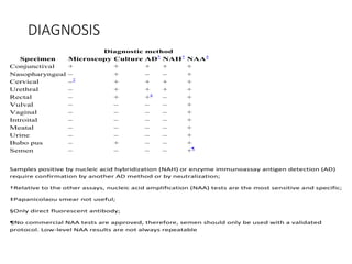
Chlamydia
By
Benson Musinguzi
Scientific Classification
• Kingdom: Bacteria
• Phylum: Chlamydiae
• Class: Chlamydiae
• Order: Chlamydiales
• Family: Chlamydiaceae
• Genus: Chlamydia
• Species: trachomatis
Pneumoniae,
psittacci
pecorum
General characteristics of Chlamydia
•Obligate intracellular coccoid parasites
•contain DNA and RNA, and ribosomes
•lack ATP, biosynthetic pathways
•cell wall but peptidoglycan absent -
• use disulfide bonds
•non motile
Obligate Parasites
•Obligate intracellular parasites of mammals and
birds
• not transmitted by arthropods.
•Multiply in the cytoplasm of the host cell.
•generally epithelial cells
•Basophilic inclusions
Energy Parasites
•The cells can synthesize DNA, RNA and protein.
•No flavoproteins or cytochromes.
•lack of ATP-generating ability
•need to obtain ATP from the host cell.
Three Species of medical importance
C. trachomatis
C. psittaci
C. pneumoniae
C trachomatis
Trachoma
conjunctivitis
proctitis
urethritis
salpingitis
Lymphogranuloma venereum
C psittaci & C
pneumoniae
Upper respiratory infection
Bronchitis
Pneumonia
C. pneumoniae
-Pharingitis
-Bronchitis
-Pneumonia
C. psittaci
produces systemic diseases:
• psittacosis,
• ornithosis and
• pneumonitis.
Chlamydial Morphologies
Elementary body
•0.25 - 0.3 um diameter
•electron-dense nucleoid
•Released from ruptured infected cells. Human
to human
•& bird to human.
Reticulate Body
•Intracytoplasmic form 0.5 - 1.0 um
•Replication and growth. ( Inclusion body )
•without a dense center.
Distinctive properties.
•Have two distinct forms:-
•Infectious elementary bodies and
•Intracellular reticulate bodies.
•Elementary bodies attach and are internalized by susceptible
host cells.
•Once inside, they reorganize into a replicative form (the
reticulate body)
•Over a 24 hour period:
•Reticulate bodies divide and begin to reorganize back into
elementary bodies.
• 48-72 hours after infection:
• The cell is lysed and
• numerous infectious elementary bodies released.
Developmental Cycle of Chlamydia
Developmental Cycle of Chlamydia cont..
•The development is initiated by the highly infectious small
cell which is taken into the host cell by phagocytosis.
•The engulfed small cell retains its morphological integrity in
vacuoles bound by membrane derived from the surface of
the host cell,
•The small cell is reorganized into a large cell which is the
vegetative multiplying form of these organisms.
•Then, still within the membrane-bound vacuole, the large
•cell grows in size and multiplies by repeated binary fission.
•The developmental cycle is completed by the reorganization
of most of the large cells into small ones which are then
available for infection of new host cells.
VIRULENCE FACTORS
•Chlamydial polymorphic outer membrane proteins(pmp)
Auto transporters, immunogenic ie elicit pro inflamatory
cytokine responses.
•Plasmids. plasmid transcriptional activity contributes to
the regulation of chlamydial chromosomal gene
expression.
•Chlamydial type iii secretion system, are complex
arrangements of structures that are designed to
promote delivery of pathogen effector proteins after
contact with the host cell.
•Toixins, putative chlamydial cytotoxins
Epidemiology.
• Trachoma:
• prevalent in Africa and Asia,
• generally in hot and dry areas.
• The organisms are very persistent.
• Their habitat : similar to that of Neisseria and Haemophilus.
• Infection can occur via:
• swimming in unchlorinated pools,
• sharing towels or
• by passage through the birth canal.
DIAGNOSIS
Specimens
-bubo pus
-Nasopharyngeal swabs
-Rectal swabs
-Tissue samples
-Semen
-Occular and genital tract samples/swabs.
Diagnosis cont..
•Isolation in cell culture
Culture confirms the presence of viable organisms.
• Historically the “gold standard”
• Variable sensitivity (50% – 80%)
• High specificity
•Antigen detection
Nucleic Acid Amplification Tests (NAATs)
• NAATs amplify and detect organism-specific genomic or
plasmid DNA or rRNA
• A number of NAATs are commercially available. They
include
• Abbott LCx
• Artus/Qiagen RealArt PCR
• Becton Dickinson BDProbe Tec®
• Gen-Probe AmpCT, Aptima®
• Roche Amplicor®
• detect C. trachomatis and in specimen.
• Significantly more sensitive than other tests
Non-Amplification Tests:
Not Recommended
•Less expensive than culture or NAATs, but sensitivity only 50
– 75%
•Direct fluorescent antibody (DFA)
• Detects intact bacteria with a fluorescent antibody
• Variety of specimen sites
•Enzyme immunoassay (EIA)
• Detects bacterial antigens with an enzyme-labeled antibody
• Use either monoclonal or polyclonal antibodies to detect chlamydial
lipopolysaccharide (LPS).
•Nucleic acid hybridization (NA probe)
• Detects specific DNA or RNA sequences of C. trachomatis and N.
gonorrhoeae
Serology
•Rarely used for uncomplicated infections
•Comparative data between types of serologic test
are lacking
•Criteria used in LGV diagnosis
•Complement fixation titers >1:64 can support
diagnosis of LGV in the appropriate clinical context.
• Serologic test interpretation for LGV is not
standardize
DIAGNOSIS
Diagnostic method
Specimen Microscopy Culture AD*
NAH*
NAA†
Conjunctival + + + + +
Nasopharyngeal – + – – +
Cervical –‡
+ + + +
Urethral – + + + +
Rectal – + +§
– +
Vulval – – – – +
Vaginal – – – – +
Introital – – – – +
Meatal – – – – +
Urine – – – – +
Bubo pus – + – – +
Semen – – – – +¶
Samples positive by nucleic acid hybridization (NAH) or enzyme immunoassay antigen detection (AD)
require confirmation by another AD method or by neutralization;
†Relative to the other assays, nucleic acid amplification (NAA) tests are the most sensitive and specific;
‡Papanicolaou smear not useful;
§Only direct fluorescent antibody;
¶No commercial NAA tests are approved, therefore, semen should only be used with a validated
protocol. Low-level NAA results are not always repeatable
Treatment and Control.
•Chemotherapeutic:
• Tetracycline or erythromycin are drugs of choice.
•Sanitary:
• Good hygiene,
• Treatment of sexual partners and
• Quarantine of birds all reduce the incidence.
Treatment and control.
• Immunological:
• No vaccine – available since specific antibodies fail to neutralize elementary bodies
in vivo.
END

8. Chlamydia Presentation.pptx

  • 1.
  • 2.
    Scientific Classification • Kingdom:Bacteria • Phylum: Chlamydiae • Class: Chlamydiae • Order: Chlamydiales • Family: Chlamydiaceae • Genus: Chlamydia • Species: trachomatis Pneumoniae, psittacci pecorum
  • 3.
    General characteristics ofChlamydia •Obligate intracellular coccoid parasites •contain DNA and RNA, and ribosomes •lack ATP, biosynthetic pathways •cell wall but peptidoglycan absent - • use disulfide bonds •non motile
  • 4.
    Obligate Parasites •Obligate intracellularparasites of mammals and birds • not transmitted by arthropods. •Multiply in the cytoplasm of the host cell. •generally epithelial cells •Basophilic inclusions
  • 5.
    Energy Parasites •The cellscan synthesize DNA, RNA and protein. •No flavoproteins or cytochromes. •lack of ATP-generating ability •need to obtain ATP from the host cell.
  • 6.
    Three Species ofmedical importance C. trachomatis C. psittaci C. pneumoniae
  • 7.
    C trachomatis Trachoma conjunctivitis proctitis urethritis salpingitis Lymphogranuloma venereum Cpsittaci & C pneumoniae Upper respiratory infection Bronchitis Pneumonia
  • 8.
  • 9.
    C. psittaci produces systemicdiseases: • psittacosis, • ornithosis and • pneumonitis.
  • 10.
    Chlamydial Morphologies Elementary body •0.25- 0.3 um diameter •electron-dense nucleoid •Released from ruptured infected cells. Human to human •& bird to human. Reticulate Body •Intracytoplasmic form 0.5 - 1.0 um •Replication and growth. ( Inclusion body ) •without a dense center.
  • 11.
    Distinctive properties. •Have twodistinct forms:- •Infectious elementary bodies and •Intracellular reticulate bodies. •Elementary bodies attach and are internalized by susceptible host cells. •Once inside, they reorganize into a replicative form (the reticulate body) •Over a 24 hour period: •Reticulate bodies divide and begin to reorganize back into elementary bodies. • 48-72 hours after infection: • The cell is lysed and • numerous infectious elementary bodies released.
  • 12.
  • 13.
    Developmental Cycle ofChlamydia cont.. •The development is initiated by the highly infectious small cell which is taken into the host cell by phagocytosis. •The engulfed small cell retains its morphological integrity in vacuoles bound by membrane derived from the surface of the host cell, •The small cell is reorganized into a large cell which is the vegetative multiplying form of these organisms. •Then, still within the membrane-bound vacuole, the large •cell grows in size and multiplies by repeated binary fission. •The developmental cycle is completed by the reorganization of most of the large cells into small ones which are then available for infection of new host cells.
  • 14.
    VIRULENCE FACTORS •Chlamydial polymorphicouter membrane proteins(pmp) Auto transporters, immunogenic ie elicit pro inflamatory cytokine responses. •Plasmids. plasmid transcriptional activity contributes to the regulation of chlamydial chromosomal gene expression. •Chlamydial type iii secretion system, are complex arrangements of structures that are designed to promote delivery of pathogen effector proteins after contact with the host cell. •Toixins, putative chlamydial cytotoxins
  • 15.
    Epidemiology. • Trachoma: • prevalentin Africa and Asia, • generally in hot and dry areas. • The organisms are very persistent. • Their habitat : similar to that of Neisseria and Haemophilus. • Infection can occur via: • swimming in unchlorinated pools, • sharing towels or • by passage through the birth canal.
  • 16.
    DIAGNOSIS Specimens -bubo pus -Nasopharyngeal swabs -Rectalswabs -Tissue samples -Semen -Occular and genital tract samples/swabs.
  • 17.
    Diagnosis cont.. •Isolation incell culture Culture confirms the presence of viable organisms. • Historically the “gold standard” • Variable sensitivity (50% – 80%) • High specificity •Antigen detection
  • 18.
    Nucleic Acid AmplificationTests (NAATs) • NAATs amplify and detect organism-specific genomic or plasmid DNA or rRNA • A number of NAATs are commercially available. They include • Abbott LCx • Artus/Qiagen RealArt PCR • Becton Dickinson BDProbe Tec® • Gen-Probe AmpCT, Aptima® • Roche Amplicor® • detect C. trachomatis and in specimen. • Significantly more sensitive than other tests
  • 19.
    Non-Amplification Tests: Not Recommended •Lessexpensive than culture or NAATs, but sensitivity only 50 – 75% •Direct fluorescent antibody (DFA) • Detects intact bacteria with a fluorescent antibody • Variety of specimen sites •Enzyme immunoassay (EIA) • Detects bacterial antigens with an enzyme-labeled antibody • Use either monoclonal or polyclonal antibodies to detect chlamydial lipopolysaccharide (LPS). •Nucleic acid hybridization (NA probe) • Detects specific DNA or RNA sequences of C. trachomatis and N. gonorrhoeae
  • 20.
    Serology •Rarely used foruncomplicated infections •Comparative data between types of serologic test are lacking •Criteria used in LGV diagnosis •Complement fixation titers >1:64 can support diagnosis of LGV in the appropriate clinical context. • Serologic test interpretation for LGV is not standardize
  • 21.
    DIAGNOSIS Diagnostic method Specimen MicroscopyCulture AD* NAH* NAA† Conjunctival + + + + + Nasopharyngeal – + – – + Cervical –‡ + + + + Urethral – + + + + Rectal – + +§ – + Vulval – – – – + Vaginal – – – – + Introital – – – – + Meatal – – – – + Urine – – – – + Bubo pus – + – – + Semen – – – – +¶ Samples positive by nucleic acid hybridization (NAH) or enzyme immunoassay antigen detection (AD) require confirmation by another AD method or by neutralization; †Relative to the other assays, nucleic acid amplification (NAA) tests are the most sensitive and specific; ‡Papanicolaou smear not useful; §Only direct fluorescent antibody; ¶No commercial NAA tests are approved, therefore, semen should only be used with a validated protocol. Low-level NAA results are not always repeatable
  • 22.
    Treatment and Control. •Chemotherapeutic: •Tetracycline or erythromycin are drugs of choice. •Sanitary: • Good hygiene, • Treatment of sexual partners and • Quarantine of birds all reduce the incidence.
  • 23.
    Treatment and control. •Immunological: • No vaccine – available since specific antibodies fail to neutralize elementary bodies in vivo. END

Editor's Notes

  • #13 There appears to be no significant difference in morphology or developmental cycle among the various chlamydia, and a single generalized description applies to all. The development cycle may be regarded as an orderly alternation of the small and large cell type. It is initiated by the highly infectious small cell which is taken into the host cell by phagocytosis. The engulfed small cell retains its morphological integrity in vacuoles bound by membrane derived from the surface of the host cell, and there is no eclipse (period in which the parasite loses the infectious ability). Instead, without loss of individuality, the small cell is reorganized into a large cell which is the vegetative multiplying form of these organisms. Then, still within the membrane-bound vacuole, the large cell grows in size and multiplies by repeated binary fission. The developmental cycle is completed by the reorganization of most of the large cells into small ones which are then available for infection of new host cells. The time required for completion of a cycle varies from 24-48 hours, depending on the particular host/parasite system involved.