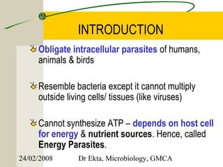
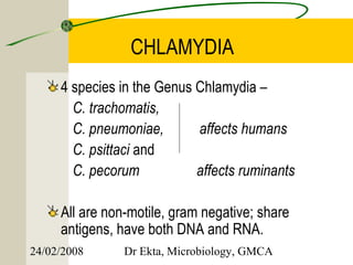
This document discusses Chlamydia, an obligate intracellular parasite. It describes the four species that infect humans (C. trachomatis, C. pneumoniae, C. psittaci, C. pecorum) and their associated diseases. C. trachomatis causes trachoma, inclusion conjunctivitis, infant pneumonia, and genital infections like lymphogranuloma venereum. The life cycle and laboratory diagnosis of Chlamydia species are also summarized.
1. CHLAMYDIA
II MBBS
24/02/2008 Dr Ekta, Microbiology, GMCA
2. KEY WORDS
Intracellular Inclusion conjunctivitis
Energy parasites Lymphogranuloma venereum
Elementary body Frei test
Reticulate body Genital chlamydiasis
Inclusion bodies – HP, LCL C. psittaci
Biotypes/ serotypes Psittacosis
Tissue culture Ornithosis
C. trachomatis C. pneumoniae
Trachoma
24/02/2008 Dr Ekta, Microbiology, GMCA
3. INTRODUCTION
Obligate intracellular parasites of humans,
animals & birds
Resemble bacteria except it cannot multiply
outside living cells/ tissues (like viruses)
Cannot synthesize ATP – depends on host cell
for energy & nutrient sources. Hence, called
Energy Parasites.
24/02/2008 Dr Ekta, Microbiology, GMCA
4. CHLAMYDIA
4 species in the Genus Chlamydia –
C. trachomatis,
C. pneumoniae, affects humans
C. psittaci and
C. pecorum affects ruminants
All are non-motile, gram negative; share
antigens, have both DNA and RNA.
24/02/2008 Dr Ekta, Microbiology, GMCA
5. Classification
C.trachomatis : eye & genital infections, infant
pneumonia, and LGV (Lymphogranuloma
Venereum) in adults
C.pneumoniae : different types of respiratory
infections.
C.psittaci : psittacosis in man, ornithosis in birds
24/02/2008 Dr Ekta, Microbiology, GMCA
6. Life cycle
Chlamydiae occur in 2 forms :
1. Elementary body – extracellular, infective form
2. Reticulate body – intracellular, growing & replicative
form
• Chlamydial microcolony within the host cell is called
Inclusion body.
• Mature inclusion body contains 100 - 500 elementary
bodies
24/02/2008 Dr Ekta, Microbiology, GMCA
8. Life cycle
C. psittaci – host cell is severely damaged, EBs
are released within 48 hrs by cell lysis
C.trachomatis – mature inclusion body appears
to be exocytosed in 72- 96 hrs.
24/02/2008 Dr Ekta, Microbiology, GMCA
9. Antigenic Properties
Three major Ags
1. Genus specific Ag – heat stable, common to all chlamydiae,
a lipopolysaccharide resembling LPS of GNB. Present in all
stages.
2. Species specific protein Ags – present at the envelope
surface, help in classifying chlamydia into species
3. Ag for Intraspecies typing – found only in some members
of a species, located on major OMP (MOMP), demonstrated
by micro- IF. Classifies species into serovars/ serotypes
24/02/2008 Dr Ekta, Microbiology, GMCA
10. Variants of Chlamydia
C. trachomatis – 2 biovars: TRIC & LGV
1. TRIC – Trachoma, Inclusion conjunctivitis
- divided into 12 serovars
2. LGV – Lymphogranuloma venereum – 3
serovars
24/02/2008 Dr Ekta, Microbiology, GMCA
11. Human diseases
Species Serotype Disease
C. trachomatis A, B, Ba, C Endemic blinding
trachoma
C. trachomatis D to K Inclusion conjunctivitis.
Genital chlamydiasis
C. trachomatis L1, L2, L3 Lymphogranuloma
venereum
C. psittaci Many serotypes Psittacosis
C. pneumoniae Acute resp. disease
24/02/2008 Dr Ekta, Microbiology, GMCA
12. Laboratory Diagnosis
4 approaches available:
1. Microscopic demonstration of inclusion or
elementary bodies
2. Isolation of chlamydia
3. Demonstration of chlamydial Ag
4. Demonstration of Abs or hypersensitivity
24/02/2008 Dr Ekta, Microbiology, GMCA
13. Microscopy
Gram negative but stained better by Giemsa, Castaneda
or Machiavello stains.
Giemsa Stain: Elementary body & the Reticulate body
stains blue in cytoplasm
Lugol’s iodine: rapid & simple screening method for
ocular infections, stains glycogen matrix of C. trachomatis
Immunoflurescence staining: more sensitive & specific,
by using monoclonal Abs. Identifies inclusion bodies as
well as extracellular elementary bodies. Used for ocular,
cervical or urethral specimens.
24/02/2008 Dr Ekta, Microbiology, GMCA
15. Culture
Yolk sac of 6 - 8 days old chick embryo.
Tissue culture – McCoy, HeLa cell lines
* C. psittaci carry the risk of laboratory infection.
24/02/2008 Dr Ekta, Microbiology, GMCA
16. Microscopic appearance
A monolayer of tissue
culture cells has been
exposed to cells of
chlamydia trachomatis.
Infected cells within the
cell sheet have a
cytoplasm with a
granular appearance.
24/02/2008 Dr Ekta, Microbiology, GMCA
17. Demonstration of antigens
Micro – IF : infected ocular or genital samples
are stained with fluorescent conjugated Ab
ELISA – best for screening large number of
specimens, detects LPS Ag
Molecular methods - PCR
24/02/2008 Dr Ekta, Microbiology, GMCA
18. Chlamydia trachomatis
Leading cause of ocular & genital infections
Pathogenicities:
1. Trachoma
2. Inclusion conjunctivitis
3. Infant pneumonia
4. Genital infections – genital chlamydiasis, LGV
24/02/2008 Dr Ekta, Microbiology, GMCA
19. Trachoma
Greek word trakkus – rough (roughness of
conjunctiva)
Caused by C. trachomatis types A, B & C.
Chronic keratoconjunctivitis
Transmitted by fingers, fomites, flies or dust
Established trachoma passes through 4 stages
(I – IV).
Infectivity is maximum in early cases, stage IV is
non infectious.
24/02/2008 Dr Ekta, Microbiology, GMCA
20. Laboratory diagnosis of Trachoma
Demonstration of
characteristic inclusion
bodies (Halberstaedter
Prowazek or HP
bodies) in conjunctival
scrapings by Giemsa.
Culture – yolk sac, cell
lines
24/02/2008 Dr Ekta, Microbiology, GMCA
21. Treatment & Control
Local application of antibiotics
Oral administration - Tetracycline or
Doxycycline for several weeks
Single dose Azithromycin
Control – mass education & chemotherapy
24/02/2008 Dr Ekta, Microbiology, GMCA
22. Inclusion Conjunctivitis
Caused by C. trachomatis types D to K
Naturally present in the genital tract
Neonatal form - “Inclusion Blenorrhoea”, develops
when the infant is in birth canal, appears 5-12 days after
birth, prevented by local application of antibiotics
Adult form – “Swimming Pool Conjunctivitis” –
associated with bathing in community swimming pools
contaminated with chlamydia from genital secretions.
24/02/2008 Dr Ekta, Microbiology, GMCA
23. Infant Pneumonia
Usually occurs around 4-16 wks of age.
Cough & wheezing. Rarely fever
Conjunctivitis often precedes pneumonia
24/02/2008 Dr Ekta, Microbiology, GMCA
24. Genital Infections
Two types :
1. Genital chlamydiasis
2. Lymphogranuloma venereum
24/02/2008 Dr Ekta, Microbiology, GMCA
25. Genital chlamydiasis
Most common STD, clinical spectrum similar to gonococcal
infections.
Men – urethritis (NGU), epididymitis, proctitis & Reiter’s syndrome
Women – acute urethral syndrome, mucopurulent cervicitis,
endometritis, salpingitis, PID, infertility, ectopic pregnancy,
premature delivery, postpartum fever.
Diagnosis – gram stained smears of urogenital exudates showing
– more than 4 neutrophils / OIF in urethritis,
– >30 / OIF in cervicitis.
Confirmatory tests – culture, micro-IF, ELISA, PCR
24/02/2008 Dr Ekta, Microbiology, GMCA
26. Lymphogranuloma venereum
Most commonly caused by L2 type
Site – regional lymph nodes
Incubation period – 3 days to 5 wks
1° lesion – small painless papulovesicular lesion
on external genitalia
2° stage – after 2 wks, lymphatic spread to
draining LNs (men – inguinal, women – intrapelvic
& pararectal)
24/02/2008 Dr Ekta, Microbiology, GMCA
27. Lymphogranuloma venereum
Nodes enlarge, suppurate, become adherent to
the skin & break down to form discharging
sinuses.
Metastatic complications – may occur, involves
joints, eyes & meninges
3° stage – chronic, lasts for several years;
scarring & lymphatic blockage
Late sequelae more distressing in women – rectal
strictures, elephantiasis of vulva (esthiomene)
24/02/2008 Dr Ekta, Microbiology, GMCA
28. Laboratory Diagnosis of LGV
Demonstration of elementary bodies in
materials aspirated from bubos (inguinal)
Isolation – cell cultures
Serology – detection of Abs
1. CFT 1: 64 or more
2. Micro- IF 1: 512 or more
Frei Test – ID test using crude chlamydial Ag,
not done now.
24/02/2008 Dr Ekta, Microbiology, GMCA
29. Chlamydia psittaci
Causes Psittacosis – disease of parrots
Shed in the droppings or nasal discharges and
aerosols are liberated.
Humans – occupational disease as in poultry
workers, pigeon farmers, petshop owners,
veterinarians; laboratory acquired infection
Infection by inhalation.
Mild influenza like syndrome to fatal pneumonia
24/02/2008 Dr Ekta, Microbiology, GMCA
30. Laboratory Diagnosis
Specimen – blood (early stages), later sputum
Demonstration of LCL (Levinthal-Cole-Lillie)
Inclusion bodies in alveolar macrophages,
mouse brain, yolk sac, cell cultures
LCL bodies - more diffuse & irrregular, not
stained by iodine
Serology – CFT, micro-IF
24/02/2008 Dr Ekta, Microbiology, GMCA
31. Chlamydia pneumoniae
Isolated in 1986 from acute respiratory diseases
in adults in Taiwan – C. psittaci strain TWAR
Later classified as a separate species.
Common cause of respiratory disease in older
children & adults.
C/F – pharyngitis, sinusitis, bronchitis &
pneumonia (atypical pneumonia)
Outbreaks in closed communities.
Reinfections are common.
24/02/2008 Dr Ekta, Microbiology, GMCA
32. Chlamydia pneumoniae
Diagnosis – Ag detection by EIA, direct IF, PCR,
as isolation is very difficult.
Treatment – clarithromycin or azithromycin
Newer findings – suspected to be associated
with atherosclerosis and its clinical effects like
coronary, carotid & cerebral arterial disease.
Evidences – detection of chlamydial Ags in
plaques, isolation of chlamydia from coronary
artery plaques, experimental induction of
atheroma in rabbits infected with chlamydia
24/02/2008 Dr Ekta, Microbiology, GMCA
33. So…What’s Chlamydia anyways?
How is it detected, treated and prevented?
If it happens to me, what do I do?
What are the possible complications?
Chlamidia
24/02/2008 Dr Ekta, Microbiology, GMCA