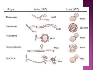
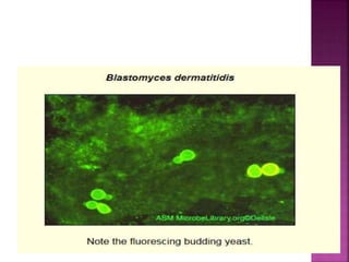
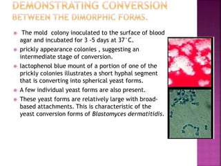
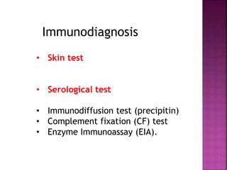
BLASTOMYCOSIS
(Blastomyces dermatitidis)
Sundar Khadka,
Microbiologist
HIV Reference Unit, NPHL
 Blastomycosis is a chronic granulomatous and
suppurative disease having a primary pulmonary
stage that is frequently followed by
dissemination to other body sites, chiefly the
skin and bone.
 Although the disease was long thought to be
restricted to the North American continent, in
recent years cases have been diagnosed in
Africa, Asia and Europe.
 Casper Gilchrist first reported the disease in
1894 from Baltimore in a patient with lesions
involving the skin.
 Clinical entity designated in his honour as
Gilchrist’s disease
 Chicago disease or North American
Blastomyces
 Thermal basis of dimorphism first described
by Hamburger in 1907
 Dermatitidis- infection of skin
 Acidic soils with rich organic debris such as
decaying vegetative matter
 rotting wood
 feces of birds and bats(providing increased
nitrogen content)
5
 General characteristics:

 Dimorphic fungi that behaves in two
different manners according to temperature
of environment :

 o Mold-like at 25 C
 § Branched hyphae are present

 o Yeast-like at 37 C
 § Broad-based yeast buds are present
 Dimorphic fungi
Macroscopic morphology
 Filamentous fungus form:
 Grown at 25 to 30oC:
 Exhibits a cottony or downy texture(covered with
fine, soft hair or feathers)
 Colonies produce white Arial hyphae on the surface
which may turn a yellowish to tan(pale brown)
colour as the colony ages. The reverse is typically
a light tan to brown.
 Moderately slow growth, usually maturing in about
2 weeks but suspect cultures should be held for 8
weeks
 Microscopic Morphology
 Filamentous fungus form
 septate hyphae
 Unbranched conidiophores of rather short, yet
varying length extend from the hyphae
 Conidia are hyaline (clear) and are produced
singly at the apex of the conidiophore or can
develop directly on the hyphae.
 Conidia are unicellular, round to pyriform (tear-
drop) in shape (~2 to 10 µm dia.)
 Conidia at the terminal end of the conidiophore
resemble a ‘lollipop’ in structure.
 Yeast form
 Grown at 37oC:
 Slow to moderate growth
 Best chance for conversion :Blood Agar or Brain-Heart
Infusion (BHI)) incubated in about 6% CO2.
 yeast form cream to tan in colour , heaped or wrinkled,
granular to verrucose (like a wart or warts).
 The yeast form of Blastomyces dermatitidis inhibited by
cycloheximide.
 Yeast Form
 Grown at 37oC: (enhanced by rich media and CO2, or in
infected tissue)
 Yeast-like cells (~8 to 15 µm dia.) exhibit a broad budding
base (4 to 5 µm dia.)
 broad‐based bud or “dumbbell-shaped” or letter 8
 The budding cell usually remains attached to the parent
cell, separating only when reaching the same size as the
parent.
 Yeast cells have thick, refractile walls.
 Older cultures may produce thick-walled
chlamydoconidia (7 to 18 µm dia.)
 Note that this organism forms a broad‐based bud,
whereas Cryptococcus neoformans is a yeast that forms a
narrow‐based bud.
 Two types
 AK type: worldwide
 K type: prevalent in Africa and smaller in size
 Portal of entry
 primary mycoses – respiratory portal; inhaled spores
 subcutaneous - inoculated skin; trauma
 cutaneous and superficial – contamination of skin
surface
•Humans are susceptible to acquiring infections by
close contact with dogs or through scratches and
bites.
 Virulence factors –
 thermal dimorphism
 toxin production
 capsules and adhesion factors
 hydrolytic enzymes
 inflammatory stimulants
 Transmission: inhalation of conidia à yeast form once in lungs
 Once inside the lungs, the change of environment (carbon dioxide
content, organic nutrients, pH and primarily the increased
temperature (37oC)), and causes the fungal spores to develop into the
yeast cell form.
 The yeast form is disseminate to other areas of the body via blood
 Symptoms may not appear for up to 120 days post infection.
 Inflammatory pyogranulomatous reactions occur at the
initial pulmonary site and at the widespread foci of
infection
 Pyogranulomatous is supparative of neutrophils followed
by granulomas of mononuclear cells.
 Mixed neutrophilic and mononuclear cells response is
distinctive of blastomycosis
 Necrosis and fibrosis may occur
 granuloma donot caseate
 Asymptomatic: 50% of infections
 Blastomycosis symptoms often mimic symptoms of upper
respiratory infections
 The incubation period is 30 to 100 days, although infection can
be asymptomatic
 When individuals are exposed to Blastomyces dermatitidis the
early symptoms may include:
 Approximately 25% of diagnosed Blastomycosis cases affect the
lungs.
 While 35% of diagnosed cases involve both the lungs and skin
 dry cough
 fever
 heavy sweating
 fatigue
 general feeling of ill health
1. Pulmonary Blastomycosis
 Acute Pulmonary: lobar or segmental consolidation,
mimics bacterial pneumonia
 Chronic Pulmonary: lobar infiltrates mimics
bronchogenic carcinoma
2. Cutaneous Blastomycosis
Skin: ulcerative lesions, subcutaneous nodules
that abscess
3.Osseous Blastomycosis
osteomyelitis
4. Disseminated Blastomycosis
 Bone / Joint: long bones, ribs, vertebrae
osteolytic
 Genitourinary Tract: prostate and epididymis
 Other organs: CNS, pericardium, adrenal gland,
GI
5. Miscellaneous types of Blastomycosis
 Extension of pulmonary
 Liver and spleen mostly affected
 Granulomatous lesions present
Skin lesions resulting from the
dissemination of the fungus from the
lungs
 Laboratory specimens depend on the
manifestation of the disease:
 pulmonary infection: sputum, BAL, Tracheal
secretions, transbronchial biopsy
 skin lesions: skin scrapings or pus, biopsy from
skin lesions
 Other specimens : biopsy of granulomatous
lesions, Exudates, urine for antigen detection
 Direct Demonstration
 KOH
 HE stain
 PAS stain
 GMS stain
 Broad based budding yeast cells
 Media:
1. Sabourauds dextrose agar,
2. BHI agar,
3. Blood agar,
4. Blood – Glucose- cysteine agar
 at 25°C and 37° C
 On Sabourauds dextrose agar25°C to 30° C:
 Initially the colony appears yeast-like at room temperature and then
develops hyphal projections, eventually becoming a fluffy white
mould.
 In microscopic Examination, B. dermatitidis has round to pyriform, 4
to 5 μm conidia attached directly to the hyphae or on short stalks.
 1.demonstrating conversion between the
dimorphic forms.(Mold to Yeast Conversion;
M to Y)
 Media: Brain Heart Infusion agar, Blood agar,
Blood-glucose- cysteine agar, Cottonseed agar,
Cysteine heart agar with rabbit blood
 2. identification using DNA probes
 3. testing for a specific exoantigen by
immunodiffusion.
 The mold colony inoculated to the surface of blood
agar and incubated for 3 -5 days at 37°C.
 prickly appearance colonies , suggesting an
intermediate stage of conversion.
 lactophenol blue mount of a portion of one of the
prickly colonies illustrates a short hyphal segment
that is converting into spherical yeast forms.
 A few individual yeast forms are also present.
 These yeast forms are relatively large with broad-
based attachments. This is characteristic of the
yeast conversion forms of Blastomyces dermatitidis.
• Skin test
• Serological test
• Immunodiffusion test (precipitin)
• Complement fixation (CF) test
• Enzyme Immunoassay (EIA).
Immunodiagnosis
• Antigen detection in urine
• Molecular Diagnosis
• DNA probes
• Intraperitoneal injection of conidia and
mycelial form produce infections in mice ,
guinea pigs, rats and hamsters
• Yeast form equally effective in causing
extensive lesions and death of mice
Animal Pathogenicity
CLS Blastomyces dermatitidis.pptx

CLS Blastomyces dermatitidis.pptx

  • 1.
  • 2.
     Blastomycosis isa chronic granulomatous and suppurative disease having a primary pulmonary stage that is frequently followed by dissemination to other body sites, chiefly the skin and bone.  Although the disease was long thought to be restricted to the North American continent, in recent years cases have been diagnosed in Africa, Asia and Europe.
  • 3.
     Casper Gilchristfirst reported the disease in 1894 from Baltimore in a patient with lesions involving the skin.  Clinical entity designated in his honour as Gilchrist’s disease  Chicago disease or North American Blastomyces  Thermal basis of dimorphism first described by Hamburger in 1907  Dermatitidis- infection of skin
  • 4.
     Acidic soilswith rich organic debris such as decaying vegetative matter  rotting wood  feces of birds and bats(providing increased nitrogen content)
  • 5.
  • 7.
     General characteristics:  Dimorphic fungi that behaves in two different manners according to temperature of environment :   o Mold-like at 25 C  § Branched hyphae are present   o Yeast-like at 37 C  § Broad-based yeast buds are present
  • 8.
     Dimorphic fungi Macroscopicmorphology  Filamentous fungus form:  Grown at 25 to 30oC:  Exhibits a cottony or downy texture(covered with fine, soft hair or feathers)  Colonies produce white Arial hyphae on the surface which may turn a yellowish to tan(pale brown) colour as the colony ages. The reverse is typically a light tan to brown.  Moderately slow growth, usually maturing in about 2 weeks but suspect cultures should be held for 8 weeks
  • 10.
     Microscopic Morphology Filamentous fungus form  septate hyphae  Unbranched conidiophores of rather short, yet varying length extend from the hyphae  Conidia are hyaline (clear) and are produced singly at the apex of the conidiophore or can develop directly on the hyphae.  Conidia are unicellular, round to pyriform (tear- drop) in shape (~2 to 10 µm dia.)  Conidia at the terminal end of the conidiophore resemble a ‘lollipop’ in structure.
  • 15.
     Yeast form Grown at 37oC:  Slow to moderate growth  Best chance for conversion :Blood Agar or Brain-Heart Infusion (BHI)) incubated in about 6% CO2.  yeast form cream to tan in colour , heaped or wrinkled, granular to verrucose (like a wart or warts).  The yeast form of Blastomyces dermatitidis inhibited by cycloheximide.
  • 16.
     Yeast Form Grown at 37oC: (enhanced by rich media and CO2, or in infected tissue)  Yeast-like cells (~8 to 15 µm dia.) exhibit a broad budding base (4 to 5 µm dia.)  broad‐based bud or “dumbbell-shaped” or letter 8  The budding cell usually remains attached to the parent cell, separating only when reaching the same size as the parent.
  • 17.
     Yeast cellshave thick, refractile walls.  Older cultures may produce thick-walled chlamydoconidia (7 to 18 µm dia.)  Note that this organism forms a broad‐based bud, whereas Cryptococcus neoformans is a yeast that forms a narrow‐based bud.
  • 18.
     Two types AK type: worldwide  K type: prevalent in Africa and smaller in size
  • 19.
     Portal ofentry  primary mycoses – respiratory portal; inhaled spores  subcutaneous - inoculated skin; trauma  cutaneous and superficial – contamination of skin surface •Humans are susceptible to acquiring infections by close contact with dogs or through scratches and bites.
  • 21.
     Virulence factors–  thermal dimorphism  toxin production  capsules and adhesion factors  hydrolytic enzymes  inflammatory stimulants
  • 22.
     Transmission: inhalationof conidia à yeast form once in lungs  Once inside the lungs, the change of environment (carbon dioxide content, organic nutrients, pH and primarily the increased temperature (37oC)), and causes the fungal spores to develop into the yeast cell form.  The yeast form is disseminate to other areas of the body via blood  Symptoms may not appear for up to 120 days post infection.
  • 23.
     Inflammatory pyogranulomatousreactions occur at the initial pulmonary site and at the widespread foci of infection  Pyogranulomatous is supparative of neutrophils followed by granulomas of mononuclear cells.  Mixed neutrophilic and mononuclear cells response is distinctive of blastomycosis  Necrosis and fibrosis may occur  granuloma donot caseate
  • 24.
     Asymptomatic: 50%of infections  Blastomycosis symptoms often mimic symptoms of upper respiratory infections  The incubation period is 30 to 100 days, although infection can be asymptomatic  When individuals are exposed to Blastomyces dermatitidis the early symptoms may include:  Approximately 25% of diagnosed Blastomycosis cases affect the lungs.  While 35% of diagnosed cases involve both the lungs and skin  dry cough  fever  heavy sweating  fatigue  general feeling of ill health
  • 25.
    1. Pulmonary Blastomycosis Acute Pulmonary: lobar or segmental consolidation, mimics bacterial pneumonia  Chronic Pulmonary: lobar infiltrates mimics bronchogenic carcinoma 2. Cutaneous Blastomycosis Skin: ulcerative lesions, subcutaneous nodules that abscess 3.Osseous Blastomycosis osteomyelitis
  • 26.
    4. Disseminated Blastomycosis Bone / Joint: long bones, ribs, vertebrae osteolytic  Genitourinary Tract: prostate and epididymis  Other organs: CNS, pericardium, adrenal gland, GI 5. Miscellaneous types of Blastomycosis  Extension of pulmonary  Liver and spleen mostly affected  Granulomatous lesions present
  • 27.
    Skin lesions resultingfrom the dissemination of the fungus from the lungs
  • 28.
     Laboratory specimensdepend on the manifestation of the disease:  pulmonary infection: sputum, BAL, Tracheal secretions, transbronchial biopsy  skin lesions: skin scrapings or pus, biopsy from skin lesions  Other specimens : biopsy of granulomatous lesions, Exudates, urine for antigen detection
  • 29.
     Direct Demonstration KOH  HE stain  PAS stain  GMS stain  Broad based budding yeast cells
  • 35.
     Media: 1. Sabouraudsdextrose agar, 2. BHI agar, 3. Blood agar, 4. Blood – Glucose- cysteine agar  at 25°C and 37° C  On Sabourauds dextrose agar25°C to 30° C:  Initially the colony appears yeast-like at room temperature and then develops hyphal projections, eventually becoming a fluffy white mould.  In microscopic Examination, B. dermatitidis has round to pyriform, 4 to 5 μm conidia attached directly to the hyphae or on short stalks.
  • 36.
     1.demonstrating conversionbetween the dimorphic forms.(Mold to Yeast Conversion; M to Y)  Media: Brain Heart Infusion agar, Blood agar, Blood-glucose- cysteine agar, Cottonseed agar, Cysteine heart agar with rabbit blood  2. identification using DNA probes  3. testing for a specific exoantigen by immunodiffusion.
  • 37.
     The moldcolony inoculated to the surface of blood agar and incubated for 3 -5 days at 37°C.  prickly appearance colonies , suggesting an intermediate stage of conversion.  lactophenol blue mount of a portion of one of the prickly colonies illustrates a short hyphal segment that is converting into spherical yeast forms.  A few individual yeast forms are also present.  These yeast forms are relatively large with broad- based attachments. This is characteristic of the yeast conversion forms of Blastomyces dermatitidis.
  • 38.
    • Skin test •Serological test • Immunodiffusion test (precipitin) • Complement fixation (CF) test • Enzyme Immunoassay (EIA). Immunodiagnosis
  • 39.
    • Antigen detectionin urine • Molecular Diagnosis • DNA probes
  • 40.
    • Intraperitoneal injectionof conidia and mycelial form produce infections in mice , guinea pigs, rats and hamsters • Yeast form equally effective in causing extensive lesions and death of mice Animal Pathogenicity