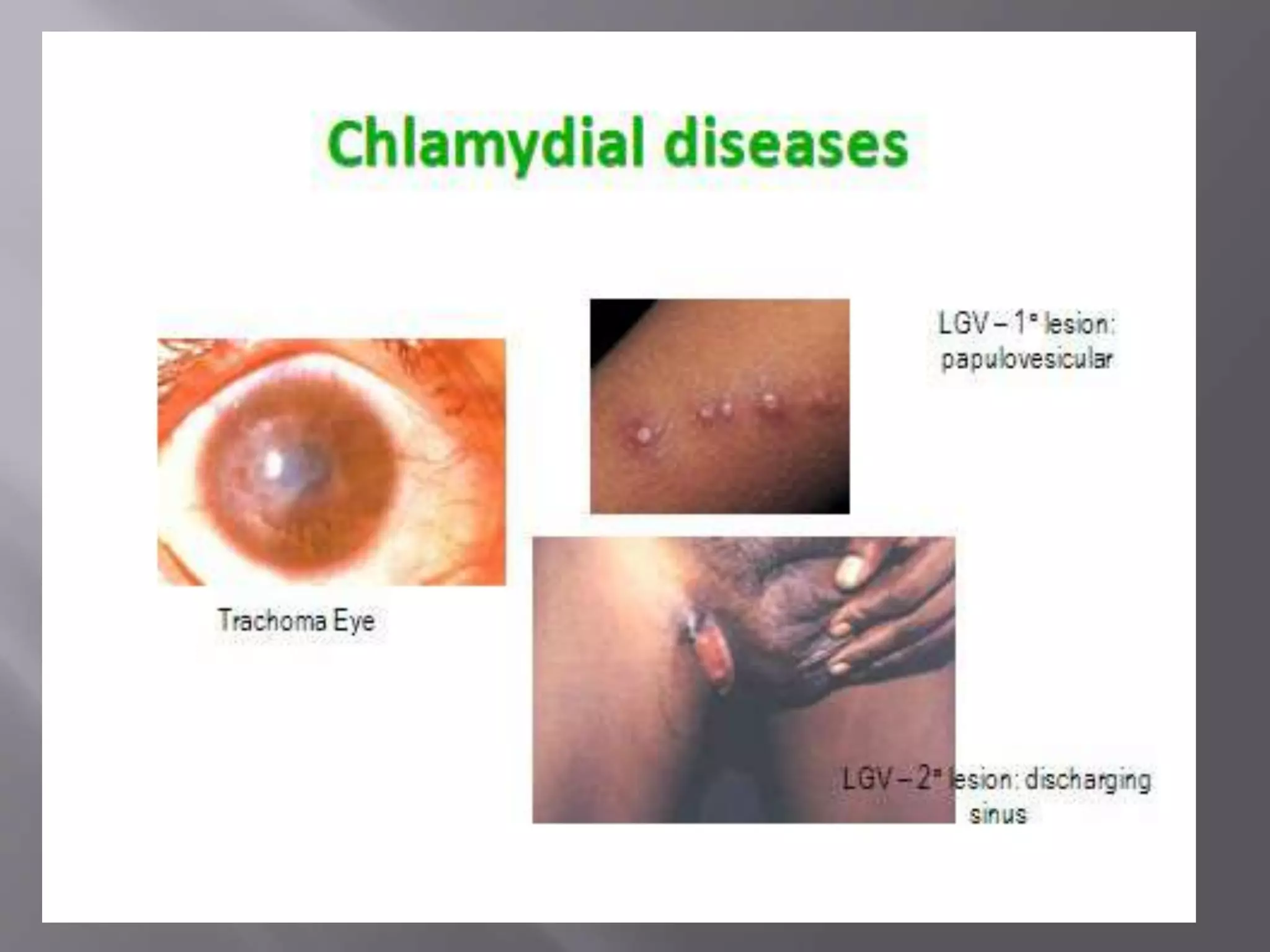
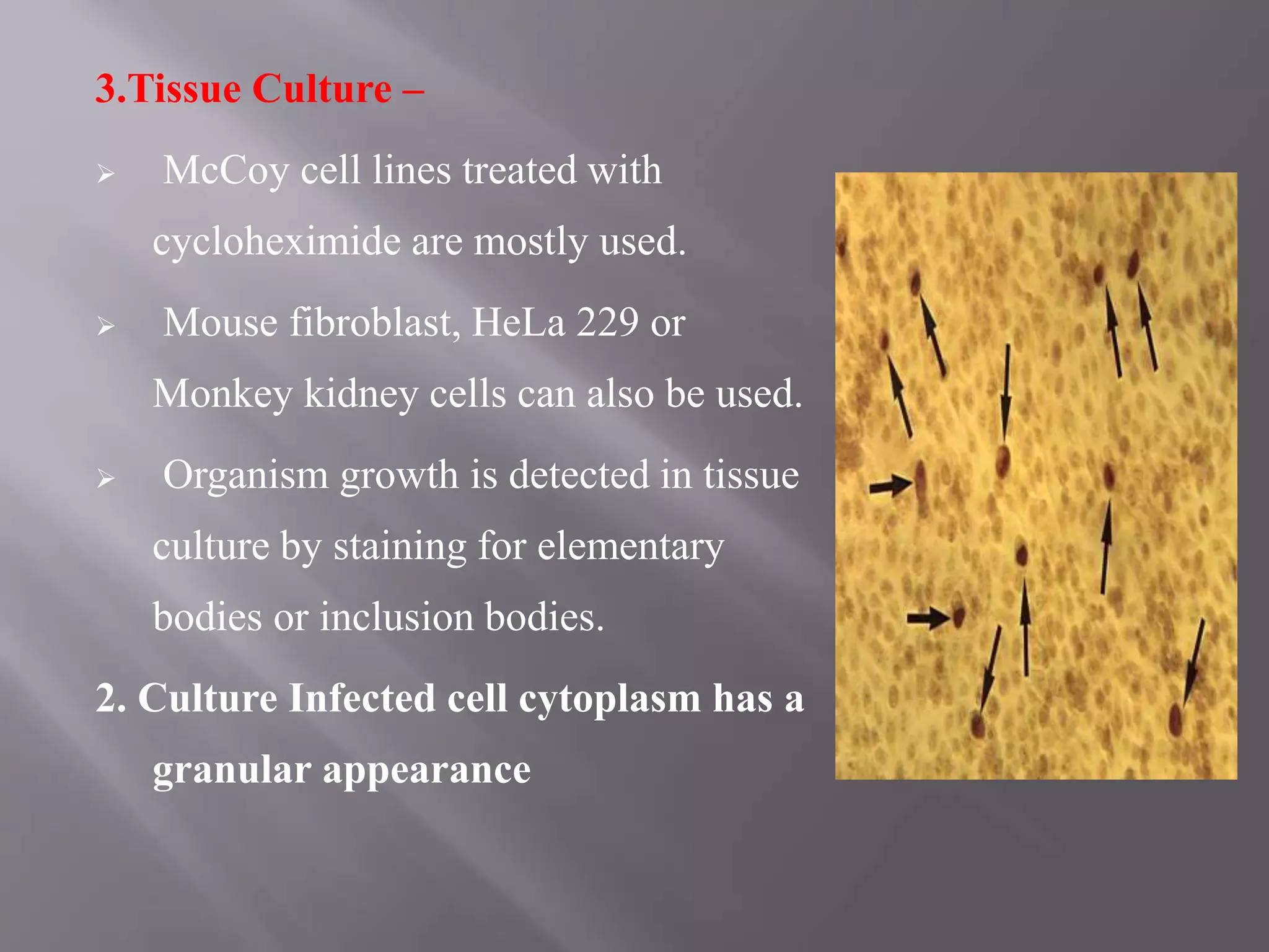
CHLAMYDIA
Dr. Manish tiwari
Department of microbiology
 Obligate intracellular parasites.
 Small, Non - motile, Gram negative. share antigens, have
both DNA and RNA, divide by binary fission.
 Have tropism for squamous epithelial cells & macrophages
of respiratory and gastrointestinal tracts.
• Causes
1. Trachoma,,
2. Lymphogranulloma Venerum ((LGV)),,
3. Psittacosis..
Can cause diverse diseases in birds and mammals.
 The name Chlamydia – Derived from characteristic
appearance of inclusion body by these agents.
 The inclusion bodies enclose the nuclei of infected cells as a
cloak or mantle(( Chlamys meaning =mantle = symbol of
authority))..
 Domain: Bacteria
 Phylum: Chlamydia
 Class: Chlamydiae
 Order: Chlamydiales
 Family: Chlamydiaceae
 Genus: Chlamydia
 Species: C. trachomatis, C ,C. pneumoniae, C. psittaci
C. pecorum affects ruminants (Only)
 Morphologically Chlamydia exist in two forms as
bellow
 Elementary Body / EB: -
 Spherical particle measuring 200- 300 nm in size.
 It is an Extracellular infective form.
 Reticulate (Initially) Body/ RB:-
 Non-infectious, and 500-1000 nm
 Intracellular, growing and replicating form.
 Elementary Body (EB) the infectious form enters the host cell
by phagocytosis.
 Enlarges its shape and size to develop into Reticulate Body
(RB), 500-1000 nm in size. RB divides repeatedly by binary
fusion to form large number of EB.
 The developing Chlamydia micro colony within host cells
known as Inclusion body each mature inclusion body contains
about 100 -500 EB.
 These newly formed EB’s, on release may infect the new cells
and the cycle continues.
 Duration of this developmental cycle is about 24- 48 hrs.
 20 serotypes, classified on the basis of
neutralization and Immunofluorescence tests.
 A, B, Ba, C, D, Da, E, F, G, H, I, Ia, J, Ja, K,
L1, L2, L2a, L2b & L3.
 Serotypes A, B, Ba, C – Responsible for
causing –
 Hyper endemic Trachoma – inflammation
of the conjunctiva and cornea and the
formation of scar tissue..
 Serotype D, Da, E, F, G, H, I, Ia, J, Ja, K – Causes –
 Inclusion Conjunctivitis,
 Non - Gonococcal Urethritis (NGU),
 Salpingitis – inflammation of fallopian tube,
 Cervicitis,
 Pneumonia of new born.
 Serotypes L1, L2, L2a, L2b, & L3 – cause
Lymphogranuloma Venereum (LGV) - A disease caused by
Chlamydia trachomatis L1 to L3;
 Transmitted by sexual contact; LGV is primarily an infection of
lymphatic's and lymph nodes.
 It gains entrance through breaks in the skin, or it can cross the
epithelial cell layer of mucous membranes.
 LGV is a sexually transmitted disease caused by C.
trachomatis serotypes L1 to L3.
 The primary lesion consist of small painless papule or
vesicle on external genitalia.
 It may ulcerate or heal spontaneously in a few days.
 The regional lymph nodes (inguinal in males and
intrapelvic &
 para - rectal in female) are enlarged, tender, and may
break
 open with the formation of sinuses.
 The enlarged inguinal lymph nodes are named bubo's.
 C. psittaci causes “Psittacosis and Ornitthosis” in birds and
man.
 Human infection occurs by inhalation of infected dried faeces.
 Psittacosis (Psittacos – parrot) is a disease of parrots.
 Disease acquired from non- psittacine birds known as
“Ornithosis” (Ornithos – birds).
 Incubation period – 1-2 wks.
 Disease may vary from mild influenza to severe illness with
pneumonia, septicemia and meningoencephalitis.
 Third commonest cause of pneumonia following S. pneumoniae
& H. influenzae.
 It is an important risk factor in cardiovascular disease, where
organism is isolated from coronary artery.
 However more studies are required to ascertain its role.
 Causes – Acute Respiratory Disease in humans.
 Has only 1 serotype.
 Serological studies shows prevalence rate – 40 -50 %.
Specie Serotype Sub species
C. trachomatis A,B,Ba, C Endemic blinding,,
Trachoma
C. trachomatis D to K Inclusion
conjunctivitis
Genital Chlamydiasis
C. trachomatis L1,, L2,, L3 Lymphogranuloma
Venereum (LGV)
C. psittaci Many serotypes Psittacosis ,,
Ornithosis
C. pneumoniae Only one serotype Acute Respiratory
Disease
C. pecorum Primary pathogen of
ruminants
 Caused by C. trachomatis serotype A, B, Ba, & C.
 Chronic Kerato--conjunctivitis.
 Major cause of blindness.
 Characterized by follicles, papillary hyperplasia, pannus
formation and in the late stage – cicauterization.
 Transmission – Eye to eye through finger, flies & fomites -
contaminated towel, clothing.
 Incubation period – 3-10 days.
 In endemic area children below 9 yrs age are mostly affected.
 Trachoma has been characterized into I – IV stages, early
stage is most infective one.
 Caused by C. trachomatis serotype D to K.
 Prevalent in sexually active young people &
spread from genital secretions to the eye by
contaminated hands contact.
 Characterized by follicular hypertrophy with
scanty no purulent discharge.
 This is formerly known as “ Swimming pool
conjunctivitis” as it was associated with
swimming in contaminated water.
 Also know as Para--trachoma..
 Neonatal form of Inclusion conjunctivitis – Inclusion Blenorrhea
 Infants acquires infection during passage through the infected
birth canal.
 It usually becomes apparent between 5-12 days after birth.
 About 20-50% infants of infected mothers develops the
infection.
 Considered benign & self limiting few may develop conjunctival
scar.
 Prevented by local antibiotic applications.
- Genital Chlamydiasis
 Chlamydia trachomatis causes “Genital Chlamydiasis”
and LGV.
 Both are sexually transmitted diseases.
 1. Genital Chlamydiasis -- Caused by D to K serotype.
 Responsible for 40% of cases of NGU, it is a STD.
In males
 Urethritis - inflammation of the urethra; results in painful
 urination.
 Epididymitis - painful inflammation of the epididymis –
 convoluted tubule in each testis, carries sperm to vas
difference.
 Proctitis - inflammation of the rectum; marked by bloody
stools and a frequent urge to defecate.
In females
 Urethritis ,Cervicitis -
 Salpingitis -inflammation of Fallopian tube (usually the result of
infection spreading from the vagina or uterus).
 Pelvic Inflammatory Disease (PID) - Inflammation of the female
pelvic organs (especially the Fallopian tubes) caused by infection
chiefly Gonococci and Chlamydia;; symptoms are abdominal
pain and fever and foul-smelling vaginal discharge.
 Infection may symptomatic or asymptomatic.
 Symptoms – Dysuria –painful or difficulty in urination, Non-
purulent discharge and frequency of urination.
 Third commonest cause of pneumonia following S. pneumoniae
& H. influenzae.
 It is an important risk factor in cardiovascular disease, where
organism is isolated from coronary artery.
 However more studies are required to ascertain its role.
 Causes – Acute Respiratory Disease in humans.
 Has only 1 serotype.
 Serological studies shows prevalence rate – 40 -50 %.
Specimen
 Scrapings or swabs - sample must contain cells
 „Urethral swab for NGU
 „End cervical swab for cervicitis
 Conjunctival swabs for ocular infections
 ™First catch urine samples in the morning
 ™Nasopharyngeal aspirate
 Bubo aspirate for LGV.
4 approaches available:
1. Microscopic demonstration of inclusion or elementary bodies.
2. Isolation of Chlamydia.
3. Demonstration of Chlamydial Ag.
4. Demonstration of Abs or hypersensitivity.
1. Microscopy
 Gram stain – Gram Negative,.
 Stain better with Castaneda, Mchiavello, or
Giemsa Stain.
 Inclusion bodies are basophilic & present in
cytoplasm.
 Inclusion bodies can be stained with -
Lugol’s iodine – because of presence of
Glycogen matrix.
 Immunofluorescence staining.
 Animal inoculation Yolk sac of 6 - 8
days old chick embryo.
 Tissue culture –
 McCoy, HeLa cell lines
 Infected cell cytoplasm has a
granular appearance
1. Animal inoculation – Mice are inoculated by intranasal or
intraperitoneal or by intracerebral inoculations.
 Mice die within 10 days and smears from various tissue
(lung, peritoneal exudates, spleen or brain) shows elementary
bodies.
2. Yolk Sac Inoculation – Yolk sac of chick embryo is
inoculated & the organism can be detected in impression
 smears stained by Giemsa stain.
3.Tissue Culture –
 McCoy cell lines treated with
cycloheximide are mostly used.
 Mouse fibroblast, HeLa 229 or
Monkey kidney cells can also be used.
 Organism growth is detected in tissue
culture by staining for elementary
bodies or inclusion bodies.
2. Culture Infected cell cytoplasm has a
granular appearance
 Tetracycline, Erythromycin and Sulfonamides.
 Vaccination not proved to be an effective or practicable
method of control.
 Treatment coupled with improved sanitation.
 Safe sexual practices.
 Treatment of patients and their sexual partners.
Chlamydia

Chlamydia

  • 1.
  • 2.
     Obligate intracellularparasites.  Small, Non - motile, Gram negative. share antigens, have both DNA and RNA, divide by binary fission.  Have tropism for squamous epithelial cells & macrophages of respiratory and gastrointestinal tracts. • Causes 1. Trachoma,, 2. Lymphogranulloma Venerum ((LGV)),, 3. Psittacosis..
  • 3.
    Can cause diversediseases in birds and mammals.  The name Chlamydia – Derived from characteristic appearance of inclusion body by these agents.  The inclusion bodies enclose the nuclei of infected cells as a cloak or mantle(( Chlamys meaning =mantle = symbol of authority))..
  • 5.
     Domain: Bacteria Phylum: Chlamydia  Class: Chlamydiae  Order: Chlamydiales  Family: Chlamydiaceae  Genus: Chlamydia  Species: C. trachomatis, C ,C. pneumoniae, C. psittaci C. pecorum affects ruminants (Only)
  • 6.
     Morphologically Chlamydiaexist in two forms as bellow  Elementary Body / EB: -  Spherical particle measuring 200- 300 nm in size.  It is an Extracellular infective form.  Reticulate (Initially) Body/ RB:-  Non-infectious, and 500-1000 nm  Intracellular, growing and replicating form.
  • 7.
     Elementary Body(EB) the infectious form enters the host cell by phagocytosis.  Enlarges its shape and size to develop into Reticulate Body (RB), 500-1000 nm in size. RB divides repeatedly by binary fusion to form large number of EB.  The developing Chlamydia micro colony within host cells known as Inclusion body each mature inclusion body contains about 100 -500 EB.  These newly formed EB’s, on release may infect the new cells and the cycle continues.  Duration of this developmental cycle is about 24- 48 hrs.
  • 9.
     20 serotypes,classified on the basis of neutralization and Immunofluorescence tests.  A, B, Ba, C, D, Da, E, F, G, H, I, Ia, J, Ja, K, L1, L2, L2a, L2b & L3.  Serotypes A, B, Ba, C – Responsible for causing –  Hyper endemic Trachoma – inflammation of the conjunctiva and cornea and the formation of scar tissue..
  • 10.
     Serotype D,Da, E, F, G, H, I, Ia, J, Ja, K – Causes –  Inclusion Conjunctivitis,  Non - Gonococcal Urethritis (NGU),  Salpingitis – inflammation of fallopian tube,  Cervicitis,  Pneumonia of new born.
  • 11.
     Serotypes L1,L2, L2a, L2b, & L3 – cause Lymphogranuloma Venereum (LGV) - A disease caused by Chlamydia trachomatis L1 to L3;  Transmitted by sexual contact; LGV is primarily an infection of lymphatic's and lymph nodes.  It gains entrance through breaks in the skin, or it can cross the epithelial cell layer of mucous membranes.
  • 12.
     LGV isa sexually transmitted disease caused by C. trachomatis serotypes L1 to L3.  The primary lesion consist of small painless papule or vesicle on external genitalia.  It may ulcerate or heal spontaneously in a few days.  The regional lymph nodes (inguinal in males and intrapelvic &  para - rectal in female) are enlarged, tender, and may break  open with the formation of sinuses.  The enlarged inguinal lymph nodes are named bubo's.
  • 13.
     C. psittacicauses “Psittacosis and Ornitthosis” in birds and man.  Human infection occurs by inhalation of infected dried faeces.  Psittacosis (Psittacos – parrot) is a disease of parrots.  Disease acquired from non- psittacine birds known as “Ornithosis” (Ornithos – birds).  Incubation period – 1-2 wks.  Disease may vary from mild influenza to severe illness with pneumonia, septicemia and meningoencephalitis.
  • 15.
     Third commonestcause of pneumonia following S. pneumoniae & H. influenzae.  It is an important risk factor in cardiovascular disease, where organism is isolated from coronary artery.  However more studies are required to ascertain its role.  Causes – Acute Respiratory Disease in humans.  Has only 1 serotype.  Serological studies shows prevalence rate – 40 -50 %.
  • 16.
    Specie Serotype Subspecies C. trachomatis A,B,Ba, C Endemic blinding,, Trachoma C. trachomatis D to K Inclusion conjunctivitis Genital Chlamydiasis C. trachomatis L1,, L2,, L3 Lymphogranuloma Venereum (LGV) C. psittaci Many serotypes Psittacosis ,, Ornithosis C. pneumoniae Only one serotype Acute Respiratory Disease C. pecorum Primary pathogen of ruminants
  • 17.
     Caused byC. trachomatis serotype A, B, Ba, & C.  Chronic Kerato--conjunctivitis.  Major cause of blindness.  Characterized by follicles, papillary hyperplasia, pannus formation and in the late stage – cicauterization.  Transmission – Eye to eye through finger, flies & fomites - contaminated towel, clothing.  Incubation period – 3-10 days.  In endemic area children below 9 yrs age are mostly affected.  Trachoma has been characterized into I – IV stages, early stage is most infective one.
  • 18.
     Caused byC. trachomatis serotype D to K.  Prevalent in sexually active young people & spread from genital secretions to the eye by contaminated hands contact.  Characterized by follicular hypertrophy with scanty no purulent discharge.  This is formerly known as “ Swimming pool conjunctivitis” as it was associated with swimming in contaminated water.  Also know as Para--trachoma..
  • 19.
     Neonatal formof Inclusion conjunctivitis – Inclusion Blenorrhea  Infants acquires infection during passage through the infected birth canal.  It usually becomes apparent between 5-12 days after birth.  About 20-50% infants of infected mothers develops the infection.  Considered benign & self limiting few may develop conjunctival scar.  Prevented by local antibiotic applications.
  • 20.
    - Genital Chlamydiasis Chlamydia trachomatis causes “Genital Chlamydiasis” and LGV.  Both are sexually transmitted diseases.  1. Genital Chlamydiasis -- Caused by D to K serotype.  Responsible for 40% of cases of NGU, it is a STD. In males  Urethritis - inflammation of the urethra; results in painful  urination.
  • 21.
     Epididymitis -painful inflammation of the epididymis –  convoluted tubule in each testis, carries sperm to vas difference.  Proctitis - inflammation of the rectum; marked by bloody stools and a frequent urge to defecate.
  • 22.
    In females  Urethritis,Cervicitis -  Salpingitis -inflammation of Fallopian tube (usually the result of infection spreading from the vagina or uterus).  Pelvic Inflammatory Disease (PID) - Inflammation of the female pelvic organs (especially the Fallopian tubes) caused by infection chiefly Gonococci and Chlamydia;; symptoms are abdominal pain and fever and foul-smelling vaginal discharge.  Infection may symptomatic or asymptomatic.  Symptoms – Dysuria –painful or difficulty in urination, Non- purulent discharge and frequency of urination.
  • 23.
     Third commonestcause of pneumonia following S. pneumoniae & H. influenzae.  It is an important risk factor in cardiovascular disease, where organism is isolated from coronary artery.  However more studies are required to ascertain its role.  Causes – Acute Respiratory Disease in humans.  Has only 1 serotype.  Serological studies shows prevalence rate – 40 -50 %.
  • 24.
    Specimen  Scrapings orswabs - sample must contain cells  „Urethral swab for NGU  „End cervical swab for cervicitis  Conjunctival swabs for ocular infections  ™First catch urine samples in the morning  ™Nasopharyngeal aspirate  Bubo aspirate for LGV.
  • 25.
    4 approaches available: 1.Microscopic demonstration of inclusion or elementary bodies. 2. Isolation of Chlamydia. 3. Demonstration of Chlamydial Ag. 4. Demonstration of Abs or hypersensitivity.
  • 26.
    1. Microscopy  Gramstain – Gram Negative,.  Stain better with Castaneda, Mchiavello, or Giemsa Stain.  Inclusion bodies are basophilic & present in cytoplasm.  Inclusion bodies can be stained with - Lugol’s iodine – because of presence of Glycogen matrix.  Immunofluorescence staining.
  • 27.
     Animal inoculationYolk sac of 6 - 8 days old chick embryo.  Tissue culture –  McCoy, HeLa cell lines  Infected cell cytoplasm has a granular appearance
  • 28.
    1. Animal inoculation– Mice are inoculated by intranasal or intraperitoneal or by intracerebral inoculations.  Mice die within 10 days and smears from various tissue (lung, peritoneal exudates, spleen or brain) shows elementary bodies. 2. Yolk Sac Inoculation – Yolk sac of chick embryo is inoculated & the organism can be detected in impression  smears stained by Giemsa stain.
  • 29.
    3.Tissue Culture – McCoy cell lines treated with cycloheximide are mostly used.  Mouse fibroblast, HeLa 229 or Monkey kidney cells can also be used.  Organism growth is detected in tissue culture by staining for elementary bodies or inclusion bodies. 2. Culture Infected cell cytoplasm has a granular appearance
  • 30.
     Tetracycline, Erythromycinand Sulfonamides.  Vaccination not proved to be an effective or practicable method of control.  Treatment coupled with improved sanitation.  Safe sexual practices.  Treatment of patients and their sexual partners.