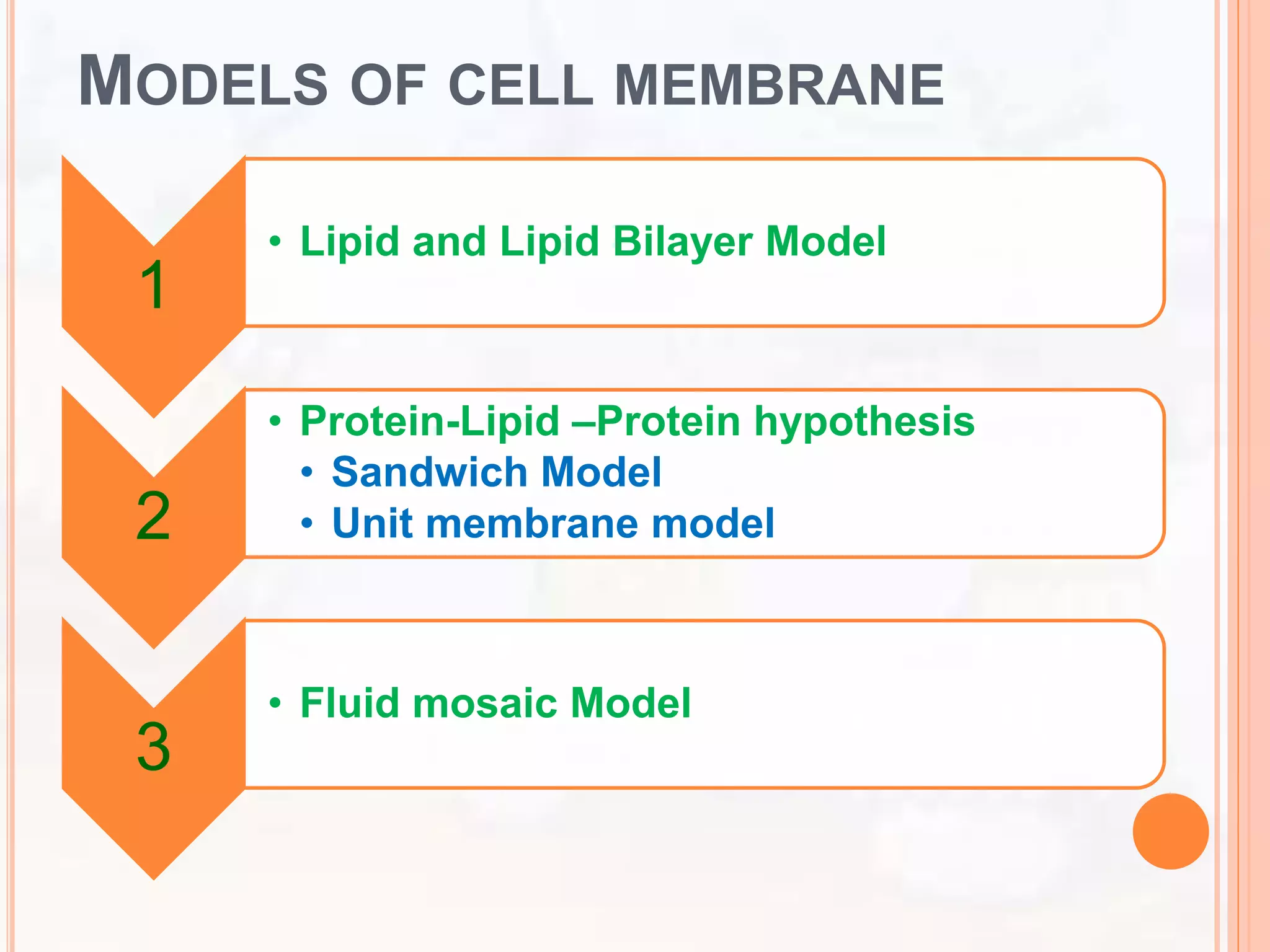
SlideShare a Scribd company logo
CELL MEMBRA
BY: Dr. Sunita Sangwan
Assistant Professor
Dept of Botany
Govt. College Bhiwani
INTRODUCTION
 Every cell, prokaryotic or eukaryotic, is surrounded by a
thin layer of outermost boundary called the plasma
membrane or cell membrane or plasma - lemma.
 It maintains the difference of the internal environment of
the cell from its external environment by controlling the
entrance and exit of the molecules and ions.
 It checks the loss of metabolically useful substances
and encourages the release of toxic metabolic
byproducts of the cell. Thus, it functions as semi-
permeable or selectively permeable membrane.
 It is about 70-100A in thickness.
 It is an important cell organelle composed of lipids and
proteins.
HISTORY
 It had been shown by Karl W. Nageli (1817-1891)
that the cell membrane is semi-permeable and is
responsible for the osmotic and other related
phenomena exhibited by living cells.
 Before 1855, he used the term zellen membrane in
his early papers.
 The term plasma membrane was used in 1855 by
him to describe the membrane as a firm protective
film that is formed by out flowing cytoplasm of an
injured cell when protein rich cell sap came in
contact with water.
MODELS OF CELL MEMBRANE
1
• Lipid and Lipid Bilayer Model
2
• Protein-Lipid –Protein hypothesis
• Sandwich Model
• Unit membrane model
3
• Fluid mosaic Model
LIPID AND LIPID BILAYER MODEL:
 This model explain the structure of plasma membrane given by
Overton, Gorter and Grendel.
 Previously only indirect information was available to explain the
structure of plasma membrane.
 In 1902, Overton observed that substances soluble in lipid could
selectively pass through the membranes. On this basis he stated
that plasma membrane is composed of a thin layer of lipid.
 Subsequently, Gorter and Grendel in 1926 observed that the
extracted from erythrocyte membranes was twice the amount
expected if a single layer was present throughout the surface area
of these cells. On this basis they stated that plasma membrane is
made up of double layer of lipid molecules.
 These models of Gorter and Grendel could not explain the proper
structure of plasma membrane but they put the foundation of future
models of membrane structure.
PROTEIN-LIPID –PROTEIN HYPOTHESIS
 This hypothesis was proposed by Davson Daniell and Robertson.
 When surface tension measurements made on the membranes, it
suggests the presence of proteins. After the existence of proteins the initial
lipid bilayer model proposed by Gorter and Grendel was modified. It was
suggested that surface tension of cells is much lower than what one would
expect if only lipids were involved.
 It may also be observed that if protein is added to model lipid water system,
surface tension is lowered. This suggested indirectly the presence of
proteins. On this basis Davson and Danielli proposed that plasma
membrane contained a lipid bilayer with protein on both surfaces.
 Initially they supposed that proteins existed as covalently bonded globular
structures bound to the polar ends of lipids. Subsequently they developed
the model in which the protein appears to be smeared over the hydrophilic
ends of the lipid bilayer. This model makes its popularity for a long time.
 With the availability of electron microscope later, fine structure of plasma
membrane could be studied. Definite plasma membrane of 6 nm to 10 nm
(10nm = 100 Å; 1 nm = 10_6mm) thickness was observed on surface of all
cells, and plasma membranes of two adjacent cells were found to be
separated by a space, 1-15nm wide.
DANIELLI AND DAVSON MODEL (SANDWICH
MODEL)
 Harvey and Coley (1931) and Danielli and Harvey (1935) studied surface
tension of cell membrane and on the basis of their observation they
pointed out the existence of protein molecules adsorbed on the surface of
lipid droplets which reduce the surface tension of droplets.
 This conclusion led James Danielli and Hugh Davson in 1935 to suggest
bimolecular leaflet model of cell membrane. Danielli and Davson model
was the first attempt to describe membrane structure in terms of
molecules and to relate the structure to biological and chemical
properties.
 According to bimolecular model of Danielli and Davson, plasma
membrane consists of two layers of phospholipid molecules (a
bimolecular leaflet) in which phospholipid molecules are arranged in such
a way that hydrophilic heads of the phospholipid molecules face outside
and hydrophobic non-polar lipid chains are associated in the inner region
of leaflet.
 The hypothesis also suggested that the polar ends of lipid molecules are
associated with monomolecular layer of globular proteins.
 The plasma membrane would thus consist of a double layer of
phospholipid molecules sandwiched between two essentially continuous
layers of protein.
PROBLEMS WITH THE DAVSON-DANIELLI
MODEL
By the end of the 1960s, new evidence cast doubts
on the viability of the Davson-Daniell model.
 The amount and type of membrane proteins vary
greatly between different cells.
 It was unclear hoe the protein in the model would
permit the membrane to change the shape without
bonds being broken.
 Membrane proteins are largely hydrophobic and
therefore should not be found where the model
positioned them: in the aqueous cytoplasm and
extracellular environment.
……
 This basic model has been modified from time to time. Danielli
(1938) suggested the presence of two types of proteins;
tangentially arranged in contact with the lipid and globular
proteins on the outer surface. Again Davson and Danielli
(1943) and Danielli (1954) considered proteins to be in the
form of a folded P-chain.
 Perhaps, these units form micelles of membranes indicated in
recent electron micrographs. Membrane models are usually
postulated to contain protein lined polar pores of about 7 Å
diameter which probably permit the passage of small ions and
water molecules across the membrane.
 In still other variations the proteins are thought to be in coiled
or globular form on both sides of lipid layers [Fig. 2.3 (A), (B),
(C)] or they are thought to be asymmetrical, with a folded P-
chain on one side and globular proteins on the other [Fig. 2.3
(D)]. Models with globular proteins on both the sides or with
folded P-proteins on both the surfaces and helical proteins
extending into the pores are also suggested.
UNIT MEMBRANE MODEL
 In 1950 J. David Robertson studied the cell membranes from
electron micrographs of sectioned material. The preparations
involved usual fixing in solutions of osmium tetraoxide and
potassium permanganate (KMnO4), and dehydrating in solvents
such as acetone before sectioning.
 In late 1950s Robertson summarized a large number of ultra-
structural data obtained by him and some other workers and
concluded that the plasma membrane and the membranes of all
cell organalles were similar in structure. Although the similarity is
not resolved by light microscopy, it is clearly seen in electron
micrographs.
 This conclusion led Robertson in 1953 to propose unit membrane
hypothesis according to which all biological membranes show
generalised unit membrane construction.
 The unit membrane model visualises cell membrane as a
trilaminar and indicates structure consisting of two dark
osmiophilic layers separated by a light osmiophilic layer.
 The physical appearance of this trilaminar model has led
to the term unit membrane. The unit membrane concept
implies a trilaminar appearance with a bimolecular lipid
layer between two protein layers.
 Each dense osmiophilic band is made up of protein (20
Å) and the polar groups of phospholipids (5 A) and is
thus 25 Å thick.
 The clear Osmiophilic zone 35 A in thickness is a
bimolecular layer of lipids without the polar groups.
 In other words, the unit membrane is 75 Å thick with a
35 Å thick phospholipid layer between two 20 Å thick
protein layers.
 The plasma membrane surrounding the cell is thicker at
the free surfaces of the cell than where it is in contact
with other cells.
 In unit membrane model the protein layers are
assymetrical. On the outer surface it is mucoprotein
while on the inner surface it is non-mucoid protein.
FLUID MOSAIC MODEL:
 The fluid mosaic model of cell membrane was proposed in
1972 by S.J. Singer and G.L. Nicolson.
 According to this model, the cell membranes have been
visualised as mosaics of lipids and proteins. The lipids are
thought to be arranged primarily in a bilayer in which
peripheral and integral proteins are embedded.
 Membrane proteins are not fixed within the lipid layer but are
free to move laterally like icebergs floating in a sea of lipids.
This picture has inspired Singer and Nicolson to coin fluid
mosaic model.
 Singer and Nicolson considered the lipoprotein association to
be hydrophobic and fluidity of the membrane results due to
hydrophobic interaction. It should be noted that phospholipids
and many intrinsic proteins are amphipatic molecules, i.e.,
both hydrophilic and hydrophobic groups occur within the
same molecule.
cont…
 The globular proteins of the membrane are of two
different types: extrinsic (peripheral protein) and
intrinsic (integral proteins).
 Because of rapid movement of lipid and protein
molecules, the fluid mosaic model is different from the
static picture of the membrane in Danielli and Davson
model. The proteins of the membrane are concerned
with the enzymatic activities, transport of molecules
and with receptor function. The lipid bilayer acts as the
permeability barrier.
 The fluidity of lipid is supported by many indirect
studies based on x-ray diffraction, differential thermal
analysis and electron spin resonance (ESR)
techniques.
cont…
DYNAMIC PROPERTIES OF LIPID BILAYER
1. The rapid motion involving flexing within each lipid
molecule is possible.
2. A rapid lateral diffusion of lipid is possible.
3. Slow motion of lipid molecule from one side of the
bilayer to the other is also possible.
4. The lipid molecules might rotate about their axis.
5. The fluid mosaic model of cell membrane is now widely
accepted as it is presumed to apply to membranes of
all types regardless of their varying characteristics and
differences in lipids protein ratio.
6. In fact this model can account for the molecular
organisation and ultrastructure of membranes in-terms
of their chemical composition.
The possible movements of phospholipids in a
membrane. The types of movements in which
membrane
 phospholipids can engage and the approximate time scales over
which they occur.
 Whereas phospholipids move from one leaflet to another at a
very slow rate, they diffuse laterally within a leaflet rapidly.
 Lipids lacking polar groups, such as cholesterol, can move
across the bilayer quite rapidly.
Lateral movement occurs
107 times per second.
Flip-flopping across the
Membrane is rare
( once per month).
ULTRA-STRUCTURE OF CELL
MEMBRANE
COMPOSITION OF CELL
MEMBRANE
MEMBRANE FLUIDITY
 The structure of the fatty acid tails of the phospholipids is important in determining the properties of the
membrane, and in particular, how fluid it is.
 Saturated fatty acids have no double bonds (are saturated with hydrogens), so they are relatively
straight. Unsaturated fatty acids, on the other hand, contain one or more double bonds, often resulting in a bend
or kink. (You can see an example of a bent, unsaturated tail in the diagram of phospholipid structure that appears
earlier in this article.) The saturated and unsaturated fatty acid tails of phospholipids behave differently as
temperature drops:
 At cooler temperatures, the straight tails of saturated fatty acids can pack tightly together, making a dense and
fairly rigid membrane.
 Phospholipids with unsaturated fatty acid tails cannot pack together as tightly because of the bent structure of the
tails. Because of this, a membrane containing unsaturated phospholipids will stay fluid at lower temperatures than
a membrane made of saturated ones.
 Most cell membranes contain a mixture of phospholipids, some with two saturated (straight) tails and others with
one saturated and one unsaturated (bent) tail. Many organisms—fish are one example—can adjust
physiologically to cold environments by changing the proportion of unsaturated fatty acids in their membranes.
For more information about saturated and unsaturated fatty acids, see the article on lipids.
 In addition to phospholipids, animals have an additional membrane component that helps to maintain
fluidity. Cholesterol, another type of lipid that is embedded among the phospholipids of the membrane, helps to
minimize the effects of temperature on fluidity.
 Image credit: "Cholesterol," by BorisTM (public domain).
 At low temperatures, cholesterol increases fluidity by keeping phospholipids from packing tightly together, while
at high temperatures, it actually reduces fluidity^{3,4}3,4start superscript, 3, comma, 4, end superscript. In this
way, cholesterol expands the range of temperatures at which a membrane maintains a functional, healthy fluidity.
 As temperatures cool, membranes switch from a fluid
state to a solid state
 The temperature at which a membrane solidifies
depends on the types of lipids
 Membranes rich in unsaturated fatty acids are more fluid
than those rich in saturated fatty acids
 Membranes must be fluid to work properly; they are
usually about as fluid as salad oil
 The steroid cholesterol has different effects on
membrane fluidity at different temperatures
 At warm temperatures (such as 37°C), cholesterol
restrains movement of phospholipids
 At cool temperatures, it maintains fluidity by preventing
tight packing
FIGURE 7.8
Fluid
Unsaturated hydrocarbon
tails
Viscous
Saturated hydrocarbon tails
(a) Unsaturated versus saturated hydrocarbon tails
(b) Cholesterol within the animal
cell membrane
Cholesterol

More Related Content

What's hot

TCA CYCLE
TCA CYCLETCA CYCLE
TCA CYCLE
patel sahebb
 
GLYCOGENESIS
GLYCOGENESISGLYCOGENESIS
GLYCOGENESIS
YESANNA
 
Enzymes definitions, types & classification
Enzymes   definitions, types & classificationEnzymes   definitions, types & classification
Enzymes definitions, types & classification
JasmineJuliet
 
Chemiosmotic theory
Chemiosmotic theoryChemiosmotic theory
Chemiosmotic theory
Dr.M.Prasad Naidu
 
Enzymes
EnzymesEnzymes
Cell membrane transduction 1
Cell membrane transduction 1Cell membrane transduction 1
Gluconeogenesis
GluconeogenesisGluconeogenesis
Gluconeogenesis
Zuli Shingala
 
Determination of primary structure of proteins
Determination of primary structure of proteinsDetermination of primary structure of proteins
Determination of primary structure of proteins
Pradeep Singh Narwat
 
Mechanism of action of enzymes- By Hurnaum Karishma (Student SSR Medical Coll...
Mechanism of action of enzymes- By Hurnaum Karishma (Student SSR Medical Coll...Mechanism of action of enzymes- By Hurnaum Karishma (Student SSR Medical Coll...
Mechanism of action of enzymes- By Hurnaum Karishma (Student SSR Medical Coll...
Namrata Chhabra
 
Enzyme structure, classification _ and _mechanism of action _ _
  Enzyme structure, classification _ and _mechanism of action _ _  Enzyme structure, classification _ and _mechanism of action _ _
Enzyme structure, classification _ and _mechanism of action _ _
Muhammad Talha Hayat
 
Oxidative phosphorylation
Oxidative phosphorylationOxidative phosphorylation
Oxidative phosphorylation
devadevi666
 
Red cell membrane
Red cell membraneRed cell membrane
Red cell membrane
Hoor Rounaq
 
GOLGI APPARATUS .pdf
GOLGI APPARATUS .pdfGOLGI APPARATUS .pdf
GOLGI APPARATUS .pdf
AzkaJavaid6
 
Membrane proteins
Membrane proteinsMembrane proteins
Membrane proteins
Lovnish Thakur
 
Enzyme regulation
Enzyme regulationEnzyme regulation
Enzyme regulation
Purnima Kartha
 
Active site of enzyme
Active site of enzymeActive site of enzyme
Active site of enzyme
avinash tiwari
 
Proteoglycans
Proteoglycans Proteoglycans
Proteoglycans
Shruthi Shree Gandhi
 
Membrane Dynamics:Properties of biological membrane (plasma membrane)
Membrane Dynamics:Properties of biological membrane (plasma membrane)Membrane Dynamics:Properties of biological membrane (plasma membrane)
Membrane Dynamics:Properties of biological membrane (plasma membrane)
Manju Chhetri
 

What's hot (20)

TCA CYCLE
TCA CYCLETCA CYCLE
TCA CYCLE
 
GLYCOGENESIS
GLYCOGENESISGLYCOGENESIS
GLYCOGENESIS
 
Enzymes definitions, types & classification
Enzymes   definitions, types & classificationEnzymes   definitions, types & classification
Enzymes definitions, types & classification
 
Chemiosmotic theory
Chemiosmotic theoryChemiosmotic theory
Chemiosmotic theory
 
Enzymes
EnzymesEnzymes
Enzymes
 
Cell membrane transduction 1
Cell membrane transduction 1Cell membrane transduction 1
Cell membrane transduction 1
 
Classification of protein
Classification of proteinClassification of protein
Classification of protein
 
Gluconeogenesis
GluconeogenesisGluconeogenesis
Gluconeogenesis
 
Determination of primary structure of proteins
Determination of primary structure of proteinsDetermination of primary structure of proteins
Determination of primary structure of proteins
 
Mechanism of action of enzymes- By Hurnaum Karishma (Student SSR Medical Coll...
Mechanism of action of enzymes- By Hurnaum Karishma (Student SSR Medical Coll...Mechanism of action of enzymes- By Hurnaum Karishma (Student SSR Medical Coll...
Mechanism of action of enzymes- By Hurnaum Karishma (Student SSR Medical Coll...
 
Enzyme structure, classification _ and _mechanism of action _ _
  Enzyme structure, classification _ and _mechanism of action _ _  Enzyme structure, classification _ and _mechanism of action _ _
Enzyme structure, classification _ and _mechanism of action _ _
 
Oxidative phosphorylation
Oxidative phosphorylationOxidative phosphorylation
Oxidative phosphorylation
 
Red cell membrane
Red cell membraneRed cell membrane
Red cell membrane
 
GOLGI APPARATUS .pdf
GOLGI APPARATUS .pdfGOLGI APPARATUS .pdf
GOLGI APPARATUS .pdf
 
Lysosomes
LysosomesLysosomes
Lysosomes
 
Membrane proteins
Membrane proteinsMembrane proteins
Membrane proteins
 
Enzyme regulation
Enzyme regulationEnzyme regulation
Enzyme regulation
 
Active site of enzyme
Active site of enzymeActive site of enzyme
Active site of enzyme
 
Proteoglycans
Proteoglycans Proteoglycans
Proteoglycans
 
Membrane Dynamics:Properties of biological membrane (plasma membrane)
Membrane Dynamics:Properties of biological membrane (plasma membrane)Membrane Dynamics:Properties of biological membrane (plasma membrane)
Membrane Dynamics:Properties of biological membrane (plasma membrane)
 

Similar to cell membrane

Plasma membrane - The Nature protection from the outside world
Plasma membrane - The Nature protection from the outside worldPlasma membrane - The Nature protection from the outside world
Plasma membrane - The Nature protection from the outside world
Rohit Mondal
 
Cell membrane theories
Cell membrane theoriesCell membrane theories
Cell membrane theories
EvelinJoseph4
 
MODELS OF CELL MEMBRANE & CELL MEMBRANE ARCHITECHTURE.pptx
MODELS OF CELL MEMBRANE & CELL MEMBRANE ARCHITECHTURE.pptxMODELS OF CELL MEMBRANE & CELL MEMBRANE ARCHITECHTURE.pptx
MODELS OF CELL MEMBRANE & CELL MEMBRANE ARCHITECHTURE.pptx
Adharsh Adler
 
Cell membrane
Cell membrane Cell membrane
Cell membrane
AliGoraya8
 
2.1CELLULAR MEMBRANES intro and history.pptx
2.1CELLULAR MEMBRANES intro and history.pptx2.1CELLULAR MEMBRANES intro and history.pptx
2.1CELLULAR MEMBRANES intro and history.pptx
DrApurvaPatel1
 
Cell membrane
Cell membraneCell membrane
Cell membrane
anupisal
 
Cell Membarne ppt.pptx
Cell Membarne ppt.pptxCell Membarne ppt.pptx
Cell Membarne ppt.pptx
PrabhatSingh628463
 
cell Membrane.pptx
cell Membrane.pptxcell Membrane.pptx
cell Membrane.pptx
JohamSarfrazAli1
 
Cytology
CytologyCytology
Cytology
shahzadebaujiti
 
Cell membrane report
Cell membrane reportCell membrane report
Cell membrane report
Intesar Aba-Conding
 
Cell membrane
Cell membraneCell membrane
Cell membrane
Harshraj Shinde
 
cellmembrane-150926043715-lva1-app6891.pdf
cellmembrane-150926043715-lva1-app6891.pdfcellmembrane-150926043715-lva1-app6891.pdf
cellmembrane-150926043715-lva1-app6891.pdf
RuchikaMaurya4
 
chemical composition of plasma membrane
chemical composition of plasma membranechemical composition of plasma membrane
chemical composition of plasma membrane
Basharat Sangal
 
7. membrane structure
7. membrane structure7. membrane structure
7. membrane structure
sophiespyrou
 
Plasma membrane
Plasma membranePlasma membrane
Plasma membrane
Akanksha Golchha
 
cell wall cell membrane.pptx
cell wall cell membrane.pptxcell wall cell membrane.pptx
cell wall cell membrane.pptx
AKHILRDONGA
 
MIBCC 101(CELL Membrane - Structure,composition and Functions).pdf
MIBCC 101(CELL Membrane - Structure,composition and Functions).pdfMIBCC 101(CELL Membrane - Structure,composition and Functions).pdf
MIBCC 101(CELL Membrane - Structure,composition and Functions).pdf
AmarnathUpadhyay7
 
Cell organeels By KK Sahu Sir
Cell organeels By KK Sahu SirCell organeels By KK Sahu Sir
Cell organeels By KK Sahu Sir
KAUSHAL SAHU
 
IB Biology 1.3 Slides: Membrane Structure
IB Biology 1.3 Slides: Membrane StructureIB Biology 1.3 Slides: Membrane Structure
IB Biology 1.3 Slides: Membrane Structure
Jacob Cedarbaum
 
Plasma membrane.pptx
Plasma membrane.pptxPlasma membrane.pptx

Similar to cell membrane (20)

Plasma membrane - The Nature protection from the outside world
Plasma membrane - The Nature protection from the outside worldPlasma membrane - The Nature protection from the outside world
Plasma membrane - The Nature protection from the outside world
 
Cell membrane theories
Cell membrane theoriesCell membrane theories
Cell membrane theories
 
MODELS OF CELL MEMBRANE & CELL MEMBRANE ARCHITECHTURE.pptx
MODELS OF CELL MEMBRANE & CELL MEMBRANE ARCHITECHTURE.pptxMODELS OF CELL MEMBRANE & CELL MEMBRANE ARCHITECHTURE.pptx
MODELS OF CELL MEMBRANE & CELL MEMBRANE ARCHITECHTURE.pptx
 
Cell membrane
Cell membrane Cell membrane
Cell membrane
 
2.1CELLULAR MEMBRANES intro and history.pptx
2.1CELLULAR MEMBRANES intro and history.pptx2.1CELLULAR MEMBRANES intro and history.pptx
2.1CELLULAR MEMBRANES intro and history.pptx
 
Cell membrane
Cell membraneCell membrane
Cell membrane
 
Cell Membarne ppt.pptx
Cell Membarne ppt.pptxCell Membarne ppt.pptx
Cell Membarne ppt.pptx
 
cell Membrane.pptx
cell Membrane.pptxcell Membrane.pptx
cell Membrane.pptx
 
Cytology
CytologyCytology
Cytology
 
Cell membrane report
Cell membrane reportCell membrane report
Cell membrane report
 
Cell membrane
Cell membraneCell membrane
Cell membrane
 
cellmembrane-150926043715-lva1-app6891.pdf
cellmembrane-150926043715-lva1-app6891.pdfcellmembrane-150926043715-lva1-app6891.pdf
cellmembrane-150926043715-lva1-app6891.pdf
 
chemical composition of plasma membrane
chemical composition of plasma membranechemical composition of plasma membrane
chemical composition of plasma membrane
 
7. membrane structure
7. membrane structure7. membrane structure
7. membrane structure
 
Plasma membrane
Plasma membranePlasma membrane
Plasma membrane
 
cell wall cell membrane.pptx
cell wall cell membrane.pptxcell wall cell membrane.pptx
cell wall cell membrane.pptx
 
MIBCC 101(CELL Membrane - Structure,composition and Functions).pdf
MIBCC 101(CELL Membrane - Structure,composition and Functions).pdfMIBCC 101(CELL Membrane - Structure,composition and Functions).pdf
MIBCC 101(CELL Membrane - Structure,composition and Functions).pdf
 
Cell organeels By KK Sahu Sir
Cell organeels By KK Sahu SirCell organeels By KK Sahu Sir
Cell organeels By KK Sahu Sir
 
IB Biology 1.3 Slides: Membrane Structure
IB Biology 1.3 Slides: Membrane StructureIB Biology 1.3 Slides: Membrane Structure
IB Biology 1.3 Slides: Membrane Structure
 
Plasma membrane.pptx
Plasma membrane.pptxPlasma membrane.pptx
Plasma membrane.pptx
 

More from Sunita Sangwan

1cell wall.pptx
1cell wall.pptx1cell wall.pptx
1cell wall.pptx
Sunita Sangwan
 
Enzyme inhibition - Competitive, Non- Competitive, Uncompetitive, Allosteric
Enzyme inhibition - Competitive, Non- Competitive, Uncompetitive, Allosteric Enzyme inhibition - Competitive, Non- Competitive, Uncompetitive, Allosteric
Enzyme inhibition - Competitive, Non- Competitive, Uncompetitive, Allosteric
Sunita Sangwan
 
Enzyme mechanism of action & kinetics
Enzyme mechanism of action & kineticsEnzyme mechanism of action & kinetics
Enzyme mechanism of action & kinetics
Sunita Sangwan
 
Charcterstics of enzymes
Charcterstics of enzymesCharcterstics of enzymes
Charcterstics of enzymes
Sunita Sangwan
 
Nomenclature of enzymes
Nomenclature of enzymesNomenclature of enzymes
Nomenclature of enzymes
Sunita Sangwan
 
Photosynthesis - Introduction
Photosynthesis - IntroductionPhotosynthesis - Introduction
Photosynthesis - Introduction
Sunita Sangwan
 
Seed dormancy
Seed dormancy Seed dormancy
Seed dormancy
Sunita Sangwan
 
Growth and development
 Growth and development Growth and development
Growth and development
Sunita Sangwan
 
Nomenclature of enzymes
Nomenclature of enzymesNomenclature of enzymes
Nomenclature of enzymes
Sunita Sangwan
 
Photosynthesis - dark reaction
Photosynthesis - dark reactionPhotosynthesis - dark reaction
Photosynthesis - dark reaction
Sunita Sangwan
 
Photosynthesis light phase
Photosynthesis light phasePhotosynthesis light phase
Photosynthesis light phase
Sunita Sangwan
 
Nutrient uptake
Nutrient uptakeNutrient uptake
Nutrient uptake
Sunita Sangwan
 
Mineral nutrition
Mineral nutritionMineral nutrition
Mineral nutrition
Sunita Sangwan
 
Physiology of stomata
Physiology of stomataPhysiology of stomata
Physiology of stomata
Sunita Sangwan
 
Transpiration and factors affecting transpiration
Transpiration and factors affecting transpirationTranspiration and factors affecting transpiration
Transpiration and factors affecting transpiration
Sunita Sangwan
 
Transport of water
Transport of waterTransport of water
Transport of water
Sunita Sangwan
 
Absorption of water
Absorption of waterAbsorption of water
Absorption of water
Sunita Sangwan
 
DPD, Water potential, Plasmolyses & Imbibition
DPD, Water potential, Plasmolyses & ImbibitionDPD, Water potential, Plasmolyses & Imbibition
DPD, Water potential, Plasmolyses & Imbibition
Sunita Sangwan
 
Diffusion, osmosis
Diffusion, osmosisDiffusion, osmosis
Diffusion, osmosis
Sunita Sangwan
 
properties of water
properties of waterproperties of water
properties of water
Sunita Sangwan
 

More from Sunita Sangwan (20)

1cell wall.pptx
1cell wall.pptx1cell wall.pptx
1cell wall.pptx
 
Enzyme inhibition - Competitive, Non- Competitive, Uncompetitive, Allosteric
Enzyme inhibition - Competitive, Non- Competitive, Uncompetitive, Allosteric Enzyme inhibition - Competitive, Non- Competitive, Uncompetitive, Allosteric
Enzyme inhibition - Competitive, Non- Competitive, Uncompetitive, Allosteric
 
Enzyme mechanism of action & kinetics
Enzyme mechanism of action & kineticsEnzyme mechanism of action & kinetics
Enzyme mechanism of action & kinetics
 
Charcterstics of enzymes
Charcterstics of enzymesCharcterstics of enzymes
Charcterstics of enzymes
 
Nomenclature of enzymes
Nomenclature of enzymesNomenclature of enzymes
Nomenclature of enzymes
 
Photosynthesis - Introduction
Photosynthesis - IntroductionPhotosynthesis - Introduction
Photosynthesis - Introduction
 
Seed dormancy
Seed dormancy Seed dormancy
Seed dormancy
 
Growth and development
 Growth and development Growth and development
Growth and development
 
Nomenclature of enzymes
Nomenclature of enzymesNomenclature of enzymes
Nomenclature of enzymes
 
Photosynthesis - dark reaction
Photosynthesis - dark reactionPhotosynthesis - dark reaction
Photosynthesis - dark reaction
 
Photosynthesis light phase
Photosynthesis light phasePhotosynthesis light phase
Photosynthesis light phase
 
Nutrient uptake
Nutrient uptakeNutrient uptake
Nutrient uptake
 
Mineral nutrition
Mineral nutritionMineral nutrition
Mineral nutrition
 
Physiology of stomata
Physiology of stomataPhysiology of stomata
Physiology of stomata
 
Transpiration and factors affecting transpiration
Transpiration and factors affecting transpirationTranspiration and factors affecting transpiration
Transpiration and factors affecting transpiration
 
Transport of water
Transport of waterTransport of water
Transport of water
 
Absorption of water
Absorption of waterAbsorption of water
Absorption of water
 
DPD, Water potential, Plasmolyses & Imbibition
DPD, Water potential, Plasmolyses & ImbibitionDPD, Water potential, Plasmolyses & Imbibition
DPD, Water potential, Plasmolyses & Imbibition
 
Diffusion, osmosis
Diffusion, osmosisDiffusion, osmosis
Diffusion, osmosis
 
properties of water
properties of waterproperties of water
properties of water
 

Recently uploaded

The Roman Empire A Historical Colossus.pdf
The Roman Empire A Historical Colossus.pdfThe Roman Empire A Historical Colossus.pdf
The Roman Empire A Historical Colossus.pdf
kaushalkr1407
 
Unit 8 - Information and Communication Technology (Paper I).pdf
Unit 8 - Information and Communication Technology (Paper I).pdfUnit 8 - Information and Communication Technology (Paper I).pdf
Unit 8 - Information and Communication Technology (Paper I).pdf
Thiyagu K
 
The approach at University of Liverpool.pptx
The approach at University of Liverpool.pptxThe approach at University of Liverpool.pptx
The approach at University of Liverpool.pptx
Jisc
 
Polish students' mobility in the Czech Republic
Polish students' mobility in the Czech RepublicPolish students' mobility in the Czech Republic
Polish students' mobility in the Czech Republic
Anna Sz.
 
How libraries can support authors with open access requirements for UKRI fund...
How libraries can support authors with open access requirements for UKRI fund...How libraries can support authors with open access requirements for UKRI fund...
How libraries can support authors with open access requirements for UKRI fund...
Jisc
 
Palestine last event orientationfvgnh .pptx
Palestine last event orientationfvgnh .pptxPalestine last event orientationfvgnh .pptx
Palestine last event orientationfvgnh .pptx
RaedMohamed3
 
aaaaaaaaaaaaaaaaaaaaaaaaaaaaaaaaaaaaaaaaa
aaaaaaaaaaaaaaaaaaaaaaaaaaaaaaaaaaaaaaaaaaaaaaaaaaaaaaaaaaaaaaaaaaaaaaaaaaaaaaaaaa
aaaaaaaaaaaaaaaaaaaaaaaaaaaaaaaaaaaaaaaaa
siemaillard
 
Model Attribute Check Company Auto Property
Model Attribute  Check Company Auto PropertyModel Attribute  Check Company Auto Property
Model Attribute Check Company Auto Property
Celine George
 
The geography of Taylor Swift - some ideas
The geography of Taylor Swift - some ideasThe geography of Taylor Swift - some ideas
The geography of Taylor Swift - some ideas
GeoBlogs
 
Ethnobotany and Ethnopharmacology ......
Ethnobotany and Ethnopharmacology ......Ethnobotany and Ethnopharmacology ......
Ethnobotany and Ethnopharmacology ......
Ashokrao Mane college of Pharmacy Peth-Vadgaon
 
How to Split Bills in the Odoo 17 POS Module
How to Split Bills in the Odoo 17 POS ModuleHow to Split Bills in the Odoo 17 POS Module
How to Split Bills in the Odoo 17 POS Module
Celine George
 
Language Across the Curriculm LAC B.Ed.
Language Across the  Curriculm LAC B.Ed.Language Across the  Curriculm LAC B.Ed.
Language Across the Curriculm LAC B.Ed.
Atul Kumar Singh
 
How to Break the cycle of negative Thoughts
How to Break the cycle of negative ThoughtsHow to Break the cycle of negative Thoughts
How to Break the cycle of negative Thoughts
Col Mukteshwar Prasad
 
Welcome to TechSoup New Member Orientation and Q&A (May 2024).pdf
Welcome to TechSoup   New Member Orientation and Q&A (May 2024).pdfWelcome to TechSoup   New Member Orientation and Q&A (May 2024).pdf
Welcome to TechSoup New Member Orientation and Q&A (May 2024).pdf
TechSoup
 
Synthetic Fiber Construction in lab .pptx
Synthetic Fiber Construction in lab .pptxSynthetic Fiber Construction in lab .pptx
Synthetic Fiber Construction in lab .pptx
Pavel ( NSTU)
 
Template Jadual Bertugas Kelas (Boleh Edit)
Template Jadual Bertugas Kelas (Boleh Edit)Template Jadual Bertugas Kelas (Boleh Edit)
Template Jadual Bertugas Kelas (Boleh Edit)
rosedainty
 
Unit 2- Research Aptitude (UGC NET Paper I).pdf
Unit 2- Research Aptitude (UGC NET Paper I).pdfUnit 2- Research Aptitude (UGC NET Paper I).pdf
Unit 2- Research Aptitude (UGC NET Paper I).pdf
Thiyagu K
 
Instructions for Submissions thorugh G- Classroom.pptx
Instructions for Submissions thorugh G- Classroom.pptxInstructions for Submissions thorugh G- Classroom.pptx
Instructions for Submissions thorugh G- Classroom.pptx
Jheel Barad
 
The Challenger.pdf DNHS Official Publication
The Challenger.pdf DNHS Official PublicationThe Challenger.pdf DNHS Official Publication
The Challenger.pdf DNHS Official Publication
Delapenabediema
 
Chapter 3 - Islamic Banking Products and Services.pptx
Chapter 3 - Islamic Banking Products and Services.pptxChapter 3 - Islamic Banking Products and Services.pptx
Chapter 3 - Islamic Banking Products and Services.pptx
Mohd Adib Abd Muin, Senior Lecturer at Universiti Utara Malaysia
 

Recently uploaded (20)

The Roman Empire A Historical Colossus.pdf
The Roman Empire A Historical Colossus.pdfThe Roman Empire A Historical Colossus.pdf
The Roman Empire A Historical Colossus.pdf
 
Unit 8 - Information and Communication Technology (Paper I).pdf
Unit 8 - Information and Communication Technology (Paper I).pdfUnit 8 - Information and Communication Technology (Paper I).pdf
Unit 8 - Information and Communication Technology (Paper I).pdf
 
The approach at University of Liverpool.pptx
The approach at University of Liverpool.pptxThe approach at University of Liverpool.pptx
The approach at University of Liverpool.pptx
 
Polish students' mobility in the Czech Republic
Polish students' mobility in the Czech RepublicPolish students' mobility in the Czech Republic
Polish students' mobility in the Czech Republic
 
How libraries can support authors with open access requirements for UKRI fund...
How libraries can support authors with open access requirements for UKRI fund...How libraries can support authors with open access requirements for UKRI fund...
How libraries can support authors with open access requirements for UKRI fund...
 
Palestine last event orientationfvgnh .pptx
Palestine last event orientationfvgnh .pptxPalestine last event orientationfvgnh .pptx
Palestine last event orientationfvgnh .pptx
 
aaaaaaaaaaaaaaaaaaaaaaaaaaaaaaaaaaaaaaaaa
aaaaaaaaaaaaaaaaaaaaaaaaaaaaaaaaaaaaaaaaaaaaaaaaaaaaaaaaaaaaaaaaaaaaaaaaaaaaaaaaaa
aaaaaaaaaaaaaaaaaaaaaaaaaaaaaaaaaaaaaaaaa
 
Model Attribute Check Company Auto Property
Model Attribute  Check Company Auto PropertyModel Attribute  Check Company Auto Property
Model Attribute Check Company Auto Property
 
The geography of Taylor Swift - some ideas
The geography of Taylor Swift - some ideasThe geography of Taylor Swift - some ideas
The geography of Taylor Swift - some ideas
 
Ethnobotany and Ethnopharmacology ......
Ethnobotany and Ethnopharmacology ......Ethnobotany and Ethnopharmacology ......
Ethnobotany and Ethnopharmacology ......
 
How to Split Bills in the Odoo 17 POS Module
How to Split Bills in the Odoo 17 POS ModuleHow to Split Bills in the Odoo 17 POS Module
How to Split Bills in the Odoo 17 POS Module
 
Language Across the Curriculm LAC B.Ed.
Language Across the  Curriculm LAC B.Ed.Language Across the  Curriculm LAC B.Ed.
Language Across the Curriculm LAC B.Ed.
 
How to Break the cycle of negative Thoughts
How to Break the cycle of negative ThoughtsHow to Break the cycle of negative Thoughts
How to Break the cycle of negative Thoughts
 
Welcome to TechSoup New Member Orientation and Q&A (May 2024).pdf
Welcome to TechSoup   New Member Orientation and Q&A (May 2024).pdfWelcome to TechSoup   New Member Orientation and Q&A (May 2024).pdf
Welcome to TechSoup New Member Orientation and Q&A (May 2024).pdf
 
Synthetic Fiber Construction in lab .pptx
Synthetic Fiber Construction in lab .pptxSynthetic Fiber Construction in lab .pptx
Synthetic Fiber Construction in lab .pptx
 
Template Jadual Bertugas Kelas (Boleh Edit)
Template Jadual Bertugas Kelas (Boleh Edit)Template Jadual Bertugas Kelas (Boleh Edit)
Template Jadual Bertugas Kelas (Boleh Edit)
 
Unit 2- Research Aptitude (UGC NET Paper I).pdf
Unit 2- Research Aptitude (UGC NET Paper I).pdfUnit 2- Research Aptitude (UGC NET Paper I).pdf
Unit 2- Research Aptitude (UGC NET Paper I).pdf
 
Instructions for Submissions thorugh G- Classroom.pptx
Instructions for Submissions thorugh G- Classroom.pptxInstructions for Submissions thorugh G- Classroom.pptx
Instructions for Submissions thorugh G- Classroom.pptx
 
The Challenger.pdf DNHS Official Publication
The Challenger.pdf DNHS Official PublicationThe Challenger.pdf DNHS Official Publication
The Challenger.pdf DNHS Official Publication
 
Chapter 3 - Islamic Banking Products and Services.pptx
Chapter 3 - Islamic Banking Products and Services.pptxChapter 3 - Islamic Banking Products and Services.pptx
Chapter 3 - Islamic Banking Products and Services.pptx
 

cell membrane

  • 1. CELL MEMBRA BY: Dr. Sunita Sangwan Assistant Professor Dept of Botany Govt. College Bhiwani
  • 2. INTRODUCTION  Every cell, prokaryotic or eukaryotic, is surrounded by a thin layer of outermost boundary called the plasma membrane or cell membrane or plasma - lemma.  It maintains the difference of the internal environment of the cell from its external environment by controlling the entrance and exit of the molecules and ions.  It checks the loss of metabolically useful substances and encourages the release of toxic metabolic byproducts of the cell. Thus, it functions as semi- permeable or selectively permeable membrane.  It is about 70-100A in thickness.  It is an important cell organelle composed of lipids and proteins.
  • 3. HISTORY  It had been shown by Karl W. Nageli (1817-1891) that the cell membrane is semi-permeable and is responsible for the osmotic and other related phenomena exhibited by living cells.  Before 1855, he used the term zellen membrane in his early papers.  The term plasma membrane was used in 1855 by him to describe the membrane as a firm protective film that is formed by out flowing cytoplasm of an injured cell when protein rich cell sap came in contact with water.
  • 4.
  • 5. MODELS OF CELL MEMBRANE
  • 6. MODELS OF CELL MEMBRANE 1 • Lipid and Lipid Bilayer Model 2 • Protein-Lipid –Protein hypothesis • Sandwich Model • Unit membrane model 3 • Fluid mosaic Model
  • 7. LIPID AND LIPID BILAYER MODEL:  This model explain the structure of plasma membrane given by Overton, Gorter and Grendel.  Previously only indirect information was available to explain the structure of plasma membrane.  In 1902, Overton observed that substances soluble in lipid could selectively pass through the membranes. On this basis he stated that plasma membrane is composed of a thin layer of lipid.  Subsequently, Gorter and Grendel in 1926 observed that the extracted from erythrocyte membranes was twice the amount expected if a single layer was present throughout the surface area of these cells. On this basis they stated that plasma membrane is made up of double layer of lipid molecules.  These models of Gorter and Grendel could not explain the proper structure of plasma membrane but they put the foundation of future models of membrane structure.
  • 8.
  • 9. PROTEIN-LIPID –PROTEIN HYPOTHESIS  This hypothesis was proposed by Davson Daniell and Robertson.  When surface tension measurements made on the membranes, it suggests the presence of proteins. After the existence of proteins the initial lipid bilayer model proposed by Gorter and Grendel was modified. It was suggested that surface tension of cells is much lower than what one would expect if only lipids were involved.  It may also be observed that if protein is added to model lipid water system, surface tension is lowered. This suggested indirectly the presence of proteins. On this basis Davson and Danielli proposed that plasma membrane contained a lipid bilayer with protein on both surfaces.  Initially they supposed that proteins existed as covalently bonded globular structures bound to the polar ends of lipids. Subsequently they developed the model in which the protein appears to be smeared over the hydrophilic ends of the lipid bilayer. This model makes its popularity for a long time.  With the availability of electron microscope later, fine structure of plasma membrane could be studied. Definite plasma membrane of 6 nm to 10 nm (10nm = 100 Å; 1 nm = 10_6mm) thickness was observed on surface of all cells, and plasma membranes of two adjacent cells were found to be separated by a space, 1-15nm wide.
  • 10. DANIELLI AND DAVSON MODEL (SANDWICH MODEL)  Harvey and Coley (1931) and Danielli and Harvey (1935) studied surface tension of cell membrane and on the basis of their observation they pointed out the existence of protein molecules adsorbed on the surface of lipid droplets which reduce the surface tension of droplets.  This conclusion led James Danielli and Hugh Davson in 1935 to suggest bimolecular leaflet model of cell membrane. Danielli and Davson model was the first attempt to describe membrane structure in terms of molecules and to relate the structure to biological and chemical properties.  According to bimolecular model of Danielli and Davson, plasma membrane consists of two layers of phospholipid molecules (a bimolecular leaflet) in which phospholipid molecules are arranged in such a way that hydrophilic heads of the phospholipid molecules face outside and hydrophobic non-polar lipid chains are associated in the inner region of leaflet.  The hypothesis also suggested that the polar ends of lipid molecules are associated with monomolecular layer of globular proteins.  The plasma membrane would thus consist of a double layer of phospholipid molecules sandwiched between two essentially continuous layers of protein.
  • 11.
  • 12. PROBLEMS WITH THE DAVSON-DANIELLI MODEL By the end of the 1960s, new evidence cast doubts on the viability of the Davson-Daniell model.  The amount and type of membrane proteins vary greatly between different cells.  It was unclear hoe the protein in the model would permit the membrane to change the shape without bonds being broken.  Membrane proteins are largely hydrophobic and therefore should not be found where the model positioned them: in the aqueous cytoplasm and extracellular environment.
  • 13. ……  This basic model has been modified from time to time. Danielli (1938) suggested the presence of two types of proteins; tangentially arranged in contact with the lipid and globular proteins on the outer surface. Again Davson and Danielli (1943) and Danielli (1954) considered proteins to be in the form of a folded P-chain.  Perhaps, these units form micelles of membranes indicated in recent electron micrographs. Membrane models are usually postulated to contain protein lined polar pores of about 7 Å diameter which probably permit the passage of small ions and water molecules across the membrane.  In still other variations the proteins are thought to be in coiled or globular form on both sides of lipid layers [Fig. 2.3 (A), (B), (C)] or they are thought to be asymmetrical, with a folded P- chain on one side and globular proteins on the other [Fig. 2.3 (D)]. Models with globular proteins on both the sides or with folded P-proteins on both the surfaces and helical proteins extending into the pores are also suggested.
  • 14. UNIT MEMBRANE MODEL  In 1950 J. David Robertson studied the cell membranes from electron micrographs of sectioned material. The preparations involved usual fixing in solutions of osmium tetraoxide and potassium permanganate (KMnO4), and dehydrating in solvents such as acetone before sectioning.  In late 1950s Robertson summarized a large number of ultra- structural data obtained by him and some other workers and concluded that the plasma membrane and the membranes of all cell organalles were similar in structure. Although the similarity is not resolved by light microscopy, it is clearly seen in electron micrographs.  This conclusion led Robertson in 1953 to propose unit membrane hypothesis according to which all biological membranes show generalised unit membrane construction.  The unit membrane model visualises cell membrane as a trilaminar and indicates structure consisting of two dark osmiophilic layers separated by a light osmiophilic layer.
  • 15.  The physical appearance of this trilaminar model has led to the term unit membrane. The unit membrane concept implies a trilaminar appearance with a bimolecular lipid layer between two protein layers.  Each dense osmiophilic band is made up of protein (20 Å) and the polar groups of phospholipids (5 A) and is thus 25 Å thick.  The clear Osmiophilic zone 35 A in thickness is a bimolecular layer of lipids without the polar groups.  In other words, the unit membrane is 75 Å thick with a 35 Å thick phospholipid layer between two 20 Å thick protein layers.  The plasma membrane surrounding the cell is thicker at the free surfaces of the cell than where it is in contact with other cells.  In unit membrane model the protein layers are assymetrical. On the outer surface it is mucoprotein while on the inner surface it is non-mucoid protein.
  • 16.
  • 17. FLUID MOSAIC MODEL:  The fluid mosaic model of cell membrane was proposed in 1972 by S.J. Singer and G.L. Nicolson.  According to this model, the cell membranes have been visualised as mosaics of lipids and proteins. The lipids are thought to be arranged primarily in a bilayer in which peripheral and integral proteins are embedded.  Membrane proteins are not fixed within the lipid layer but are free to move laterally like icebergs floating in a sea of lipids. This picture has inspired Singer and Nicolson to coin fluid mosaic model.  Singer and Nicolson considered the lipoprotein association to be hydrophobic and fluidity of the membrane results due to hydrophobic interaction. It should be noted that phospholipids and many intrinsic proteins are amphipatic molecules, i.e., both hydrophilic and hydrophobic groups occur within the same molecule. cont…
  • 18.  The globular proteins of the membrane are of two different types: extrinsic (peripheral protein) and intrinsic (integral proteins).  Because of rapid movement of lipid and protein molecules, the fluid mosaic model is different from the static picture of the membrane in Danielli and Davson model. The proteins of the membrane are concerned with the enzymatic activities, transport of molecules and with receptor function. The lipid bilayer acts as the permeability barrier.  The fluidity of lipid is supported by many indirect studies based on x-ray diffraction, differential thermal analysis and electron spin resonance (ESR) techniques. cont…
  • 19.
  • 20.
  • 21. DYNAMIC PROPERTIES OF LIPID BILAYER 1. The rapid motion involving flexing within each lipid molecule is possible. 2. A rapid lateral diffusion of lipid is possible. 3. Slow motion of lipid molecule from one side of the bilayer to the other is also possible. 4. The lipid molecules might rotate about their axis. 5. The fluid mosaic model of cell membrane is now widely accepted as it is presumed to apply to membranes of all types regardless of their varying characteristics and differences in lipids protein ratio. 6. In fact this model can account for the molecular organisation and ultrastructure of membranes in-terms of their chemical composition.
  • 22. The possible movements of phospholipids in a membrane. The types of movements in which membrane  phospholipids can engage and the approximate time scales over which they occur.  Whereas phospholipids move from one leaflet to another at a very slow rate, they diffuse laterally within a leaflet rapidly.  Lipids lacking polar groups, such as cholesterol, can move across the bilayer quite rapidly.
  • 23. Lateral movement occurs 107 times per second. Flip-flopping across the Membrane is rare ( once per month).
  • 26.
  • 27.
  • 28.
  • 29.
  • 30.
  • 31.
  • 32.
  • 33.
  • 34.
  • 35.
  • 36.
  • 37.
  • 38.
  • 39.
  • 40.
  • 41.
  • 42.
  • 43.
  • 44.
  • 45.
  • 46.
  • 47.
  • 48.
  • 49.
  • 50.
  • 51.
  • 52.
  • 53.
  • 54.
  • 55.
  • 56.
  • 57.
  • 58.
  • 59.
  • 60.
  • 61.
  • 62.
  • 63.
  • 64.
  • 65.
  • 66.
  • 67.
  • 68.
  • 69.
  • 70.
  • 71.
  • 72.
  • 73.
  • 74.
  • 75.
  • 76.
  • 77.
  • 78.
  • 79.
  • 80.
  • 81.
  • 82.
  • 83.
  • 84.
  • 85.
  • 86.
  • 87.
  • 88.
  • 89.
  • 90.
  • 91.
  • 92.
  • 93.
  • 94.
  • 95.
  • 96.
  • 97.
  • 98.
  • 99. MEMBRANE FLUIDITY  The structure of the fatty acid tails of the phospholipids is important in determining the properties of the membrane, and in particular, how fluid it is.  Saturated fatty acids have no double bonds (are saturated with hydrogens), so they are relatively straight. Unsaturated fatty acids, on the other hand, contain one or more double bonds, often resulting in a bend or kink. (You can see an example of a bent, unsaturated tail in the diagram of phospholipid structure that appears earlier in this article.) The saturated and unsaturated fatty acid tails of phospholipids behave differently as temperature drops:  At cooler temperatures, the straight tails of saturated fatty acids can pack tightly together, making a dense and fairly rigid membrane.  Phospholipids with unsaturated fatty acid tails cannot pack together as tightly because of the bent structure of the tails. Because of this, a membrane containing unsaturated phospholipids will stay fluid at lower temperatures than a membrane made of saturated ones.  Most cell membranes contain a mixture of phospholipids, some with two saturated (straight) tails and others with one saturated and one unsaturated (bent) tail. Many organisms—fish are one example—can adjust physiologically to cold environments by changing the proportion of unsaturated fatty acids in their membranes. For more information about saturated and unsaturated fatty acids, see the article on lipids.  In addition to phospholipids, animals have an additional membrane component that helps to maintain fluidity. Cholesterol, another type of lipid that is embedded among the phospholipids of the membrane, helps to minimize the effects of temperature on fluidity.  Image credit: "Cholesterol," by BorisTM (public domain).  At low temperatures, cholesterol increases fluidity by keeping phospholipids from packing tightly together, while at high temperatures, it actually reduces fluidity^{3,4}3,4start superscript, 3, comma, 4, end superscript. In this way, cholesterol expands the range of temperatures at which a membrane maintains a functional, healthy fluidity.
  • 100.  As temperatures cool, membranes switch from a fluid state to a solid state  The temperature at which a membrane solidifies depends on the types of lipids  Membranes rich in unsaturated fatty acids are more fluid than those rich in saturated fatty acids  Membranes must be fluid to work properly; they are usually about as fluid as salad oil  The steroid cholesterol has different effects on membrane fluidity at different temperatures  At warm temperatures (such as 37°C), cholesterol restrains movement of phospholipids  At cool temperatures, it maintains fluidity by preventing tight packing
  • 101. FIGURE 7.8 Fluid Unsaturated hydrocarbon tails Viscous Saturated hydrocarbon tails (a) Unsaturated versus saturated hydrocarbon tails (b) Cholesterol within the animal cell membrane Cholesterol