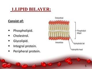
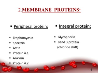
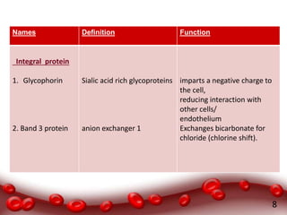
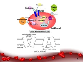
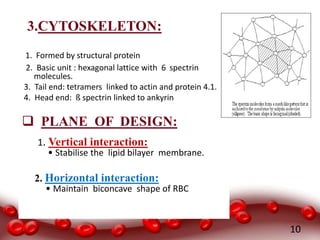
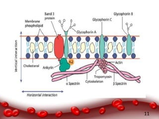
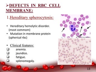
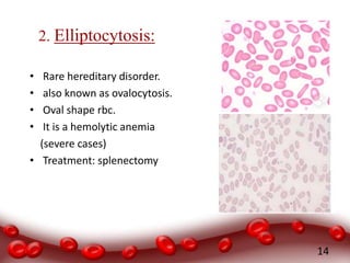
RED CELL
MEMBRANE
CLINICAL LAB SCIENCES
3RD YR 5TH SEMESTER
CELL MEMBRANE OF RBC
2
RBC CELL MEMBRANE:
CONSIST OF:
 PROTEIN 50%
 PHOSPHOLIPID 20%
 CHOLESTROL MOLECULES 20%
 CARBOHYDRATE 10%
3
THREE BASIC COMPONENTS OF RBC
CELL MEMBRANE
 Lipid bilayer.
 Integral membrane protein.
 A membrane cytoskeleton.
4
1.LIPID BILAYER:
Consist of:
 Phospholipid.
 Cholestrol.
 Glycolipid.
 Integral protein.
 Peripheral protein.
5
2.MEMBRANE PROTEINS:
 Peripheral protein:
 Trophomyosin
 Spectrin
 Actin
 Protein 4.1
 Ankyrin
 Protein 4.2
 Integral protein:
 Glycophorin
 Band 3 protein
(chloride shift)
6
7
Names Definition Function
Peripheral protein
1. Spectrin
2. Actin
3. Ankyrin
4. Protein 4.1
5. Protein 4.2
6. Trophomyosin
cytoskeletal protein that lines
the intracellular side of the
plasma membrane
Abundant protein in cell
membrane
are a family of adaptor protein
is a major structural element.
is an ATP-binding protein
Heterodimeric protein
Responsible for biconcave shape
of RBC
participates in more protein-
protein interactions
Interacts with band 3 protein and
spectrin to achieve linkage
between bilayer and
skeleton.
Stabilises actin-spectrin
interactions.
regulate the association of
protein 3 with ankyrin.
Stabilizing the actin filaments.
8
Names Definition Function
Integral protein
1. Glycophorin
2. Band 3 protein
Sialic acid rich glycoproteins
anion exchanger 1
imparts a negative charge to
the cell,
reducing interaction with
other cells/
endothelium
Exchanges bicarbonate for
chloride (chlorine shift).
9
3.CYTOSKELETON:
1. Formed by structural protein
2. Basic unit : hexagonal lattice with 6 spectrin
molecules.
3. Tail end: tetramers linked to actin and protein 4.1.
4. Head end: ß spectrin linked to ankyrin
 PLANE OF DESIGN:
1. Vertical interaction:
• Stabilise the lipid bilayer membrane.
2. Horizontal interaction:
• Maintain biconcave shape of RBC
10
11
12
DEFECTS IN RBC CELL
MEMBRANE:
1.Hereditary spherocytosis:
• Hereditary hemolytic disorder.
(most commom)
• Mutation in membrane protein
(spherical rbc)
• Clinical features:
 anemia.
 jaundice.
 fatigue.
 splenomegaly.
13
2. Elliptocytosis:
• Rare hereditary disorder.
• also known as ovalocytosis.
• Oval shape rbc.
• It is a hemolytic anemia
(severe cases)
• Treatment: splenectomy
14
15
16

Red cell membrane

  • 1.
    RED CELL MEMBRANE CLINICAL LABSCIENCES 3RD YR 5TH SEMESTER
  • 2.
  • 3.
    RBC CELL MEMBRANE: CONSISTOF:  PROTEIN 50%  PHOSPHOLIPID 20%  CHOLESTROL MOLECULES 20%  CARBOHYDRATE 10% 3
  • 4.
    THREE BASIC COMPONENTSOF RBC CELL MEMBRANE  Lipid bilayer.  Integral membrane protein.  A membrane cytoskeleton. 4
  • 5.
    1.LIPID BILAYER: Consist of: Phospholipid.  Cholestrol.  Glycolipid.  Integral protein.  Peripheral protein. 5
  • 6.
    2.MEMBRANE PROTEINS:  Peripheralprotein:  Trophomyosin  Spectrin  Actin  Protein 4.1  Ankyrin  Protein 4.2  Integral protein:  Glycophorin  Band 3 protein (chloride shift) 6
  • 7.
    7 Names Definition Function Peripheralprotein 1. Spectrin 2. Actin 3. Ankyrin 4. Protein 4.1 5. Protein 4.2 6. Trophomyosin cytoskeletal protein that lines the intracellular side of the plasma membrane Abundant protein in cell membrane are a family of adaptor protein is a major structural element. is an ATP-binding protein Heterodimeric protein Responsible for biconcave shape of RBC participates in more protein- protein interactions Interacts with band 3 protein and spectrin to achieve linkage between bilayer and skeleton. Stabilises actin-spectrin interactions. regulate the association of protein 3 with ankyrin. Stabilizing the actin filaments.
  • 8.
    8 Names Definition Function Integralprotein 1. Glycophorin 2. Band 3 protein Sialic acid rich glycoproteins anion exchanger 1 imparts a negative charge to the cell, reducing interaction with other cells/ endothelium Exchanges bicarbonate for chloride (chlorine shift).
  • 9.
  • 10.
    3.CYTOSKELETON: 1. Formed bystructural protein 2. Basic unit : hexagonal lattice with 6 spectrin molecules. 3. Tail end: tetramers linked to actin and protein 4.1. 4. Head end: ß spectrin linked to ankyrin  PLANE OF DESIGN: 1. Vertical interaction: • Stabilise the lipid bilayer membrane. 2. Horizontal interaction: • Maintain biconcave shape of RBC 10
  • 11.
  • 12.
  • 13.
    DEFECTS IN RBCCELL MEMBRANE: 1.Hereditary spherocytosis: • Hereditary hemolytic disorder. (most commom) • Mutation in membrane protein (spherical rbc) • Clinical features:  anemia.  jaundice.  fatigue.  splenomegaly. 13
  • 14.
    2. Elliptocytosis: • Rarehereditary disorder. • also known as ovalocytosis. • Oval shape rbc. • It is a hemolytic anemia (severe cases) • Treatment: splenectomy 14
  • 15.
  • 16.