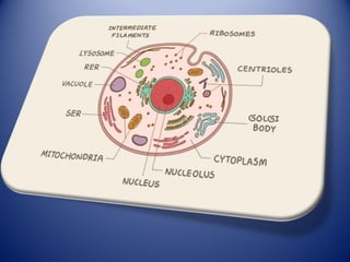
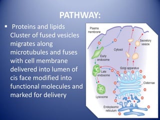
2. What is Golgi Apparatus ?
Also known as Golgi complex, Golgi body or
Golgi. Membrane bound organelles, which are
sac-like.
Found in cytoplasm of most eukaryotic cells
and absent in prokaryotes and mammalian
RBCs.
Ranges from one to several within a cell. In
plant cells several small Golgi complex –
dictyosomes.
It is responsible for transporting, modifying
and packaging proteins and lipids into vesicles.
3. DISCOVERY
The Golgi complex was discovered
by an Italian physician Camillo Golgi
in 1898 during an investigation of
the nervous system.
Its electron microscopic structure
was described by Dalton and Felix in
1954.
4. LOCATION
The Golgi apparatus is specially extensive in
the secretory cells.
The Golgi is located right near the nucleus. It's
called a perinuclear body, and it's actually
right near the endoplasmic reticulum as well.
5.
6. Structure of Golgi Apparatus
Is made up of
several stack of
parallel, flattened
sac or cisternae.
Many peripheral
tubules and
vesicles.
7. CISTERNAE
Golgi apparatus is made up of approx. 4- 8
cisternae usually equally spaced in stark
separated from each other by thin layer of
intercisternal cytoplasm .
Golgi complex has a distinct polarity ,the two
poles are cis and trans face responsible for
receiving and shipping departments.
Forming (cis)face-convex side of
stack,Maturing(trans)face- concave side of stack.
8. Tubules & vesicles
• Tubules: - small, round tubules
formed from the periphery of
the cisternae . And few get
enlarged at the end to form
vesicles.
• Vesicles:-lie near the end and
concave surface of the golgi
complex .
• Types of vesicles:-smooth
vesicles and coated vesicles
• Golgi matrix:-all golgi elements
filled with a fluid.
9. PATHWAY:
Proteins and lipids
Cluster of fused vesicles
migrates along
microtubules and fuses
with cell membrane
delivered into lumen of
cis face modified into
functional molecules and
marked for delivery
10. The vesicular transport model
• In this model, the Golgi cisternae are stable and
distinct suborganelles with different resident
proteins.
• Cargo proteins are transported from one cisterna
to the next by anterograde COPI vesicles,
whereas the Golgi proteins are excluded from
them and remain in the cisternae.
• Cargo proteins reach the TGN and exit by clathrin-
coated vesicles or other carriers..
11. The cisternal maturation model
• In this model, the Golgi cisternae themselves
function as the cargo carriers.
• They are transient compartments that are
newly formed by homotypic fusion of COPII
vesicles, progress from cis to trans, and break
down at the TGN stage.
• The Golgi proteins recycle back from later to
earlier cisternae by retrograde COPI vesicles,
and the nature of the cisternae gradually
changes as they progress.
15. SYNTHESIS
It is also major site of carbohydrate synthesis .
Includes synthesis of glycoasaminoglycans
(GAGs).
Golgi attaches to polysaccharides and protein
to form proteoglycans.
16. SULFHATION
• Golgi involves in the sulfation of ceratain molecules
passing through lumen via sulphotransferases that
gain sulphur molecule from a donor called PAPS .
• Sulfation is generally performed in trans – Golgi
network
• Sulfation occurs in GAGs of proteoglycans as well as
core protein
• Level of sulfation is very important to proteoglycans.
17. APOPTOSIS
• Golgi has a putative role in apoptosis
• A newly characterized protein (Golgi anti-
apoptotic protein) almost exclusively resides
the Golgi and protects cells from apoptosis
• As yet it is an undefined mechanism
18. PHOSPHORYLATION
Phosphorylation of molecules
require energy in the form of
ATP .
The ATP is imported into the
lumen of Golgi which is
utilized by casein kinase 1
and casein kinase 2 .
19. Protein glycosylation within Golgi
• Protein processing within golgi involves the
modification and synthesis of carbohydrate
portions of glycoprotein.
• One of the major aspects of this processing is the
modification of N-linked oligosaccharides that
were added to protein in ER .
20. LIPID AND POLYSACCHARIDE
METABOLISM
• Ceramide sphingomyelin glycolipid
Phosphorylcholine group is transferred from
phosphatidylcholine to ceramide.
• When one or more sugar residues or added
then alternatively a variety of different
glycolipids can be synthesized
21. Vesicular transport from ER to Golgi
bodies
• Membrane – enclosed
transport vesicles
transport proteins from
one membrane-enclosed
compartment to another.
• Proteins doesn’t move
across the lipid bilayer
instead only move
between topologically
equivalent compartment
22. Protein Sorting and Export from the
Golgi Apparatus
• Secretory pathway
involves sorting of
proteins into different
kinds of transport
vesicles.
• They bud from the
trans Golgi network
and deliver their
contents to the
appropriate cellular
locations.
23. Transport from the Golgi apparatus
Transport from golgi takes place by two pathways.
1. Constitutive secretory pathway
2. Regulated secretory pathway
24. Constitutive secretory pathway
Proteins are secreted from a cell continuously,
regardless of external signals or factors.
Proteins are stored in vesicles in the Golgi and
move directly to the cell surface and fuse with
the PM and release the soluble proteins.
25. Regulated secretory pathway:-
• A distinct regulated secretory pathway in which
specific proteins are secreted in response to
environmental signals.
• Proteins are sorted in trans Golgi network and
packed into secretory vesicles. These secretory
vesicles are usually larger than other transport
vesicles.
• And stored until specific signals are received ,
then fuses with plasma membrane to release the
proteins.
26.
27. Selective transport of proteins to
lysosomes
• The process of protein sorting in the Golgi
lysosomes through selective transport of
proteins.
• Then proteins are modified by ‘mannose
phosphorylation in cis Golgi network.
• These phosphorylated mannose molecules are
specifically recognized by a ‘mannose-6-
phosphate receptor’ in trans Golgi network.
28. SUMMARY AND CONCLUSION
• GA is made of a stack of membrane bound
sacs. It receives most of the substances from
Endoplasmic Reticulum.
• It produces vesicles which carry secretions on
to the cell surface.
• Basically, it functions as a “post office”- where
the proteins and lipids are taken, labelled and
transported to other locations within the
body.