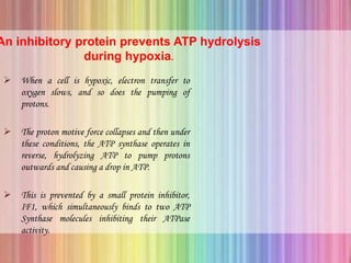
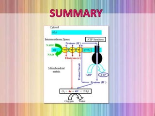
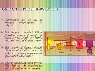
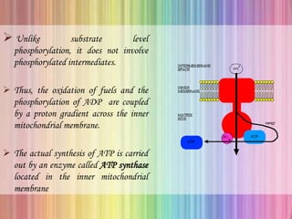
Oxidative phosphorylation occurs in the mitochondria, where ATP is produced through the transfer of electrons from NADH or FADH2 to O2, creating a proton gradient that drives ATP synthesis via ATP synthase. The process is regulated by cellular energy demands, with ADP levels influencing the rate of respiration and ATP regeneration. Under hypoxic conditions, a protein inhibitor named IF1 prevents ATP hydrolysis, maintaining ATP production by blocking ATP synthase from reversing its function.
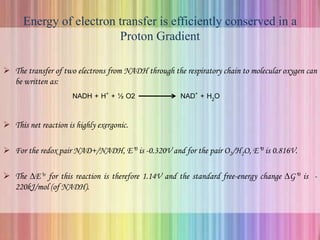
OXIDATIVE PHOSPHORYLATION
Mitochondriaare the site of
oxidative phosphorylation in
eukaryotes.
It is the process in which ATP is
formed as a result of transfer of
electrons from NADH or FADH2
to O2 by a series of electron carriers.
This transfer of electrons through
the inner mitochondrial membrane
leads to the pumping of protons out
of the mitochondrial matrix.
ATP is synthesized when protons
flow back to the mitochondrial
matrix through an enzyme complex.
3.
Unlike substratelevel
phosphorylation, it does not involve
phosphorylated intermediates.
Thus, the oxidation of fuels and the
phosphorylation of ADP are coupled
by a proton gradient across the inner
mitochondrial membrane.
The actual synthesis of ATP is carried
out by an enzyme called ATP synthase
located in the inner mitochondrial
membrane
4.
Electron - TransferReactions in Mitochondria
The discovery in 1948 by Eugene Kennedy
and Albert Lehninger that mitochondria
are the site of oxidative phosphorylation in
eukaryotes marked the beginning of the
modern phase of studies in biological energy
transduction.
Oxidative phosphorylation begins with the
entry of electrons into the respiratory chain.
Most of these electrons arise from the
action of dehydrogenases that collect
electrons from catabolic pathways and
funnel into universal electron acceptors –
nicotinamide nucleotides or flavin
nucleotides.
Energy of electrontransfer is efficiently conserved in a
Proton Gradient
The transfer of two electrons from NADH through the respiratory chain to molecular oxygen can
be written as:
NADH + H+ + ½ O2 NAD+ + H2O
This net reaction is highly exergonic.
For the redox pair NAD+/NADH, E’0 is -0.320V and for the pair O2/H2O, E’0 is 0.816V.
The ∆E’o for this reaction is therefore 1.14V and the standard free-energy change ∆G’0 is -
220kJ/mol (of NADH).
7.
In activelyrespiring mitochondria,
the actions of many dehydrogenases
keep the actual [NADH]/[NAD]
ratio well above unity, and the real
free energy change for the reaction is
substantially greater (more negative)
than -220 kJ/mol.
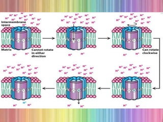
Much of this energy is used to
pump protons out of the matrix.
For each pair of electrons
transferred to O2, four protons are
pumped out by Complex I, four by
Complex III and two by Complex
IV.
8.
CHEMIOSMOTIC MODEL
PeterMitchell proposed that electron transport
and ATP synthesis are coupled by a proton
gradient across the inner mitochondrial
membrane.
The transfer of electrons through the
respiratory chain leads to the pumping of
protons from the matrix to the cytoplasmic side
of the inner mitochondrial membrane.
The H+ concentration becomes lower in the
matrix, and an electric field with the matrix
side negative is generated.
9.
Protons thenflow back into the matrix
to equalize the distribution.
This flow of protons drives the synthesis
of ATP by ATP synthase.
The pH gradient and membrane
potential constitute a proton-motive
force that is used to drive ATP
synthesis.
10.
Proton Motive Force
The energy rich unequal distribution of
protons is called proton-motive force.
The proton- motive force can be thought
of as being composed of two components
: a chemical gradient and a charge
gradient.
The chemical gradient for protons is the
pH gradient and the charge gradient is
created by the positive charge on the
unequally distributed protons forming
the chemical gradient.
11.
ATP SYNTHESIS
o ATPsynthase, a large enzyme complex of the inner
mitochondrial membrane catalyzes the formation of ATP from
ADP and Pi, accompanied by the flow of protons from the P to
the N side of the membrane.
12.
ATP SYNTHASE
Itis also called COMPLEX V.
It is a large, complex enzyme that looks
like a ball on a stick.
Much of the “stick” part, called the F0
subunit, is embedded in the inner
mitochondrial membrane.
The 85-A0 diameter ball, called the F1
subunit, protrudes into the mitochondrial
matrix.
The F1 subunit contains the catalytic
activity of the synthase.
13.
F1 Subunit
Itconsists of five types of polypeptide chains
(α3, β3, γ, δ, ε).
The α and β subunits, which make up the bulk
of the F1, are arranged alternately in the
hexameric ring.
Only the β subunit participate directly in
catalysis.
Below the α and β subunit is a central stalk
consisting of the γ and ε proteins.
The γ subunit includes a long helical coiled coil
that extends into the center of the α3, β3
hexamer.
The three β subunits is crucial for understanding
the mechanism of ATP synthesis.
F1 in cross
section
14.
F0 Subunit
Itis a hydrophobic segment that
spans the inner mitochondrial
membrane.
F0 contains the proton channel of
the complex.
This channel consists of a ring
comprising from 10 to 14c subunits
that are embedded in the membrane.
A single a subunit binds to the
outside of the ring.
15.
The F0and F1 subunits are connected in
two ways: by the central γε stalk and by
an exterior column.
The exterior column consists of one a
subunit, two b subunits and the δ
subunit.
We can think of the enzyme as consisting
of a moving part and a stationary part :
the moving unit or rotor consists of the c
ring and the γε stalk and the stationary
unit or stator is composed of the
remainder of the molecule.
Rotation of relative to &
16.
Role of protongradient is not to form ATP but to release
it from the synthase.
The newly synthesized ATP does
not leave the surface of the
enzyme, it is the proton gradient
that causes the enzyme to release
the ATP formed on the surface.
For the continued synthesis of
ATP, the enzyme must cycle
between a form that binds ATP
very tightly and a form that
releases ATP.
17.
Binding – ChangeMechanism
• Paul Boyer proposed the binding
change mechanism for proton driven
ATP synthesis.
• This proposal states that a β subunit
can perform three sequential steps in
the function of ATP synthesis by
changing conformation.
• Interaction with the γ subunit make
the three β subunits unequivalent.
• One β subunit can be in the L or loose
conformation. This conformation bind
ADP and Pi.
18.
A secondsubunit can be in the T or
tight conformation. This
conformation binds ATP with great
avidity so that it will convert bound
ADP and Pi into ATP.
The final subunit will be in the O or
open form where ATP is released
and ADP and Pi binds to the O –
form subunit.
The rotation of the γ subunit drives
the interconversion of these three
forms and ATP is synthesized.
20.
Proton flow aroundthe c ring powers ATP
synthesis
The streaming of protons through the F0 “pore”
causes the cylinder of c subunits and the attached γ
subunit to rotate along its long axis which is
perpendicular to the plane of the membrane.
The γ comes in contact with the β subunit which
forces the three β subunit to interact in such a way
that one subunit assumes the β – empty
conformation, its neighbor assumes β-ADP form
and the other neighbor in β-ATP form.
Thus one complete rotation of the γ subunit causes
each β subunit to cycle through all three of its
possible conformations and for each rotation three
ATP are synthesized and released from the enzyme
surface.
22.
ADENINE NUCLEOTIDE TRANSLOCASE
ATP and ADP do not diffuse freely across the inner
mitochondrial membrane.
A specific transport protein called Adenine
nucleotide translocase (ATP- ADP Translocase)
enables the ADP to enter into the mitochondrial
matrix and ATP to cytosol.
ATP4-
matrix + ADP3-
cytoplasm ADP3-
matrix + ATP4-
cytoplasm
This antiporter moves four negative charges out for
every three moved in, its activity is favored by the
transmembrane electrochemical gradient, which gives
the matrix a net negative charge.
23.
PHOSPHATE TRANSLOCASE
• Thistransport process is also favored by
the transmembrane proton gradient.
• This promotes symport of one H2PO4-
and one H+ into the matrix.
• The process requires movement of one
proton from the P to the N side of the
inner membrane, consuming some of the
energy of electron transfer.
25.
ATP SYNTHASOME
Acomplex of the ATP Synthase
and both translocase is called the
ATP Synthasome.
This can be isolated from the
mitochondria by gentle dissection
with detergents, suggesting that
the functions of these three
proteins are very tightly regulated.
27.
REGULATION OF OXIDATIVEPHOSPHORYLATION
Oxidative phosphorylation is regulated by
cellular energy needs.
o The intracellular [ADP] and the mass action ratio
([ADP]/[ATP][Pi]) are measures of cells energy status.
o Normally this ratio is very high, so the ATP-ADP system is
almost fully phosphorylated.
o When the rate of some energy requiring process increases, the rate
of breaking down of ATP to ADP and Pi increases, lowering the
mass-action ratio.
o When more ADP is available for oxidative phosphorylation, the
rate of respiration increases, causing regeneration of ATP.
28.
An inhibitory proteinprevents ATP hydrolysis
during hypoxia.
When a cell is hypoxic, electron transfer to
oxygen slows, and so does the pumping of
protons.
The proton motive force collapses and then under
these conditions, the ATP synthase operates in
reverse, hydrolyzing ATP to pump protons
outwards and causing a drop in ATP.
This is prevented by a small protein inhibitor,
IF1, which simultaneously binds to two ATP
Synthase molecules inhibiting their ATPase
activity.