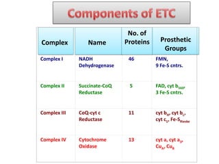
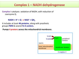
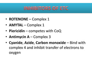
• ETC is the transfer of electrons from NADH
and FADH2 to oxygen via multiple carriers.
• The electrons derieved from NADH and FADH2
combine with O2, and the energy released
from these oxidation/reduction reactions is
used to derieve the synthesis of ATP from ADP.
• This transfer of electrons is done by multiple
carriers which constitute the ELECTRON
TRASPORT CHAIN.
Complex Name
No. of
Proteins Prosthetic
Groups
Complex I NADH
Dehydrogenase
46 FMN,
9 Fe-S cntrs.
Complex II Succinate-CoQ
Reductase
5 FAD, cyt b560,
3 Fe-S cntrs.
Complex III CoQ-cyt c
Reductase
11 cyt bH, cyt bL,
cyt c1, Fe-SRieske
Complex IV Cytochrome
Oxidase
13 cyt a, cyt a3,
CuA, CuB
inner mitochondrial
membrane
matrix
NAD+
NADH
Complex I
FMN peripheral
domain
membrane domain
Complex I catalyzes oxidation of NADH, with reduction of
coenzyme Q.
NADH + H+ + Q  NAD+ + QH2
It includes at least 46 proteins, along with prosthetic
groups FMN & several Fe-S centers.
Pumps 4 protons across the mitochondrial membrane.
The initial electron transfers are:
NADH + H+ + FMN  NAD+ + FMNH2
FMNH2 + (Fe-S)ox  FMNH· + (Fe-S)red + H+
After Fe-S is reoxidized by transfer of the electron to the next iron-sulfur center in
the pathway:
FMNH· + (Fe-S)ox  FMN + (Fe-S)red + H+
Iron-sulfur centers are arranged as a wire, providing a pathway for e- transfer from
FMN through the protein
 FMN
A B
 FMN
Peripheral domain of a bacterial Complex I
membrane
domain

PDB 2FUG
 N2
N2, the last Fe-S center in the chain,
passes e- one at a time to the mobile
lipid redox carrier coenzyme Q.
A proposed binding site for CoQ is
close to N2 at the interface of
peripheral & membrane domains.
Coenzyme Q accepts 2e- and picks up
2H+ to yield the fully reduced QH2.
• It is a benzoquinone linked to a number of
isoprene units.
• Coenzyme Q (CoQ, Q, Ubiquione) is very hydrophobic. It
dissolves in the hydrocarbon core of a membrane.
• 3 redox states-
1. Fully oxidised- Ubiquinone Q
2. Partially oxidised- Semiquinone
3. Fully reduced- Ubiquione
• Only electron carrier that is not a protein
bound prosthetic group.
• Succinate Dehydrogenase of the
Krebs Cycle is also called complex II or
Succinate-CoQ Reductase.
• Inner mitochondrial membrane
bound protein.
• FAD is the initial e- acceptor.
•FAD is reduced to FADH2 during
oxidation of succinate to fumarate.
•FADH2 is then reoxidized by transfer of
electrons through a series of 3 iron-
sulfur centers to CoQ, yielding QH2.
•It does not pump any proton during
transport of electron across the inner
mitochondrial membrane.
COO-
C
C
COO-
H H
H H
COO-
C
C
COO-
H
H
Q QH2
via FAD
succinate fumarate
Succinate Dehydrogenase
(Complex II)
• X-ray crystallographic
analysis of E. coli complex II
indicates a linear
arrangement of electron
carriers within complex II,
consistent with the predicted
sequence of electron
transfers:
FAD  FeScenter 1  FeScenter 2  FeScenter 3  CoQ
• Complex III accepts electrons from coenzyme QH2 that is
generated by electron transfer in complexes I & II.
• Concominantly, it releases two protons into
transmembrane space.
• Within complex 3,the released electrons are transferred
to an iron sulfur center and then to two b-type
cytochromes or cytochrome c1.
• Finally the two electrons are transferred to two molecules
of the oxidised form of cytochrome c. two additional
protons are translocated from mitochondrial matrix
across the intermembrane space. This transfer of protons
involves the proton motive Q cycle.
• Cytochromes are proteins with heme prosthetic groups.
They absorb light at characteristic wavelengths.
• It carries electron one at a time to complex 4.
• Major respiratory Cytochromes- b, c or a.
• In ETC-
 Two a type cyt i.e. cyt a and a3.
 Two b type cyt i.e. cyt b1 and b2.
 Two c type cyt i.e. cyt c and c1.
• Cytochrome c is a key regulator of programme cell
death in mammalian cells.
• It catalyses the transfer of electrons from reduced
cyt c to molecular oxygen.
• Contains 13 subunits
• 2 heme groups i.e. heme a & heme a3
• 3 copper ions arranged as 2 copper centers
designated as Cua & Cub.
• Cua contain 2 copper ions linked by 2 bridging
disulfide residues.
• Cub is coordinated by 3 histidine residues.
• Two protons per pair of electron are pumped
across the membrane and another two protons
are transferred to molecular oxygen to form
water.
• Metal centers of cytochrome oxidase
(complex IV):
• heme a & heme a3,
• CuA (2 adjacent Cu atoms) & CuB.
• O2 reacts at a binuclear center consisting
of heme a3 and CuB.
•Electrons enter complex IV one at a time
from cyt c to CuA.
•They then pass via cyt a to the binuclear
center where the chemical reaction takes
place.
heme a3
CuB
binuclear center
His ligands
PDB 1OCC
e- transfer: cyt c → CuA → cyt a → heme a3/CuB → O2
• Mitochondrial ATP synthase consist of two
multisubunit components F0 and F1 which are
linked by a slender stalk.
• F0 is a elecrically driven motor that spans the lipid
bilayer foming a channel through which protons
can cross the membrane.
• F0 provides channel for protons.
• F1 harvest the free energy derieved from proton
movement down the electrochemical gradient by
catalyzing the synthesis of ATP.
• F1 Phosphorylates ADP to ATP.
• Proposed by PETER MITCHELL in 1961.
• This hypothesis couples electron transport to ATP generation.
• Mitchell suggested that ATP is generated by use of energy
stored in the form of proton gradient across biological
membranes rather than by direct chemical transfer of high
energy groups.
• Complex 1 and 4 appear as proton pump which transport
protons across the membrane due to conformational change
induced by electron transfer.
• In Complex 3 protons are carried across the membrane by
Ubiquione.
• Complex 1 and 3 pump four protons per pair of electrons.
• Complex 4 pumps two protons per pair of electrons transported
and other two protons are combined with oxygen to form
water.
PATHWAY NADH FADH2 ATP
GLYCOLYSIS 2 0 2
KREBS CYCLE 8 2 2
TOTAL 10 2 4
TOTAL ATP 25 3 4
1 NADH
10 H+ X 1 ATP = 2.5 ATP
4 H+
1 FADH2
6 H+ X 1 ATP = 1.5 ATP
4 H+
Pyruvate dehydrogenase
NADH ……………………………….2.5 ATP
Krebs
3 NADH X 2.5 ATP/NADH ……….7.5 ATP
FADH2 X 1.5 ATP / FADH2……….1.5 ATP
GTP X 1 ATP / GTP ……………..1.0 ATP
(from a separate reaction)
Total
…………….12.5 ATP
(Per glucose = X 2 = 25 ATP)
•NADH made in cytosol
•Can’t get into matrix of mitochondrion
•2 mechanisms
1. In muscle and brain
Glycerol phosphate shuttle
2. In liver and heart
Malate / aspartate shuttle
Glycerol phosphate shuttle
 In muscle and brain
 Each NADH converted to FADH2 inside mitochondrion
 FADH2 enters later in the electron transport chain
 Produces 1.5 ATP
 Gycerol phosphate shuttle
 2 NADH per glucose - 2 FADH2
 2 FADH2 X 1.5 ATP / FADH2……….3.0 ATP
 2 ATP in glycoysis ……………………2.0 ATP
 From pyruvate and Krebs
 12.5 ATP X 2 per glucose ……………..25.0 ATP
Total = 30.0 ATP/ glucose
• In liver and heart
• NADH oxidized while reducing oxaloacetate to
malate
– Malate dehydrogenase
• Malate crosses membrane
• Malate – Aspartate Shuttle
– 2 NADH per glucose - 2 NADH
– 2 NADH X 2.5 ATP / NADH…………5.0 ATP
– 2 ATP from glycolysis………………..2.0 ATP
– From pyruvate and Krebs
• 12.5 ATP X 2 per glucose ……………..25.0 ATP
Total = 32.0 ATP/ glucose
• ROTENONE – Complex 1
• AMYTAL – Complex 1
• Piericidin – competes with CoQ
• Antimycin A – Complex 3
• Cyanide, Azide, Carbon monoxide – Bind with
complex 4 and inhibit transfer of electrons to
oxygen
• 2,4 Dinitrophenol
• Dicoumarol
• Carbonyl cyanide p-
flouromethoxyphenylhydrazone(FCCP)
Electron transport chain

Electron transport chain

  • 2.
    • ETC isthe transfer of electrons from NADH and FADH2 to oxygen via multiple carriers. • The electrons derieved from NADH and FADH2 combine with O2, and the energy released from these oxidation/reduction reactions is used to derieve the synthesis of ATP from ADP. • This transfer of electrons is done by multiple carriers which constitute the ELECTRON TRASPORT CHAIN.
  • 5.
    Complex Name No. of ProteinsProsthetic Groups Complex I NADH Dehydrogenase 46 FMN, 9 Fe-S cntrs. Complex II Succinate-CoQ Reductase 5 FAD, cyt b560, 3 Fe-S cntrs. Complex III CoQ-cyt c Reductase 11 cyt bH, cyt bL, cyt c1, Fe-SRieske Complex IV Cytochrome Oxidase 13 cyt a, cyt a3, CuA, CuB
  • 6.
    inner mitochondrial membrane matrix NAD+ NADH Complex I FMNperipheral domain membrane domain Complex I catalyzes oxidation of NADH, with reduction of coenzyme Q. NADH + H+ + Q  NAD+ + QH2 It includes at least 46 proteins, along with prosthetic groups FMN & several Fe-S centers. Pumps 4 protons across the mitochondrial membrane.
  • 7.
    The initial electrontransfers are: NADH + H+ + FMN  NAD+ + FMNH2 FMNH2 + (Fe-S)ox  FMNH· + (Fe-S)red + H+ After Fe-S is reoxidized by transfer of the electron to the next iron-sulfur center in the pathway: FMNH· + (Fe-S)ox  FMN + (Fe-S)red + H+ Iron-sulfur centers are arranged as a wire, providing a pathway for e- transfer from FMN through the protein  FMN A B  FMN Peripheral domain of a bacterial Complex I membrane domain  PDB 2FUG  N2 N2, the last Fe-S center in the chain, passes e- one at a time to the mobile lipid redox carrier coenzyme Q. A proposed binding site for CoQ is close to N2 at the interface of peripheral & membrane domains. Coenzyme Q accepts 2e- and picks up 2H+ to yield the fully reduced QH2.
  • 8.
    • It isa benzoquinone linked to a number of isoprene units. • Coenzyme Q (CoQ, Q, Ubiquione) is very hydrophobic. It dissolves in the hydrocarbon core of a membrane. • 3 redox states- 1. Fully oxidised- Ubiquinone Q 2. Partially oxidised- Semiquinone 3. Fully reduced- Ubiquione • Only electron carrier that is not a protein bound prosthetic group.
  • 9.
    • Succinate Dehydrogenaseof the Krebs Cycle is also called complex II or Succinate-CoQ Reductase. • Inner mitochondrial membrane bound protein. • FAD is the initial e- acceptor. •FAD is reduced to FADH2 during oxidation of succinate to fumarate. •FADH2 is then reoxidized by transfer of electrons through a series of 3 iron- sulfur centers to CoQ, yielding QH2. •It does not pump any proton during transport of electron across the inner mitochondrial membrane. COO- C C COO- H H H H COO- C C COO- H H Q QH2 via FAD succinate fumarate Succinate Dehydrogenase (Complex II)
  • 10.
    • X-ray crystallographic analysisof E. coli complex II indicates a linear arrangement of electron carriers within complex II, consistent with the predicted sequence of electron transfers: FAD  FeScenter 1  FeScenter 2  FeScenter 3  CoQ
  • 11.
    • Complex IIIaccepts electrons from coenzyme QH2 that is generated by electron transfer in complexes I & II. • Concominantly, it releases two protons into transmembrane space. • Within complex 3,the released electrons are transferred to an iron sulfur center and then to two b-type cytochromes or cytochrome c1. • Finally the two electrons are transferred to two molecules of the oxidised form of cytochrome c. two additional protons are translocated from mitochondrial matrix across the intermembrane space. This transfer of protons involves the proton motive Q cycle.
  • 12.
    • Cytochromes areproteins with heme prosthetic groups. They absorb light at characteristic wavelengths. • It carries electron one at a time to complex 4. • Major respiratory Cytochromes- b, c or a. • In ETC-  Two a type cyt i.e. cyt a and a3.  Two b type cyt i.e. cyt b1 and b2.  Two c type cyt i.e. cyt c and c1. • Cytochrome c is a key regulator of programme cell death in mammalian cells.
  • 13.
    • It catalysesthe transfer of electrons from reduced cyt c to molecular oxygen. • Contains 13 subunits • 2 heme groups i.e. heme a & heme a3 • 3 copper ions arranged as 2 copper centers designated as Cua & Cub. • Cua contain 2 copper ions linked by 2 bridging disulfide residues. • Cub is coordinated by 3 histidine residues. • Two protons per pair of electron are pumped across the membrane and another two protons are transferred to molecular oxygen to form water.
  • 14.
    • Metal centersof cytochrome oxidase (complex IV): • heme a & heme a3, • CuA (2 adjacent Cu atoms) & CuB. • O2 reacts at a binuclear center consisting of heme a3 and CuB. •Electrons enter complex IV one at a time from cyt c to CuA. •They then pass via cyt a to the binuclear center where the chemical reaction takes place. heme a3 CuB binuclear center His ligands PDB 1OCC e- transfer: cyt c → CuA → cyt a → heme a3/CuB → O2
  • 15.
    • Mitochondrial ATPsynthase consist of two multisubunit components F0 and F1 which are linked by a slender stalk. • F0 is a elecrically driven motor that spans the lipid bilayer foming a channel through which protons can cross the membrane. • F0 provides channel for protons. • F1 harvest the free energy derieved from proton movement down the electrochemical gradient by catalyzing the synthesis of ATP. • F1 Phosphorylates ADP to ATP.
  • 17.
    • Proposed byPETER MITCHELL in 1961. • This hypothesis couples electron transport to ATP generation. • Mitchell suggested that ATP is generated by use of energy stored in the form of proton gradient across biological membranes rather than by direct chemical transfer of high energy groups. • Complex 1 and 4 appear as proton pump which transport protons across the membrane due to conformational change induced by electron transfer. • In Complex 3 protons are carried across the membrane by Ubiquione. • Complex 1 and 3 pump four protons per pair of electrons. • Complex 4 pumps two protons per pair of electrons transported and other two protons are combined with oxygen to form water.
  • 19.
    PATHWAY NADH FADH2ATP GLYCOLYSIS 2 0 2 KREBS CYCLE 8 2 2 TOTAL 10 2 4 TOTAL ATP 25 3 4 1 NADH 10 H+ X 1 ATP = 2.5 ATP 4 H+ 1 FADH2 6 H+ X 1 ATP = 1.5 ATP 4 H+
  • 20.
    Pyruvate dehydrogenase NADH ……………………………….2.5ATP Krebs 3 NADH X 2.5 ATP/NADH ……….7.5 ATP FADH2 X 1.5 ATP / FADH2……….1.5 ATP GTP X 1 ATP / GTP ……………..1.0 ATP (from a separate reaction) Total …………….12.5 ATP (Per glucose = X 2 = 25 ATP)
  • 21.
    •NADH made incytosol •Can’t get into matrix of mitochondrion •2 mechanisms 1. In muscle and brain Glycerol phosphate shuttle 2. In liver and heart Malate / aspartate shuttle
  • 23.
    Glycerol phosphate shuttle In muscle and brain  Each NADH converted to FADH2 inside mitochondrion  FADH2 enters later in the electron transport chain  Produces 1.5 ATP  Gycerol phosphate shuttle  2 NADH per glucose - 2 FADH2  2 FADH2 X 1.5 ATP / FADH2……….3.0 ATP  2 ATP in glycoysis ……………………2.0 ATP  From pyruvate and Krebs  12.5 ATP X 2 per glucose ……………..25.0 ATP Total = 30.0 ATP/ glucose
  • 25.
    • In liverand heart • NADH oxidized while reducing oxaloacetate to malate – Malate dehydrogenase • Malate crosses membrane
  • 26.
    • Malate –Aspartate Shuttle – 2 NADH per glucose - 2 NADH – 2 NADH X 2.5 ATP / NADH…………5.0 ATP – 2 ATP from glycolysis………………..2.0 ATP – From pyruvate and Krebs • 12.5 ATP X 2 per glucose ……………..25.0 ATP Total = 32.0 ATP/ glucose
  • 27.
    • ROTENONE –Complex 1 • AMYTAL – Complex 1 • Piericidin – competes with CoQ • Antimycin A – Complex 3 • Cyanide, Azide, Carbon monoxide – Bind with complex 4 and inhibit transfer of electrons to oxygen
  • 28.
    • 2,4 Dinitrophenol •Dicoumarol • Carbonyl cyanide p- flouromethoxyphenylhydrazone(FCCP)