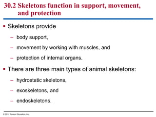
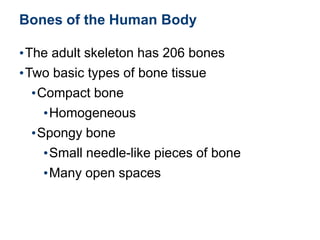
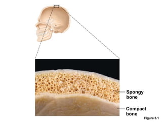
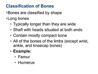
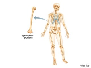
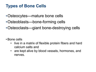
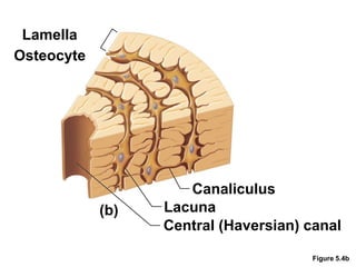
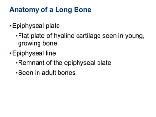
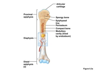
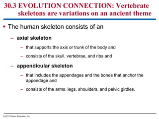
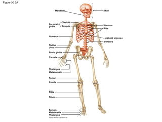
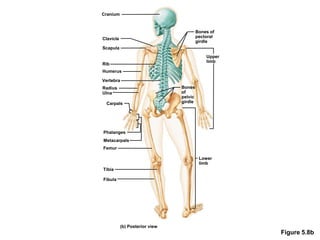
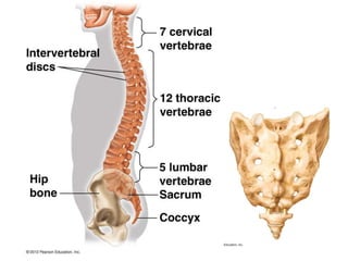
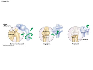
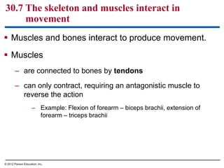
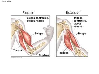
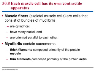
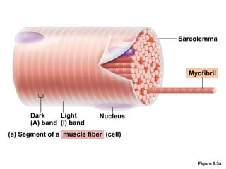
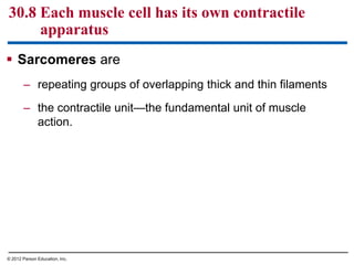
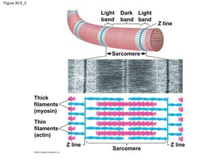
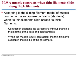
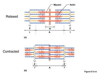
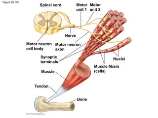
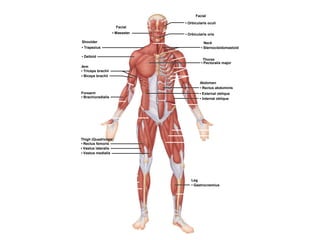
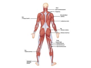
© 2012 Pearson Education, Inc. Lecture by Edward J. Zalisko
PowerPoint Lectures for
Campbell Biology: Concepts & Connections, Seventh Edition
Reece, Taylor, Simon, and Dickey
Chapter 30 How Animals Move
Figure 30.0_1
Chapter 30: Big Ideas
Movement and
Locomotion
The Vertebrate Skeleton
Muscle Contraction
and Movement
 Skeletons provide
– body support,
– movement by working with muscles, and
– protection of internal organs.
 There are three main types of animal skeletons:
– hydrostatic skeletons,
– exoskeletons, and
– endoskeletons.
30.2 Skeletons function in support, movement,
and protection
© 2012 Parson Education, Inc.
1. Hydrostatic skeletons are
– fluid held under pressure in a closed
body compartment and
– found in worms and cnidarians.
– Hydrostatic skeletons
– help protect other body parts by
cushioning them from shocks,
– give the body shape, and
– provide support for muscle action.
30.2 Skeletons function in support, movement,
and protection
© 2012 Parson Education, Inc.
2. Exoskeletons are rigid external skeletons that
consist of
– chitin and protein in arthropods and
– calcium carbonate shells in molluscs.
– Exoskeletons must be shed to permit growth.
30.2 Skeletons function in support, movement,
and protection
© 2012 Parson Education, Inc.
3. Endoskeletons consist of hard or leathery
supporting elements situated among the soft
tissues of an animal. They may be made of
– cartilage or cartilage and bone (vertebrates),
– spicules (sponges), or
– hard plates (echinoderms).
30.2 Skeletons function in support, movement,
and protection
© 2012 Parson Education, Inc.
 The vertebrate
skeletal system
provides
– structural support
– means of locomotion
30.3 EVOLUTION CONNECTION: Vertebrate
skeletons are variations on an ancient theme
© 2012 Parson Education, Inc.
Bones of the Human Body
•The adult skeleton has 206 bones
•Two basic types of bone tissue
•Compact bone
•Homogeneous
•Spongy bone
•Small needle-like pieces of bone
•Many open spaces
Figure 5.1
Spongy
bone
Compact
bone
Classification of Bones
•Bones are classified by shape
•Long bones
• Typically longer than they are wide
• Shaft with heads situated at both ends
• Contain mostly compact bone
• All of the bones of the limbs (except wrist,
ankle, and kneecap bones)
• Example:
• Femur
• Humerus
Figure 5.2a
Figure 5.3a
Distal
epiphysis
Diaphysis
Proximal
epiphysis
Articular
cartilage
Spongy bone
Epiphyseal
line
Periosteum
Compact bone
Medullary
cavity (lined
by endosteum)
(a)
Types of Bone Cells
•Osteocytes—mature bone cells
•Osteoblasts—bone-forming cells
•Osteoclasts—giant bone-destroying cells
• Bone cells
• live in a matrix of flexible protein fibers and hard
calcium salts and
• are kept alive by blood vessels, hormones, and
nerves.
Lamella
Osteocyte
Figure 5.4b
Canaliculus
Lacuna
Central (Haversian) canal
(b)
Anatomy of a Long Bone
•Epiphyseal plate
•Flat plate of hyaline cartilage seen in young,
growing bone
•Epiphyseal line
•Remnant of the epiphyseal plate
•Seen in adult bones
Figure 5.3a
Distal
epiphysis
Diaphysis
Proximal
epiphysis
Articular
cartilage
Spongy bone
Epiphyseal
line
Periosteum
Compact bone
Medullary
cavity (lined
by endosteum)
(a)
 The human skeleton consists of an
– axial skeleton
– that supports the axis or trunk of the body and
– consists of the skull, vertebrae, and ribs and
– appendicular skeleton
– that includes the appendages and the bones that anchor the
appendage and
– consists of the arms, legs, shoulders, and pelvic girdles.
30.3 EVOLUTION CONNECTION: Vertebrate
skeletons are variations on an ancient theme
© 2012 Parson Education, Inc.
Figure 30.3A
Skull
Sternum
Ribs
Vertebra
Clavicle
Scapula
Pectoral
girdle
Humerus
Radius
Ulna
Pelvic girdle
Carpals
Phalanges
Metacarpals
Femur
Patella
Tibia
Fibula
Tarsals
Metatarsals
Phalanges
Mandible
xiphoid process
Figure 5.8b
(b) Posterior view
Fibula
Tibia
Femur
Metacarpals
Phalanges
Carpals
Radius
Ulna
Vertebra
Humerus
Rib
Scapula
Clavicle
Cranium
Bones of
pectoral
girdle
Upper
limb
Bones
of
pelvic
girdle
Lower
limb
 Cartilage at the ends of bones
– cushions joints and
– reduces friction of movements.
30.4 Bones are complex living organs
© 2012 Parson Education, Inc.
Articular
cartilage
 Long bones have
– a central cavity storing
fatty yellow bone marrow
– spongy bone located at the
ends of bones containing
red bone marrow, that
produces blood cells.
30.4 Bones are complex living organs
© 2012 Parson Education, Inc.
 Osteoporosis is
– a bone disease,
– characterized by low bone
mass and structural
deterioration, and
– less likely if a person
– has high levels of calcium
in the diet,
– exercises regularly, and
– does not smoke.
30.5 CONNECTION: Healthy bones resist stress
and heal from injuries
© 2012 Parson Education, Inc.
30.6 Joints permit different types of movement
 Joints allow limited movement of bones.
 Different joints permit various movements.
– Ball-and-socket joints enable rotation in the arms and
legs.
– Hinge joints in the elbows and knees permit
movement in a single plane.
– Pivot joints enable the rotation of the forearm at the
elbow.
© 2012 Parson Education, Inc.
Figure 30.6
Head
of humerus
Humerus
Scapula
Ulna
Ball-and-socket joint Hinge joint
Ulna
Pivot joint
Radius
 Muscles and bones interact to produce movement.
 Muscles
– are connected to bones by tendons
– can only contract, requiring an antagonistic muscle to
reverse the action
– Example: Flexion of forearm – biceps brachii, extension of
forearm – triceps brachii
30.7 The skeleton and muscles interact in
movement
© 2012 Parson Education, Inc.
Figure 30.7A
Biceps contracted,
triceps relaxed
Biceps
Triceps
Tendons
Triceps
Biceps
Triceps
contracted,
biceps
relaxed
Flexion Extension
 Muscle fibers (skeletal muscle cells) are cells that
consist of bundles of myofibrils
– are cylindrical,
– have many nuclei, and
– are oriented parallel to each other.
 Myofibrils contain sacromeres
– thick filaments composed primarily of the protein
myosin
– thin filaments composed primarily of the protein actin.
30.8 Each muscle cell has its own contractile
apparatus
© 2012 Parson Education, Inc.
Figure 6.3a
Sarcolemma
Myofibril
Dark
(A) band
Light
(I) band
Nucleus
(a) Segment of a muscle fiber (cell)
 Sarcomeres are
– repeating groups of overlapping thick and thin filaments
– the contractile unit—the fundamental unit of muscle
action.
30.8 Each muscle cell has its own contractile
apparatus
© 2012 Parson Education, Inc.
Figure 30.8
Muscle
Several muscle fibers
Single muscle fiber
(cell)
Plasma membrane
Nuclei
Myofibril
Light
band
Dark
band
Light
band
Z line
Sarcomere
Sarcomere
Z lineZ line
Thick
filaments
(myosin)
Thin
filaments
(actin)
Figure 30.8_3
Sarcomere
Z lineZ line
Thick
filaments
(myosin)
Thin
filaments
(actin)
Light
band
Dark
band
Light
band
Z line
Sarcomere
 According to the sliding-filament model of muscle
contraction, a sarcomere contracts (shortens)
when its thin filaments slide across its thick
filaments.
– Contraction shortens the sarcomere without changing
the lengths of the thick and thin filaments.
– When the muscle is fully contracted, the thin filaments
overlap in the middle of the sarcomere.
30.9 A muscle contracts when thin filaments slide
along thick filaments
© 2012 Parson Education, Inc.
Figure 6.7a–b
Myosin Actin
Z H
I
Z
A I
(a)
(b)
Z
I A I
Z
Relaxed
Contracted
 A motor neuron
– carries an action potential to a muscle cell,
– releases the neurotransmitter acetylcholine (Ach) from
its synaptic terminal, and
– initiates a muscle contraction.
30.10 Motor neurons stimulate muscle contraction
© 2012 Parson Education, Inc.
Figure 6.5
 A motor unit consists of
– a neuron and
– the set of muscle fibers it controls.
 More forceful muscle contractions result when
additional motor units are activated.
30.10 Motor neurons stimulate muscle contraction
© 2012 Parson Education, Inc.
Figure 30.10C
Spinal cord
Motor neuron
cell body
Nerve
Motor neuron
axon
Synaptic
terminals
Muscle
Tendon
Muscle fibers
(cells)
Nuclei
Bone
Motor
unit 1
Motor
unit 2
Facial
• Masseter
Shoulder
• Trapezius
• Deltoid
Arm
• Triceps brachii
• Biceps brachii
Forearm
• Brachioradialis
Thigh (Quadriceps)
• Rectus femoris
• Vastus lateralis
• Vastus medialis
Facial
• Orbicularis oculi
• Orbicularis oris
Neck
• Sternocleidomastoid
Thorax
• Pectoralis major
Abdomen
• Rectus abdominis
• External oblique
• Internal oblique
Leg
• Gastrocnemius
Arm
• Triceps brachii
Leg
• Gastrocnemius
Calcaneal
(Achilles)
tendon
Neck
• Sternocleidomastoid
• Trapezius
Shoulder/Back
• Deltoid
• Latissimus dorsi
Hip
• Gluteus medius
• Gluteus maximus
Thigh
• Hamstrings:
Biceps femoris
Semitendinosus
Semimembranosus

Bio 2 Chapter 30 - Bones and Muscles

  • 1.
    © 2012 PearsonEducation, Inc. Lecture by Edward J. Zalisko PowerPoint Lectures for Campbell Biology: Concepts & Connections, Seventh Edition Reece, Taylor, Simon, and Dickey Chapter 30 How Animals Move
  • 2.
    Figure 30.0_1 Chapter 30:Big Ideas Movement and Locomotion The Vertebrate Skeleton Muscle Contraction and Movement
  • 3.
     Skeletons provide –body support, – movement by working with muscles, and – protection of internal organs.  There are three main types of animal skeletons: – hydrostatic skeletons, – exoskeletons, and – endoskeletons. 30.2 Skeletons function in support, movement, and protection © 2012 Parson Education, Inc.
  • 4.
    1. Hydrostatic skeletonsare – fluid held under pressure in a closed body compartment and – found in worms and cnidarians. – Hydrostatic skeletons – help protect other body parts by cushioning them from shocks, – give the body shape, and – provide support for muscle action. 30.2 Skeletons function in support, movement, and protection © 2012 Parson Education, Inc.
  • 5.
    2. Exoskeletons arerigid external skeletons that consist of – chitin and protein in arthropods and – calcium carbonate shells in molluscs. – Exoskeletons must be shed to permit growth. 30.2 Skeletons function in support, movement, and protection © 2012 Parson Education, Inc.
  • 6.
    3. Endoskeletons consistof hard or leathery supporting elements situated among the soft tissues of an animal. They may be made of – cartilage or cartilage and bone (vertebrates), – spicules (sponges), or – hard plates (echinoderms). 30.2 Skeletons function in support, movement, and protection © 2012 Parson Education, Inc.
  • 7.
     The vertebrate skeletalsystem provides – structural support – means of locomotion 30.3 EVOLUTION CONNECTION: Vertebrate skeletons are variations on an ancient theme © 2012 Parson Education, Inc.
  • 8.
    Bones of theHuman Body •The adult skeleton has 206 bones •Two basic types of bone tissue •Compact bone •Homogeneous •Spongy bone •Small needle-like pieces of bone •Many open spaces
  • 9.
  • 10.
    Classification of Bones •Bonesare classified by shape •Long bones • Typically longer than they are wide • Shaft with heads situated at both ends • Contain mostly compact bone • All of the bones of the limbs (except wrist, ankle, and kneecap bones) • Example: • Femur • Humerus
  • 11.
  • 12.
  • 13.
    Types of BoneCells •Osteocytes—mature bone cells •Osteoblasts—bone-forming cells •Osteoclasts—giant bone-destroying cells • Bone cells • live in a matrix of flexible protein fibers and hard calcium salts and • are kept alive by blood vessels, hormones, and nerves.
  • 14.
  • 15.
    Anatomy of aLong Bone •Epiphyseal plate •Flat plate of hyaline cartilage seen in young, growing bone •Epiphyseal line •Remnant of the epiphyseal plate •Seen in adult bones
  • 16.
  • 17.
     The humanskeleton consists of an – axial skeleton – that supports the axis or trunk of the body and – consists of the skull, vertebrae, and ribs and – appendicular skeleton – that includes the appendages and the bones that anchor the appendage and – consists of the arms, legs, shoulders, and pelvic girdles. 30.3 EVOLUTION CONNECTION: Vertebrate skeletons are variations on an ancient theme © 2012 Parson Education, Inc.
  • 18.
  • 19.
    Figure 5.8b (b) Posteriorview Fibula Tibia Femur Metacarpals Phalanges Carpals Radius Ulna Vertebra Humerus Rib Scapula Clavicle Cranium Bones of pectoral girdle Upper limb Bones of pelvic girdle Lower limb
  • 21.
     Cartilage atthe ends of bones – cushions joints and – reduces friction of movements. 30.4 Bones are complex living organs © 2012 Parson Education, Inc. Articular cartilage
  • 22.
     Long boneshave – a central cavity storing fatty yellow bone marrow – spongy bone located at the ends of bones containing red bone marrow, that produces blood cells. 30.4 Bones are complex living organs © 2012 Parson Education, Inc.
  • 23.
     Osteoporosis is –a bone disease, – characterized by low bone mass and structural deterioration, and – less likely if a person – has high levels of calcium in the diet, – exercises regularly, and – does not smoke. 30.5 CONNECTION: Healthy bones resist stress and heal from injuries © 2012 Parson Education, Inc.
  • 24.
    30.6 Joints permitdifferent types of movement  Joints allow limited movement of bones.  Different joints permit various movements. – Ball-and-socket joints enable rotation in the arms and legs. – Hinge joints in the elbows and knees permit movement in a single plane. – Pivot joints enable the rotation of the forearm at the elbow. © 2012 Parson Education, Inc.
  • 25.
  • 26.
     Muscles andbones interact to produce movement.  Muscles – are connected to bones by tendons – can only contract, requiring an antagonistic muscle to reverse the action – Example: Flexion of forearm – biceps brachii, extension of forearm – triceps brachii 30.7 The skeleton and muscles interact in movement © 2012 Parson Education, Inc.
  • 27.
    Figure 30.7A Biceps contracted, tricepsrelaxed Biceps Triceps Tendons Triceps Biceps Triceps contracted, biceps relaxed Flexion Extension
  • 28.
     Muscle fibers(skeletal muscle cells) are cells that consist of bundles of myofibrils – are cylindrical, – have many nuclei, and – are oriented parallel to each other.  Myofibrils contain sacromeres – thick filaments composed primarily of the protein myosin – thin filaments composed primarily of the protein actin. 30.8 Each muscle cell has its own contractile apparatus © 2012 Parson Education, Inc.
  • 29.
    Figure 6.3a Sarcolemma Myofibril Dark (A) band Light (I)band Nucleus (a) Segment of a muscle fiber (cell)
  • 30.
     Sarcomeres are –repeating groups of overlapping thick and thin filaments – the contractile unit—the fundamental unit of muscle action. 30.8 Each muscle cell has its own contractile apparatus © 2012 Parson Education, Inc.
  • 31.
    Figure 30.8 Muscle Several musclefibers Single muscle fiber (cell) Plasma membrane Nuclei Myofibril Light band Dark band Light band Z line Sarcomere Sarcomere Z lineZ line Thick filaments (myosin) Thin filaments (actin)
  • 32.
    Figure 30.8_3 Sarcomere Z lineZline Thick filaments (myosin) Thin filaments (actin) Light band Dark band Light band Z line Sarcomere
  • 33.
     According tothe sliding-filament model of muscle contraction, a sarcomere contracts (shortens) when its thin filaments slide across its thick filaments. – Contraction shortens the sarcomere without changing the lengths of the thick and thin filaments. – When the muscle is fully contracted, the thin filaments overlap in the middle of the sarcomere. 30.9 A muscle contracts when thin filaments slide along thick filaments © 2012 Parson Education, Inc.
  • 34.
    Figure 6.7a–b Myosin Actin ZH I Z A I (a) (b) Z I A I Z Relaxed Contracted
  • 35.
     A motorneuron – carries an action potential to a muscle cell, – releases the neurotransmitter acetylcholine (Ach) from its synaptic terminal, and – initiates a muscle contraction. 30.10 Motor neurons stimulate muscle contraction © 2012 Parson Education, Inc.
  • 36.
  • 37.
     A motorunit consists of – a neuron and – the set of muscle fibers it controls.  More forceful muscle contractions result when additional motor units are activated. 30.10 Motor neurons stimulate muscle contraction © 2012 Parson Education, Inc.
  • 38.
    Figure 30.10C Spinal cord Motorneuron cell body Nerve Motor neuron axon Synaptic terminals Muscle Tendon Muscle fibers (cells) Nuclei Bone Motor unit 1 Motor unit 2
  • 39.
    Facial • Masseter Shoulder • Trapezius •Deltoid Arm • Triceps brachii • Biceps brachii Forearm • Brachioradialis Thigh (Quadriceps) • Rectus femoris • Vastus lateralis • Vastus medialis Facial • Orbicularis oculi • Orbicularis oris Neck • Sternocleidomastoid Thorax • Pectoralis major Abdomen • Rectus abdominis • External oblique • Internal oblique Leg • Gastrocnemius
  • 40.
    Arm • Triceps brachii Leg •Gastrocnemius Calcaneal (Achilles) tendon Neck • Sternocleidomastoid • Trapezius Shoulder/Back • Deltoid • Latissimus dorsi Hip • Gluteus medius • Gluteus maximus Thigh • Hamstrings: Biceps femoris Semitendinosus Semimembranosus