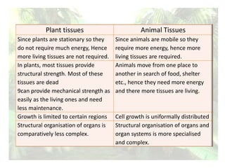
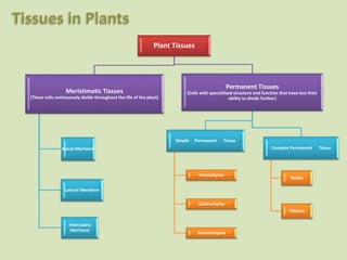
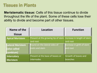
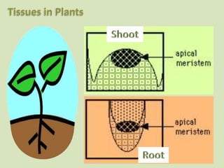
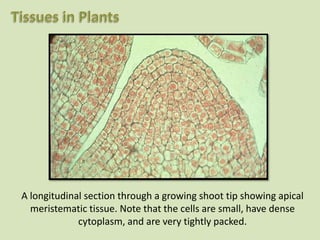
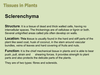
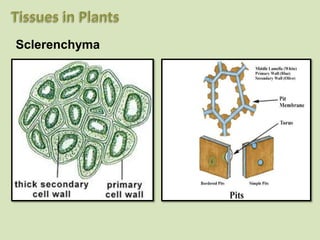
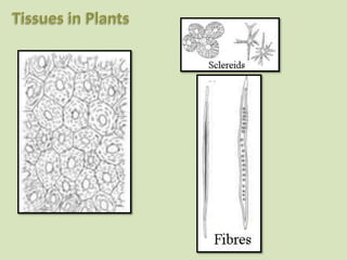
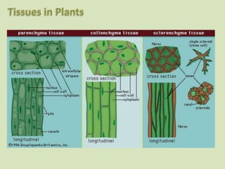
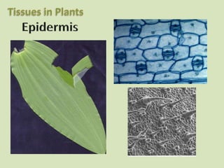
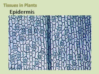
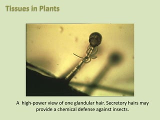
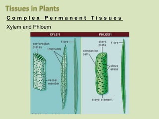
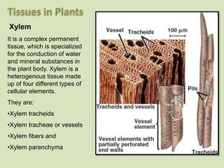
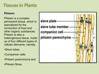
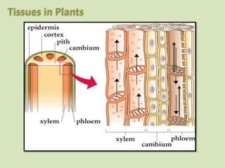
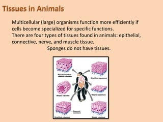
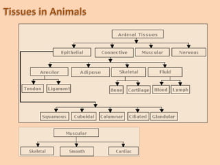
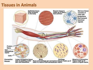
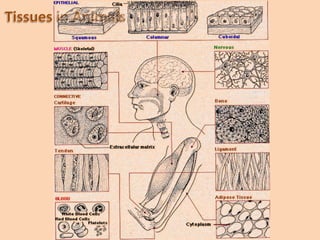
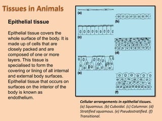
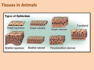
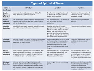
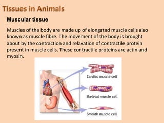
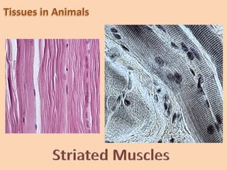
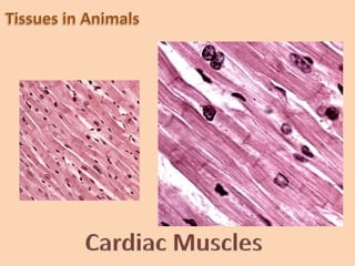
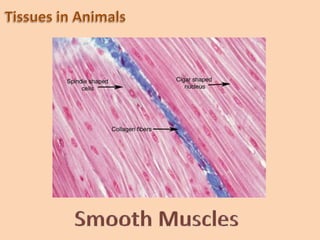
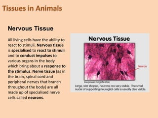
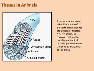
Tissue is a cellular organizational
level intermediate between cells and
a complete organism. Hence, a tissue
is an ensemble of cells, not necessarily
identical, but from the same origin,
that together carry out a specific
function.
The study of tissue is known
as histology or, in connection
with disease, histopathology.
Plant tissues Animal Tissues
Since plants are stationary so they
do not require much energy, Hence
more living tissues are not required.
Since animals are mobile so they
require more energy, hence more
living tissues are required.
In plants, most tissues provide
structural strength. Most of these
tissues are dead
9can provide mechanical strength as
easily as the living ones and need
less maintenance.
Animals move from one place to
another in search of food, shelter
etc., hence they need more energy
and there more tissues are living.
Growth is limited to certain regions Cell growth is uniformally distributed
Structural organisation of organs is
comparatively less complex.
Structural organisation of organs and
organ systems is more specialised
and complex.
Plant Tissues
Meristimatic Tissues
(These cells continuously divide throughout the life of the plant)
Apical Meristem
Lateral Meristem
Intercalary
Meristem
Permanent Tissues
(Cells with speciallised structure and function that have lost their
ability to divide further)
Simple Permanent Tissue
Parenchyma
Collenchyma
Sclerenchyma
Complex Permanent Tissue
Xylem
Phloem
Meristematic tissue: Cells of this tissue continue to divide
throughout the life of the plant. Some of these cells lose their
ability to divide and become part of other tissues.
Name of the
tissue
Location Function
Apical Meristem Present at the growing tip of stem
and root
Increase in length of stem
and root
Lateral Meristem
(also called
cambium)
Found on the lateral sides of
roots and stem
Increase in girth of stem
and root
Intercalary
Meristem
Present at the base of leaves or
internodes
Growth of leaves and
branches
A longitudinal section through a growing shoot tip showing apical
meristematic tissue. Note that the cells are small, have dense
cytoplasm, and are very tightly packed.
High power view of a longitudinal
section of the Coleus apical
meristem. The apical meristem is a
dome-shaped mass of dividing cells
at the tip of the shoot. The apical
meristem will produce the three
primary meristems: protoderm,
procambium, and ground
meristem. These three meristems
in turn will produce new cells that
will differentiate into the
epidermis, primary vascular
tissues, and ground tissues (pith
and cortex).
A longitudinal section through a root tip. The meristematic tissue is
located just above the root cap. This too is apical meristem; division
of these cells followed by cell elongation results in the root growing
in length.
It is a cross section of a dicot
stem.
Focus on the two large
vascular bundles in the center
of the slide.
The xylem tissue is stained
red.
Just above the xylem is a
layer of meristematic tissue,
the vascular cambium.
The phloem tissue is found
outside of the vascular
cambium.
This is a high-power view of a cross-section showing a lateral meristem, the vascular cambium, in the
same plant shown in previous slide. Again, the xylem tissue is stained red, and the large cells on the top
of the slide are phloem. The green brick-like cells between the xylem and phloem is the area in which
the vascular cambium is located. The new cells produced by the cambium are initially like those of the
cambium itself, but, as they grow and mature, their characteristics slowly change as they differentiate
into other tissues. The vascular cambium is a single layer of cells within this brick like region; it is
responsible for the growth in diameter of a stem. The tissues produced by the vascular cambium are
secondary tissues.
Permanent tissue: Cells of this tissue have lost their ability to
divide and they have a specialized structure to perform
specific functions.
Based on the type of cells present in the tissue, the
Permanent tissue is divided into two categories:
Simple Permanent Tissue
and
Complex Permanent Tissue.
While the simple permanent tissue consist of only one type
of cells (eg. Parenchyma),
the complex permanent tissue consists of more than one
type of cells (eg. Xylem and phloem)
S i m p l e P e r m a n e n t T i s s u e s
Parenchyma
Structure:It is the fundamental tissue composed of thin walled, living
cells whose cell wall is composed of cellulose. Small intercellular spaces
are present between the cells.
Location and function: It occurs in all soft parts of plants and is
meant for storage of food and to provide turgidity to softer parts of plants.
Parenchyma tissue in stem and roots store nutrients and water.
Types of parenchyma:
i) Chlorenchyma :Certain parenchymatous tissue contain chloroplast
and synthesize food by the process of photosynthesis.
ii) Aerenchyma: In aquatic plants parenchymatous cells have air cavities
between them to store air, such a tissue is called Aerenchyma. It provides
buoyancy to the aquatic plants so that they can float in water.
Collenchyma
Structure: This tissue is composed of somewhat elongated cells with
cell walls that are irregularly thickened at corners due to deposition of
cellulose or pectin. They may be oval, circular or polygonal. Very little
intercellular spaces are present.
Location: It occurs below the epidermis of stem and petiole (stalk of
the leaf) and around veins.
Function: This tissue provides mechanical support and flexibility and
in some cases it may possess chloroplasts to perform Photosynthesis.
The stem and leaves are able to bend easily and then come back to
their original position due to the presence of collenchyma.
Collenchyma in Transverse Section Showing
Wall Thickenings
1. Cell Wall
2. Wall Thickenings
3. Protoplasm
4. Vacuole
Sclerenchyma
Structure: It is a tissue of dead and thick walled cells, having no
intercellular spaces. The thickenings are of cellulose or lignin or both.
Several unlignified areas called pits often develop on walls.
Location: This tissue is usually found in the hard and stiff parts of the
plant like seed coat, husk of coconut, in the stem around vascular
bundles, veins of leaves and hard covering of fruits and nuts.
Function: It is the chief mechanical tissue in plants and is able to bear
push, pull, strain and shearing forces. It provides strength to plant
parts and also protects the delicate parts of the plants.
They are of two types: fibres and sclereids.
Sclerenchyma
Epidermis and Bark
The protective tissues
The epidermis usually consists of a single-layered group of cells that covers
plants leaves, flowers, roots and stems. It forms a boundary between the plant
and the external world.
Bark is formed from the meristem that appears later in the life cycle of a plant.
Woody stems and some other stem structures produce a secondary covering
called the secondary meristem or periderm or cork cambium that replaces the
epidermis as the protective covering.
The periderm replaces the epidermis, and acts as a protective covering like the
epidermis.
Cells produced on the outside by periderm form the cork. Cells of have suberin
in their walls to protect the stem from drying and pathogen attack. Older cork
cells are dead, as is the case with woody stems. As the stem grows, the cork
cambium produces new layers of cork which are impermeable to gases and
water.
A high-power view of one glandular hair. Secretory hairs may
provide a chemical defense against insects.
Another type of surface tissue, the outer bark or periderm (stained red in this slide).
Periderm is found on the surface of woody plants; it includes the cork cells on the
surface of older woody stems. The periderm replaces the epidermis in plants that have
secondary growth. The cork cells are dead; it is their waterproofed cell walls that
function as the protective outer covering of plants. Meristematic cells within the
periderm (cork cambium, the other lateral meristem) produce the cork cells.
C o m p l e x P e r m a n e n t T i s s u e s
Xylem and Phloem
It is a complex permanent
tissue, which is specialized
for the conduction of water
and mineral substances in
the plant body. Xylem is a
heterogenous tissue made
up of four different types of
cellular elements.
They are:
•Xylem tracheids
•Xylem tracheae or vessels
•Xylem fibers and
•Xylem parenchyma
Xylem
Phloem:
Phloem is a complex
permanent tissue, which is
specialized for the
conduction of food and
other organic substances.
Phloem is also a
heterogenous tissue, made
up of four different types of
cellular elements, namely,
•Sieve tubes
•Companion cells
•Phloem parenchyma and
•Phloem fibres
Multicellular (large) organisms function more efficiently if
cells become specialized for specific functions.
There are four types of tissues found in animals: epithelial,
connective, nerve, and muscle tissue.
Sponges do not have tissues.
Epithelial tissue covers the
whole surface of the body. It is
made up of cells that are
closely packed and are
composed of one or more
layers. This tissue is
specialised to form the
covering or lining of all internal
and external body surfaces.
Epithelial tissue that occurs on
surfaces on the interior of the
body is known as
endothelium.
Epithelial tissue
Cellular arrangements in epithelial tissues.
(a) Squamous. (b) Cuboidal. (c) Columnar. (d)
Stratified squamous. (e) Pseudostratified. (f)
Transitional.
Types of Epithelial Tissue
Name of
the Tissue
Structure Location Function
Simple
Squamous
Epithelium
Squamous cells have the appearance of thin, flat
plates.The nucleus is flat and elliptical.
They form the lining of cavities such
as the mouth, oesophagus, blood
vessels, heart and lungs
Protection and transportation of
substances through a selectively
permeable surface.
Simple
Stratified
Epithelium
Cells are arranged in many layers and the top layers of
this tissue may consist of dead cells that are covered
with keratin (a protein)
The mammalian skin is an example of
dry, keratinised, stratified
epithelium.
Protection and to prevent wear
and tear
Cuboidal
Epithelium
cuboidal cells are roughly square or cuboidal in shape.
Each cell has a spherical nucleus in the centre
Cuboidal epithelium is found in
glands and in the lining of the kidney
tubules as well as in the ducts of the
glands. They also constitute the
germinal epithelium which produces
the egg cells in the female ovary and
the sperm cells in the male testes.
Protection and mechanical support
Columnar
Epithelium
Cells occur in one or more layers. The cells are elongated and
column-shaped. The nuclei are elongated and are usually
located near the base of the cells.
Columnar epithelium forms the lining
of the stomach and intestines. Some
columnar cells are specialised for
sensory reception such as in the
nose, ears and the taste buds of the
tongue.
Protection, absorption of food (in
small intestine), secretion of
enzymes
Ciliated
Columnar
Epithelium
simple columnar epithelial cells, but in addition, they
posses fine hair-like outgrowths, cilia on their free
surfaces. These cilia are capable of rapid, rhythmic,
wavelike beatings in a certain direction.
Ciliated epithelium is usually found in
the air passages like the nose. It is
also found in the uterus and
Fallopian tubes of females.
This movement of the cilia in a
certain direction causes the mucus,
to move (flow or stream) in that
direction. The movement of the
cilia propel the ovum to the uterus.
Glandular
Epithelium
Columnar epithelium with goblet cells is called
glandular epithelium. Some parts of the glandular
epithelium consist of such a large number of goblet
cells that there are only a few normal epithelial cells
left.
Inner lining of stomach and salivary
glands
specialised gland cells are capable
of synthesising and secreting
certain substances such as
enzymes, hormones, milk, mucus,
sweat, wax and saliva.
Connective Tissue
It is an animal tissue that is characterized by the abundance of
extracellular components (such as fibers and intercellular substances).
The tissue derives its name from its function in connecting, supporting,
surrounding or binding cells and tissues.
Connective tissue is composed of:
•cells
•extracellular matrix
Extracellular matrix is a special feature that distinguishes connective
tissue from the other tissues of the body. This matrix may be jelly-like,
fluid, dense or rigid. The nature of matrix differs according to the
function of that particular connective tissue.
Muscular tissue
Muscles of the body are made up of elongated muscle cells also
known as muscle fibre. The movement of the body is brought
about by the contraction and relaxation of contractile protein
present in muscle cells. These contractile proteins are actin and
myosin.
Nervous Tissue
All living cells have the ability to
react to stimuli. Nervous tissue
is specialised to react to stimuli
and to conduct impulses to
various organs in the body
which bring about a response to
the stimulus. Nerve tissue (as in
the brain, spinal cord and
peripheral nerves that branch
throughout the body) are all
made up of specialised nerve
cells called neurons.
Neurons have many different shapes and
sizes. However, a typical neuron in a human
consists of four major regions: a cell body,
dendrites, an axon, and synaptic terminals.
Like all cells, the entire neuron is surrounded
by a cell membrane. The cell body is the
enlarged portion of a neuron that most
closely resembles other cells. It contains the
nucleus and other organelles (for example,
the mitochondria and endoplasmic
reticulum). The dendrites and axon are thin
cytoplasmic extensions of the neuron. The
dendrites, which branch out in treelike
fashion from the cell body, are specialized to
receive signals and transmit them toward the
cell body. The single long axon carries
signals away from the cell body.
In humans, a single axon may be as long as
1 meter (about 3 feet). Some neurons that
have cell bodies in the spinal cord have
axons that extend all the way down to the
toes.
A nerve is an enclosed,
cable-like bundle of
axons (the long, slender
projections of neurons).
A nerve provides a
common pathway for
the electrochemical
nerve impulses that are
transmitted along each
of the axons.
STUDY OF ANIMAL TISSUES

STUDY OF ANIMAL TISSUES

  • 2.
    Tissue is acellular organizational level intermediate between cells and a complete organism. Hence, a tissue is an ensemble of cells, not necessarily identical, but from the same origin, that together carry out a specific function.
  • 3.
    The study oftissue is known as histology or, in connection with disease, histopathology.
  • 4.
    Plant tissues AnimalTissues Since plants are stationary so they do not require much energy, Hence more living tissues are not required. Since animals are mobile so they require more energy, hence more living tissues are required. In plants, most tissues provide structural strength. Most of these tissues are dead 9can provide mechanical strength as easily as the living ones and need less maintenance. Animals move from one place to another in search of food, shelter etc., hence they need more energy and there more tissues are living. Growth is limited to certain regions Cell growth is uniformally distributed Structural organisation of organs is comparatively less complex. Structural organisation of organs and organ systems is more specialised and complex.
  • 5.
    Plant Tissues Meristimatic Tissues (Thesecells continuously divide throughout the life of the plant) Apical Meristem Lateral Meristem Intercalary Meristem Permanent Tissues (Cells with speciallised structure and function that have lost their ability to divide further) Simple Permanent Tissue Parenchyma Collenchyma Sclerenchyma Complex Permanent Tissue Xylem Phloem
  • 6.
    Meristematic tissue: Cellsof this tissue continue to divide throughout the life of the plant. Some of these cells lose their ability to divide and become part of other tissues. Name of the tissue Location Function Apical Meristem Present at the growing tip of stem and root Increase in length of stem and root Lateral Meristem (also called cambium) Found on the lateral sides of roots and stem Increase in girth of stem and root Intercalary Meristem Present at the base of leaves or internodes Growth of leaves and branches
  • 8.
    A longitudinal sectionthrough a growing shoot tip showing apical meristematic tissue. Note that the cells are small, have dense cytoplasm, and are very tightly packed.
  • 9.
    High power viewof a longitudinal section of the Coleus apical meristem. The apical meristem is a dome-shaped mass of dividing cells at the tip of the shoot. The apical meristem will produce the three primary meristems: protoderm, procambium, and ground meristem. These three meristems in turn will produce new cells that will differentiate into the epidermis, primary vascular tissues, and ground tissues (pith and cortex).
  • 10.
    A longitudinal sectionthrough a root tip. The meristematic tissue is located just above the root cap. This too is apical meristem; division of these cells followed by cell elongation results in the root growing in length.
  • 11.
    It is across section of a dicot stem. Focus on the two large vascular bundles in the center of the slide. The xylem tissue is stained red. Just above the xylem is a layer of meristematic tissue, the vascular cambium. The phloem tissue is found outside of the vascular cambium.
  • 12.
    This is ahigh-power view of a cross-section showing a lateral meristem, the vascular cambium, in the same plant shown in previous slide. Again, the xylem tissue is stained red, and the large cells on the top of the slide are phloem. The green brick-like cells between the xylem and phloem is the area in which the vascular cambium is located. The new cells produced by the cambium are initially like those of the cambium itself, but, as they grow and mature, their characteristics slowly change as they differentiate into other tissues. The vascular cambium is a single layer of cells within this brick like region; it is responsible for the growth in diameter of a stem. The tissues produced by the vascular cambium are secondary tissues.
  • 13.
    Permanent tissue: Cellsof this tissue have lost their ability to divide and they have a specialized structure to perform specific functions. Based on the type of cells present in the tissue, the Permanent tissue is divided into two categories: Simple Permanent Tissue and Complex Permanent Tissue. While the simple permanent tissue consist of only one type of cells (eg. Parenchyma), the complex permanent tissue consists of more than one type of cells (eg. Xylem and phloem)
  • 14.
    S i mp l e P e r m a n e n t T i s s u e s Parenchyma Structure:It is the fundamental tissue composed of thin walled, living cells whose cell wall is composed of cellulose. Small intercellular spaces are present between the cells. Location and function: It occurs in all soft parts of plants and is meant for storage of food and to provide turgidity to softer parts of plants. Parenchyma tissue in stem and roots store nutrients and water. Types of parenchyma: i) Chlorenchyma :Certain parenchymatous tissue contain chloroplast and synthesize food by the process of photosynthesis. ii) Aerenchyma: In aquatic plants parenchymatous cells have air cavities between them to store air, such a tissue is called Aerenchyma. It provides buoyancy to the aquatic plants so that they can float in water.
  • 16.
    Collenchyma Structure: This tissueis composed of somewhat elongated cells with cell walls that are irregularly thickened at corners due to deposition of cellulose or pectin. They may be oval, circular or polygonal. Very little intercellular spaces are present. Location: It occurs below the epidermis of stem and petiole (stalk of the leaf) and around veins. Function: This tissue provides mechanical support and flexibility and in some cases it may possess chloroplasts to perform Photosynthesis. The stem and leaves are able to bend easily and then come back to their original position due to the presence of collenchyma.
  • 17.
    Collenchyma in TransverseSection Showing Wall Thickenings 1. Cell Wall 2. Wall Thickenings 3. Protoplasm 4. Vacuole
  • 18.
    Sclerenchyma Structure: It isa tissue of dead and thick walled cells, having no intercellular spaces. The thickenings are of cellulose or lignin or both. Several unlignified areas called pits often develop on walls. Location: This tissue is usually found in the hard and stiff parts of the plant like seed coat, husk of coconut, in the stem around vascular bundles, veins of leaves and hard covering of fruits and nuts. Function: It is the chief mechanical tissue in plants and is able to bear push, pull, strain and shearing forces. It provides strength to plant parts and also protects the delicate parts of the plants. They are of two types: fibres and sclereids.
  • 19.
  • 22.
    Epidermis and Bark Theprotective tissues The epidermis usually consists of a single-layered group of cells that covers plants leaves, flowers, roots and stems. It forms a boundary between the plant and the external world. Bark is formed from the meristem that appears later in the life cycle of a plant. Woody stems and some other stem structures produce a secondary covering called the secondary meristem or periderm or cork cambium that replaces the epidermis as the protective covering. The periderm replaces the epidermis, and acts as a protective covering like the epidermis. Cells produced on the outside by periderm form the cork. Cells of have suberin in their walls to protect the stem from drying and pathogen attack. Older cork cells are dead, as is the case with woody stems. As the stem grows, the cork cambium produces new layers of cork which are impermeable to gases and water.
  • 25.
    A high-power viewof one glandular hair. Secretory hairs may provide a chemical defense against insects.
  • 27.
    Another type ofsurface tissue, the outer bark or periderm (stained red in this slide). Periderm is found on the surface of woody plants; it includes the cork cells on the surface of older woody stems. The periderm replaces the epidermis in plants that have secondary growth. The cork cells are dead; it is their waterproofed cell walls that function as the protective outer covering of plants. Meristematic cells within the periderm (cork cambium, the other lateral meristem) produce the cork cells.
  • 28.
    C o mp l e x P e r m a n e n t T i s s u e s Xylem and Phloem
  • 29.
    It is acomplex permanent tissue, which is specialized for the conduction of water and mineral substances in the plant body. Xylem is a heterogenous tissue made up of four different types of cellular elements. They are: •Xylem tracheids •Xylem tracheae or vessels •Xylem fibers and •Xylem parenchyma Xylem
  • 30.
    Phloem: Phloem is acomplex permanent tissue, which is specialized for the conduction of food and other organic substances. Phloem is also a heterogenous tissue, made up of four different types of cellular elements, namely, •Sieve tubes •Companion cells •Phloem parenchyma and •Phloem fibres
  • 32.
    Multicellular (large) organismsfunction more efficiently if cells become specialized for specific functions. There are four types of tissues found in animals: epithelial, connective, nerve, and muscle tissue. Sponges do not have tissues.
  • 36.
    Epithelial tissue coversthe whole surface of the body. It is made up of cells that are closely packed and are composed of one or more layers. This tissue is specialised to form the covering or lining of all internal and external body surfaces. Epithelial tissue that occurs on surfaces on the interior of the body is known as endothelium. Epithelial tissue Cellular arrangements in epithelial tissues. (a) Squamous. (b) Cuboidal. (c) Columnar. (d) Stratified squamous. (e) Pseudostratified. (f) Transitional.
  • 38.
    Types of EpithelialTissue Name of the Tissue Structure Location Function Simple Squamous Epithelium Squamous cells have the appearance of thin, flat plates.The nucleus is flat and elliptical. They form the lining of cavities such as the mouth, oesophagus, blood vessels, heart and lungs Protection and transportation of substances through a selectively permeable surface. Simple Stratified Epithelium Cells are arranged in many layers and the top layers of this tissue may consist of dead cells that are covered with keratin (a protein) The mammalian skin is an example of dry, keratinised, stratified epithelium. Protection and to prevent wear and tear Cuboidal Epithelium cuboidal cells are roughly square or cuboidal in shape. Each cell has a spherical nucleus in the centre Cuboidal epithelium is found in glands and in the lining of the kidney tubules as well as in the ducts of the glands. They also constitute the germinal epithelium which produces the egg cells in the female ovary and the sperm cells in the male testes. Protection and mechanical support Columnar Epithelium Cells occur in one or more layers. The cells are elongated and column-shaped. The nuclei are elongated and are usually located near the base of the cells. Columnar epithelium forms the lining of the stomach and intestines. Some columnar cells are specialised for sensory reception such as in the nose, ears and the taste buds of the tongue. Protection, absorption of food (in small intestine), secretion of enzymes Ciliated Columnar Epithelium simple columnar epithelial cells, but in addition, they posses fine hair-like outgrowths, cilia on their free surfaces. These cilia are capable of rapid, rhythmic, wavelike beatings in a certain direction. Ciliated epithelium is usually found in the air passages like the nose. It is also found in the uterus and Fallopian tubes of females. This movement of the cilia in a certain direction causes the mucus, to move (flow or stream) in that direction. The movement of the cilia propel the ovum to the uterus. Glandular Epithelium Columnar epithelium with goblet cells is called glandular epithelium. Some parts of the glandular epithelium consist of such a large number of goblet cells that there are only a few normal epithelial cells left. Inner lining of stomach and salivary glands specialised gland cells are capable of synthesising and secreting certain substances such as enzymes, hormones, milk, mucus, sweat, wax and saliva.
  • 39.
    Connective Tissue It isan animal tissue that is characterized by the abundance of extracellular components (such as fibers and intercellular substances). The tissue derives its name from its function in connecting, supporting, surrounding or binding cells and tissues. Connective tissue is composed of: •cells •extracellular matrix Extracellular matrix is a special feature that distinguishes connective tissue from the other tissues of the body. This matrix may be jelly-like, fluid, dense or rigid. The nature of matrix differs according to the function of that particular connective tissue.
  • 42.
    Muscular tissue Muscles ofthe body are made up of elongated muscle cells also known as muscle fibre. The movement of the body is brought about by the contraction and relaxation of contractile protein present in muscle cells. These contractile proteins are actin and myosin.
  • 47.
    Nervous Tissue All livingcells have the ability to react to stimuli. Nervous tissue is specialised to react to stimuli and to conduct impulses to various organs in the body which bring about a response to the stimulus. Nerve tissue (as in the brain, spinal cord and peripheral nerves that branch throughout the body) are all made up of specialised nerve cells called neurons.
  • 48.
    Neurons have manydifferent shapes and sizes. However, a typical neuron in a human consists of four major regions: a cell body, dendrites, an axon, and synaptic terminals. Like all cells, the entire neuron is surrounded by a cell membrane. The cell body is the enlarged portion of a neuron that most closely resembles other cells. It contains the nucleus and other organelles (for example, the mitochondria and endoplasmic reticulum). The dendrites and axon are thin cytoplasmic extensions of the neuron. The dendrites, which branch out in treelike fashion from the cell body, are specialized to receive signals and transmit them toward the cell body. The single long axon carries signals away from the cell body. In humans, a single axon may be as long as 1 meter (about 3 feet). Some neurons that have cell bodies in the spinal cord have axons that extend all the way down to the toes.
  • 50.
    A nerve isan enclosed, cable-like bundle of axons (the long, slender projections of neurons). A nerve provides a common pathway for the electrochemical nerve impulses that are transmitted along each of the axons.