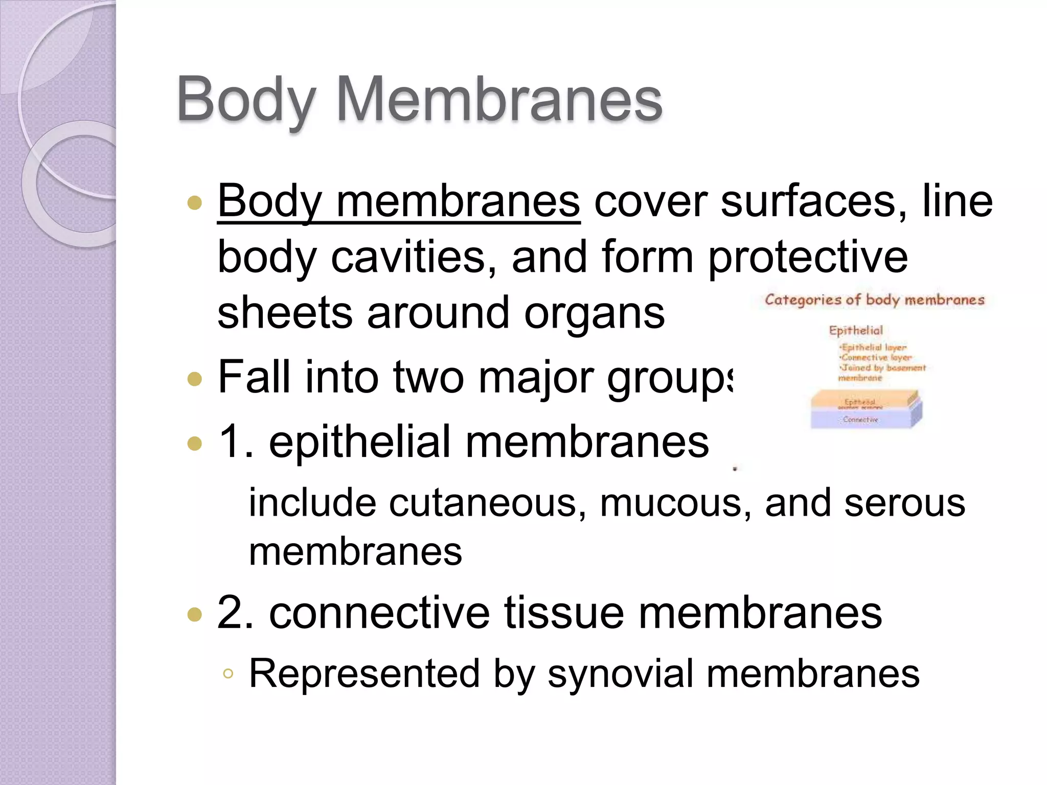
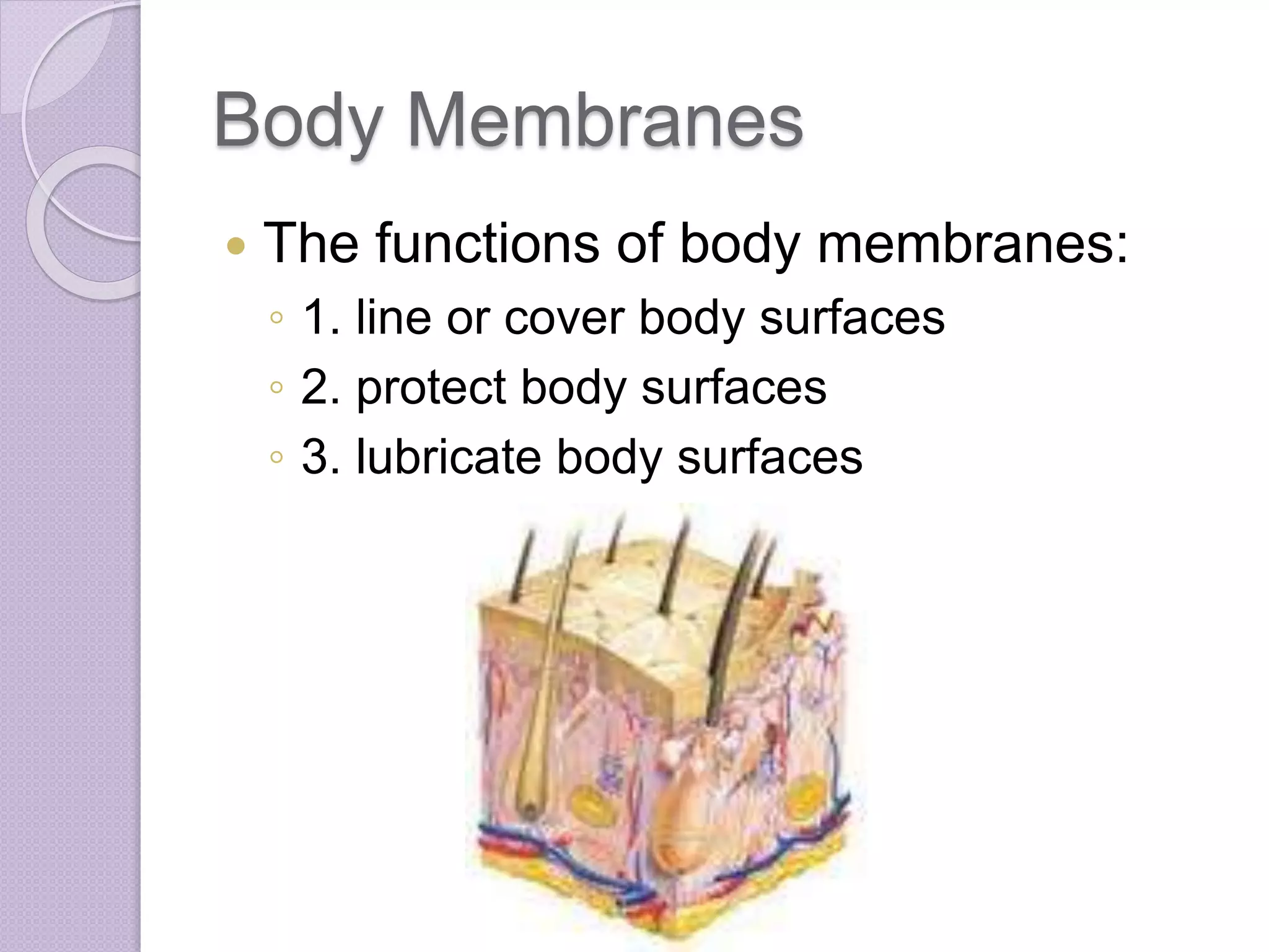
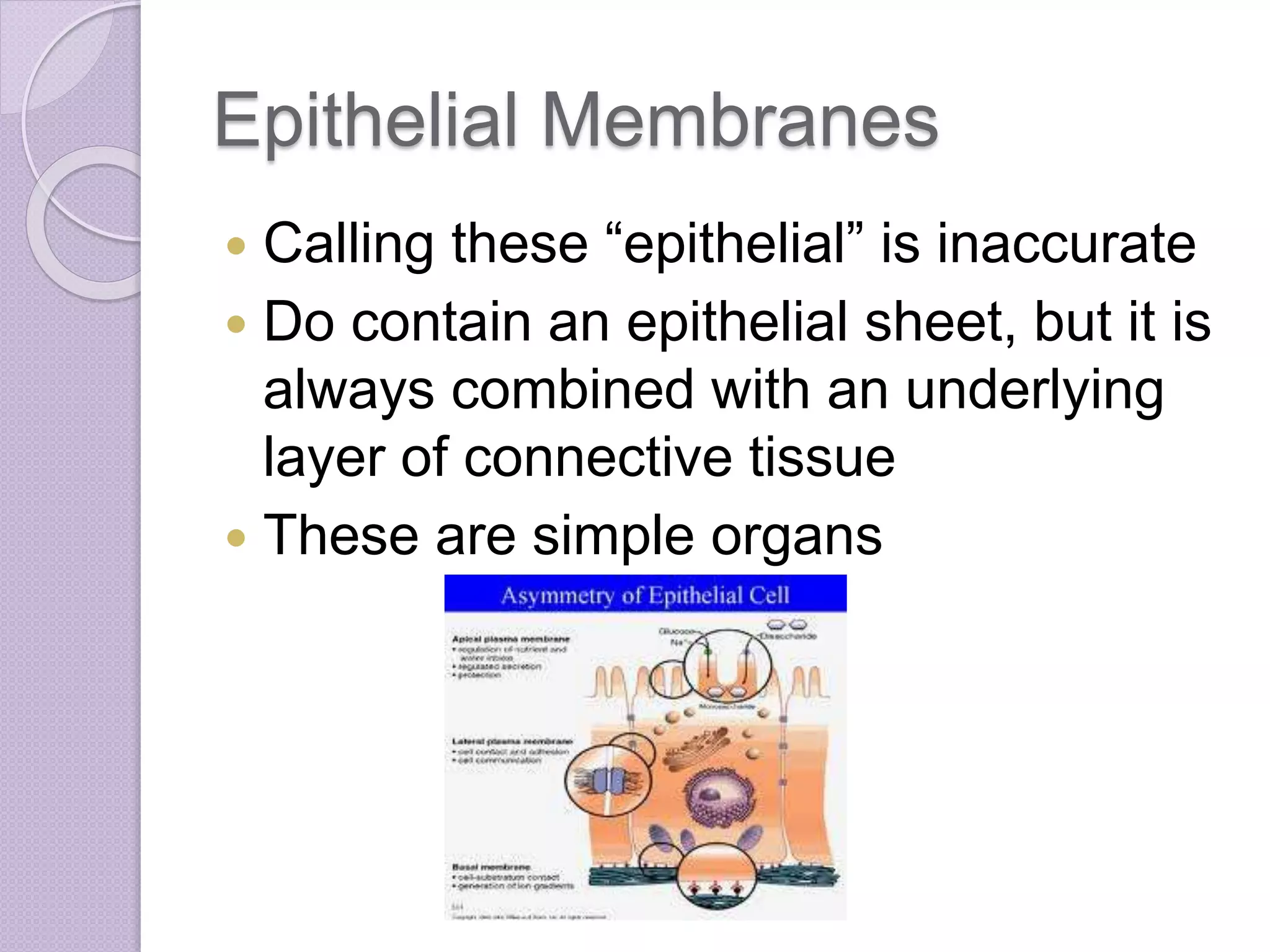
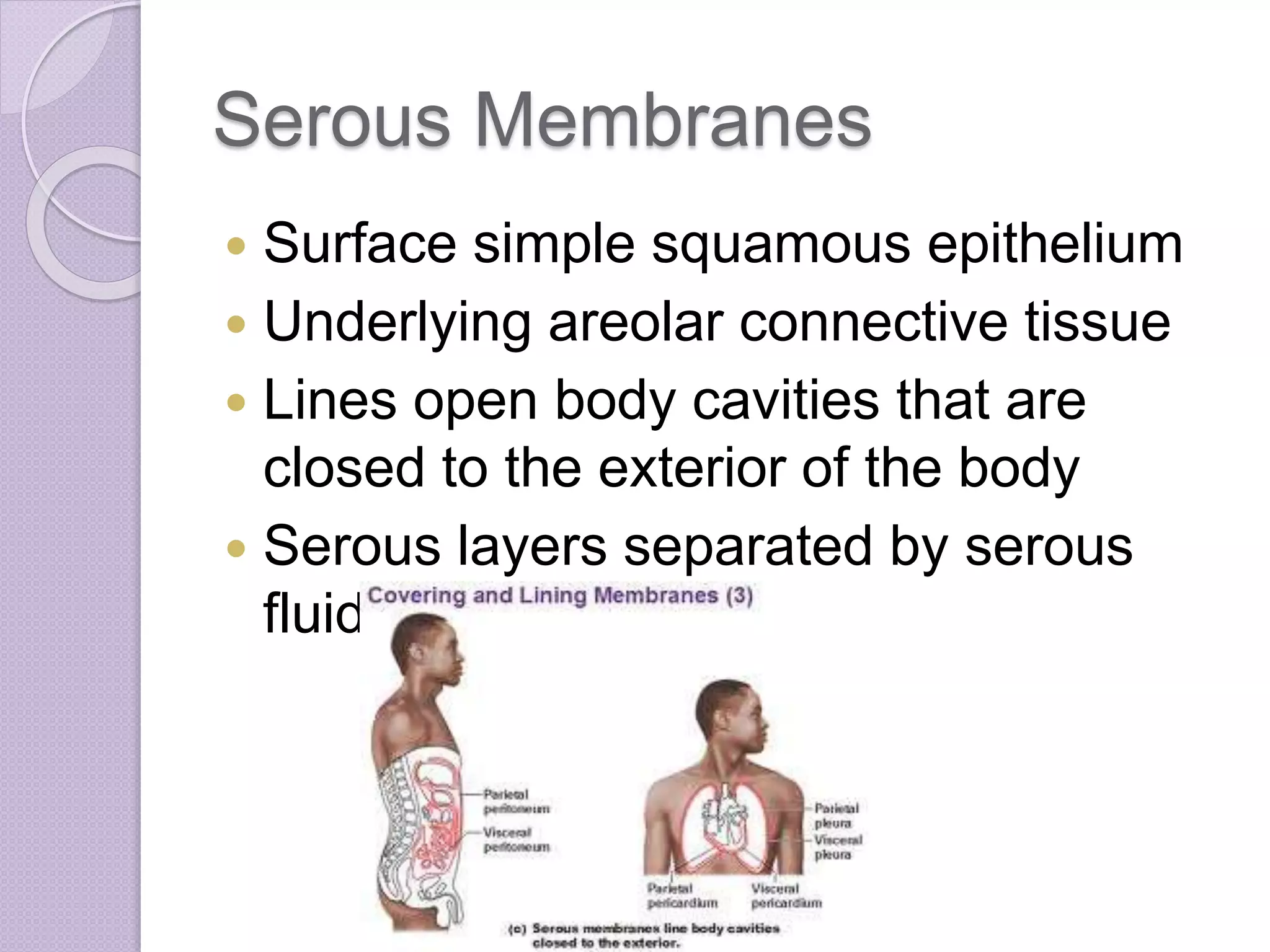
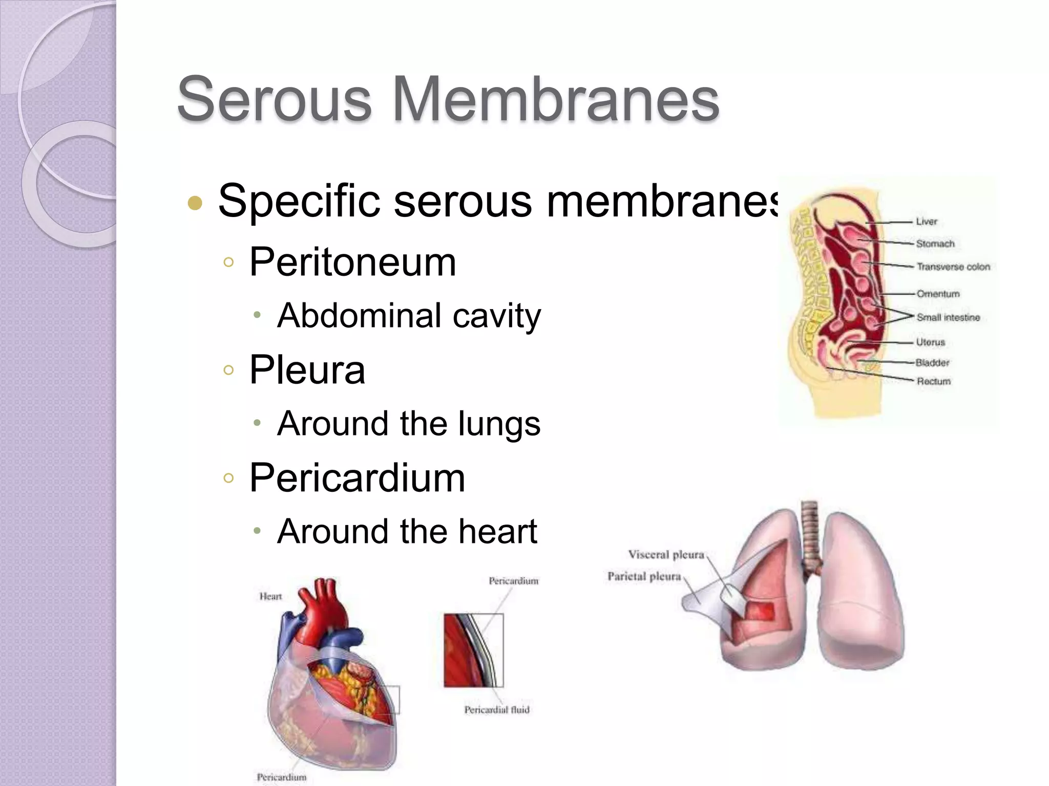
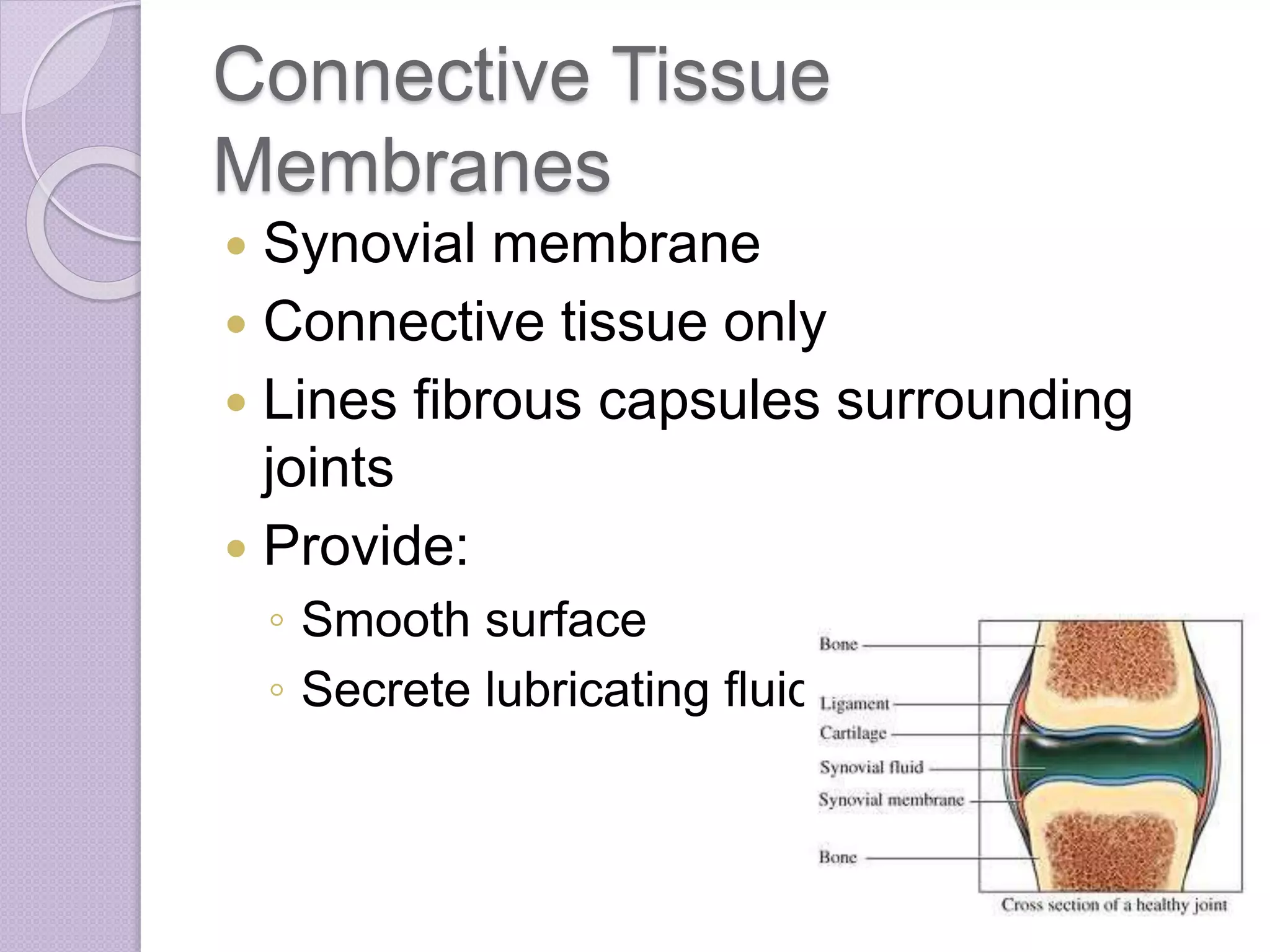
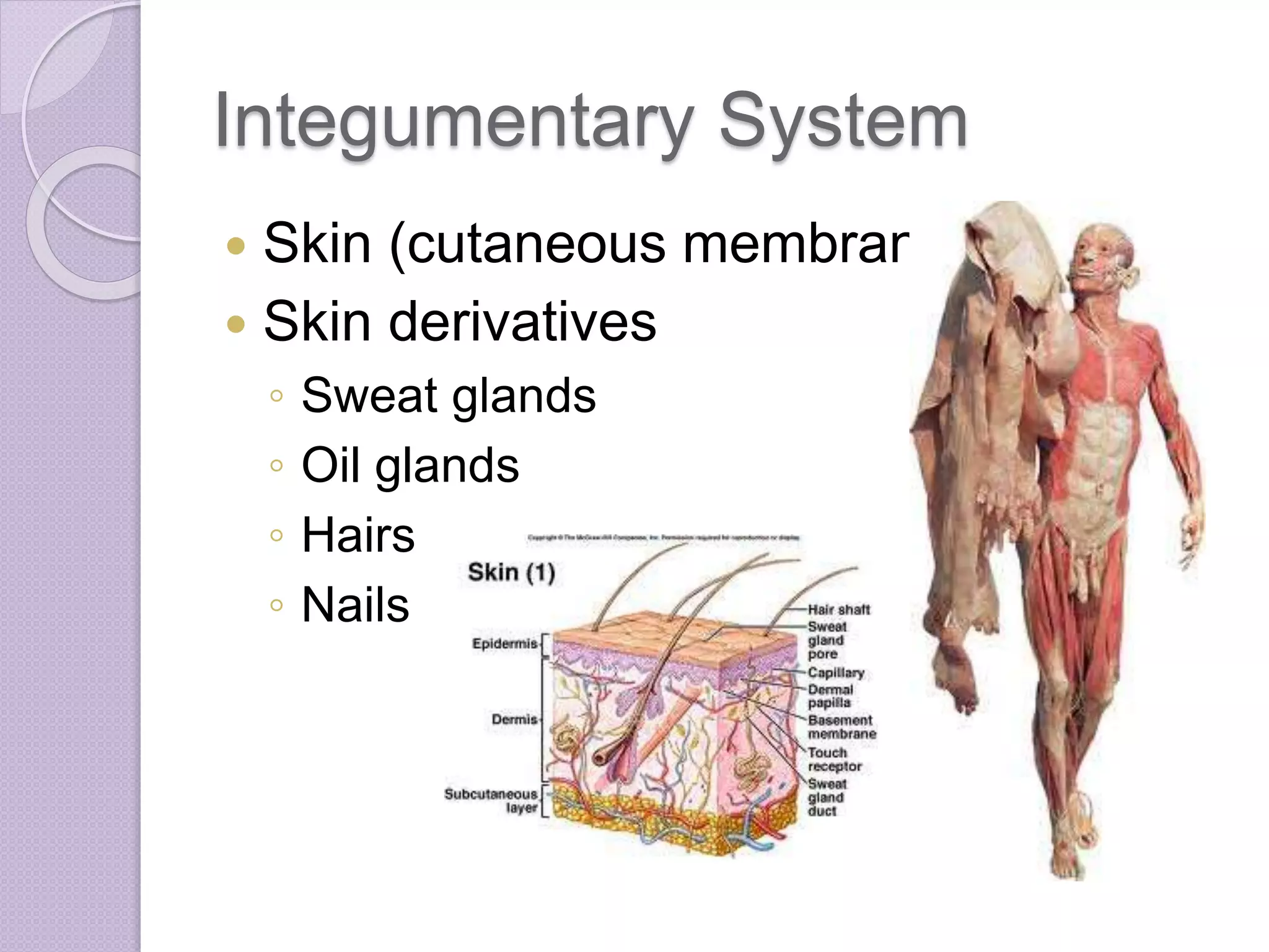
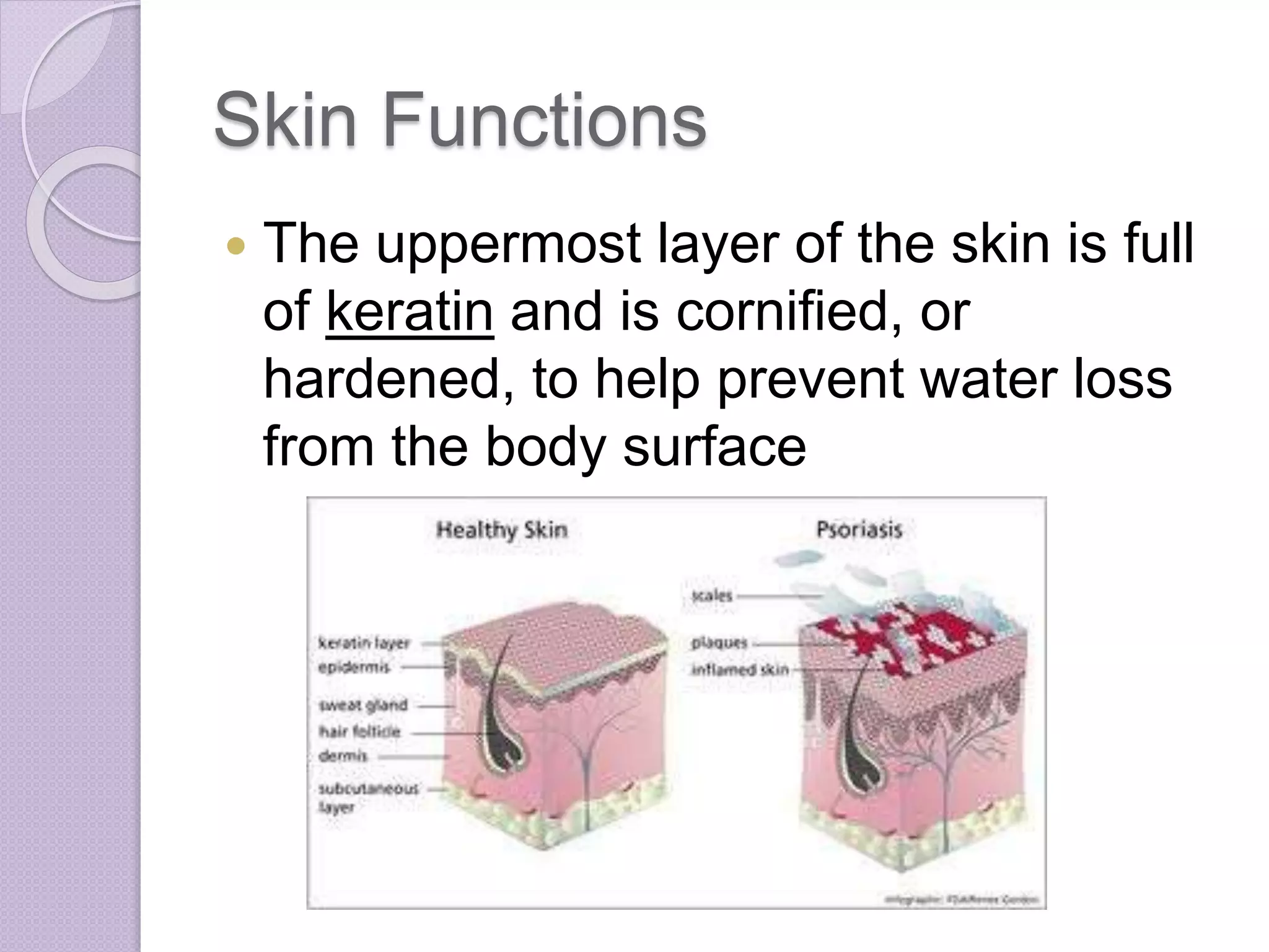
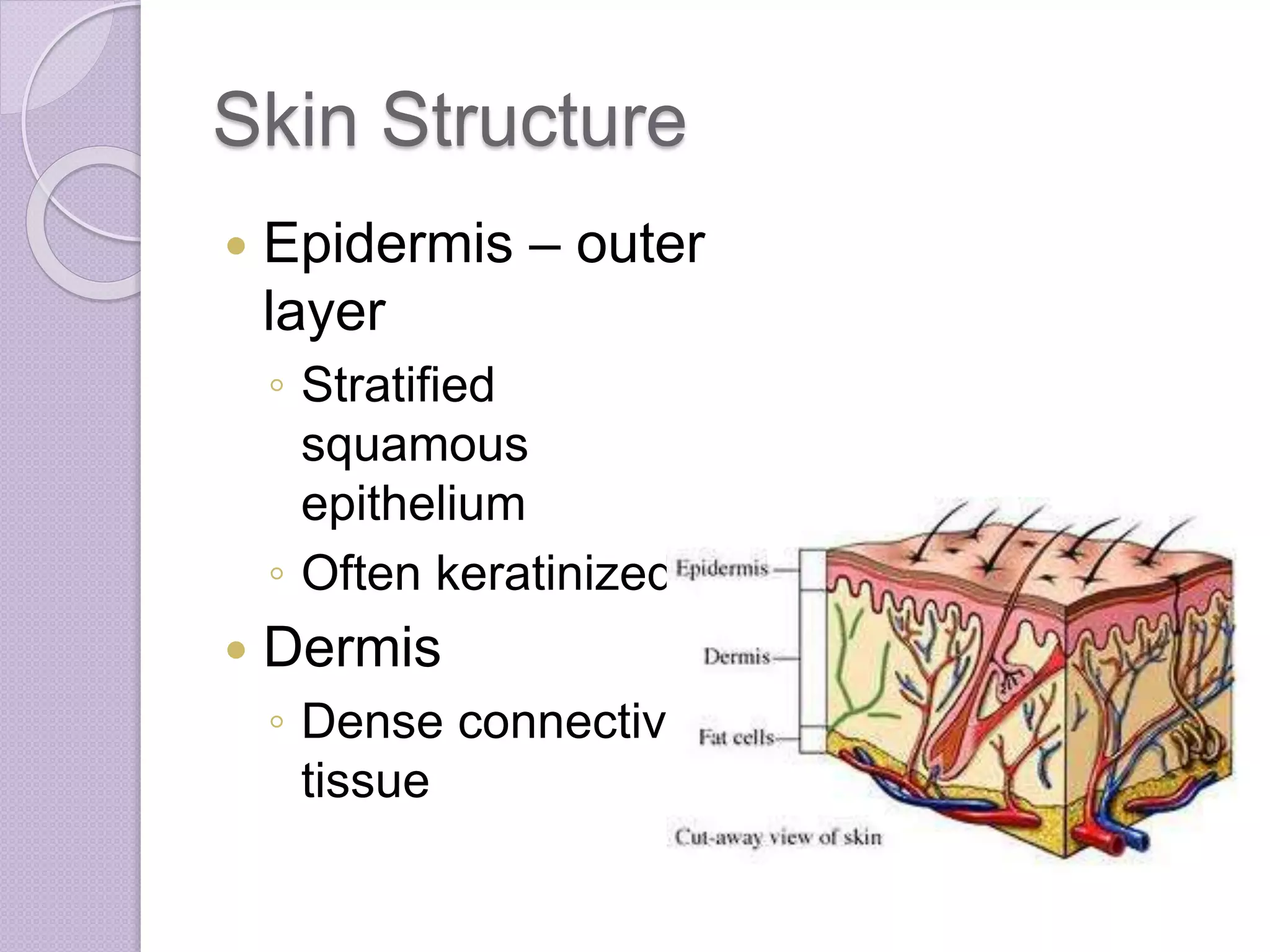
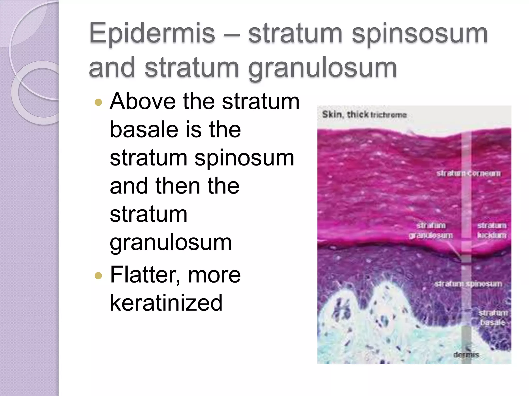
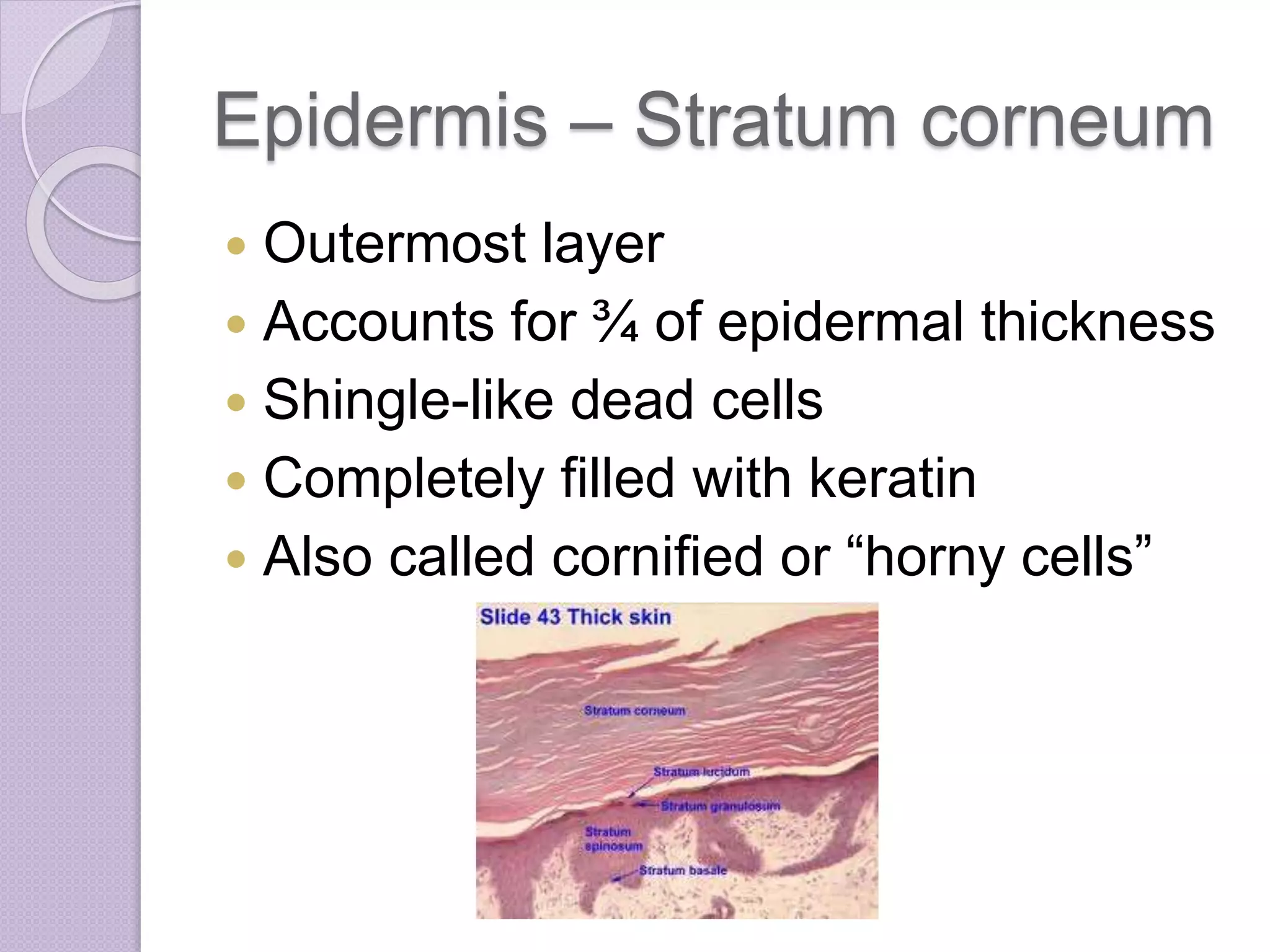
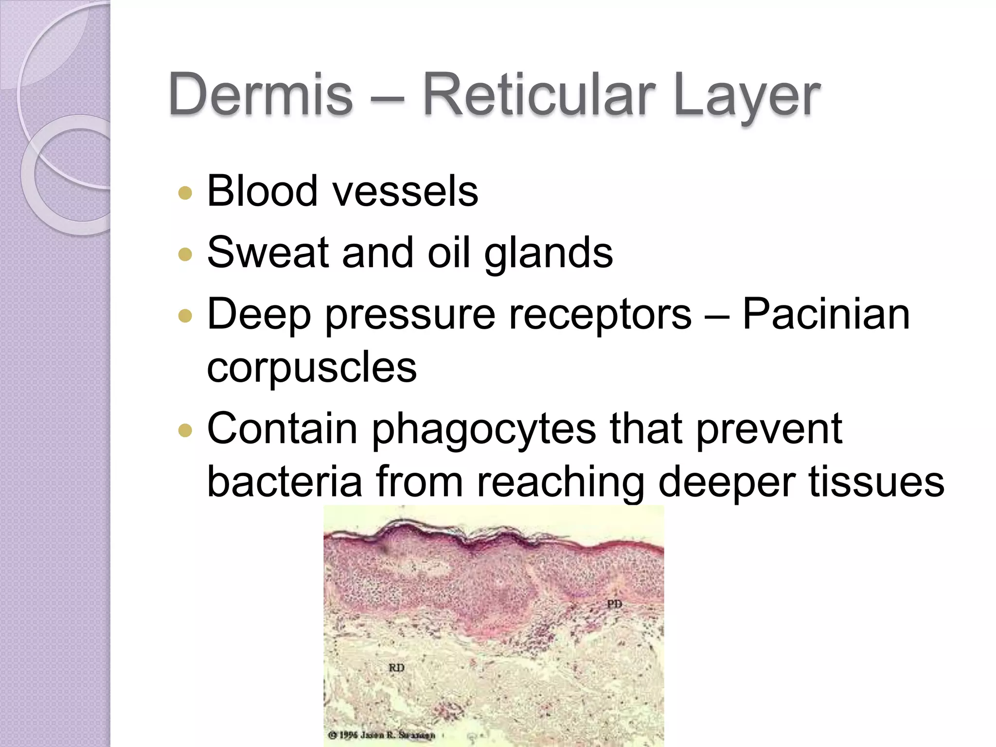
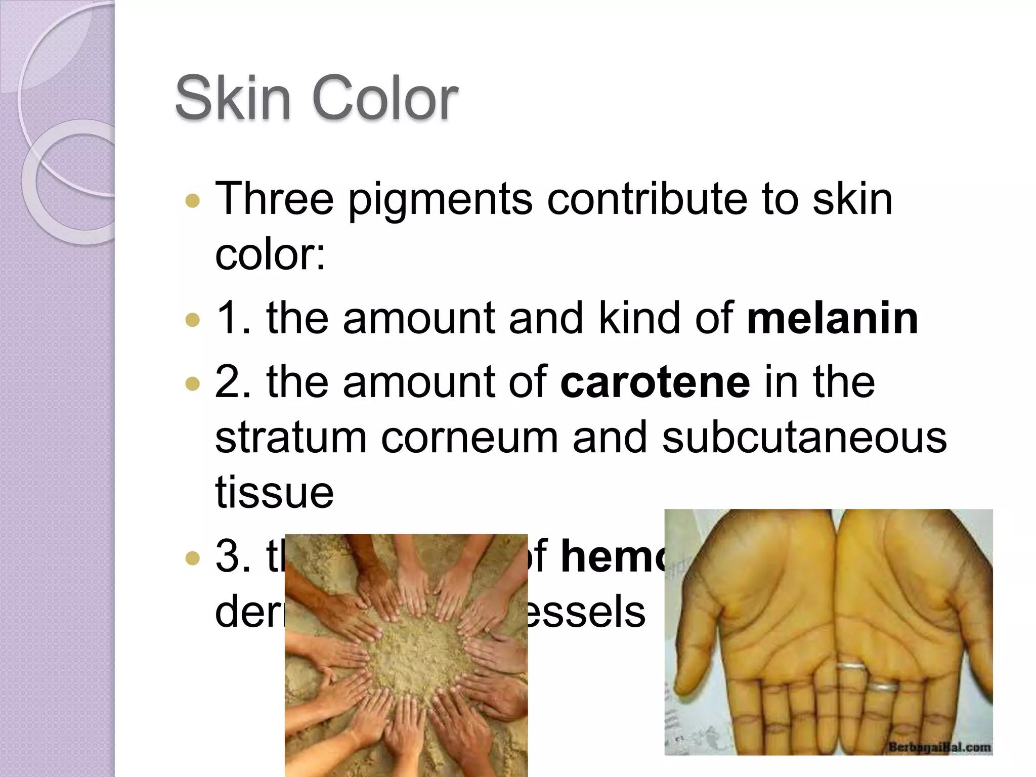
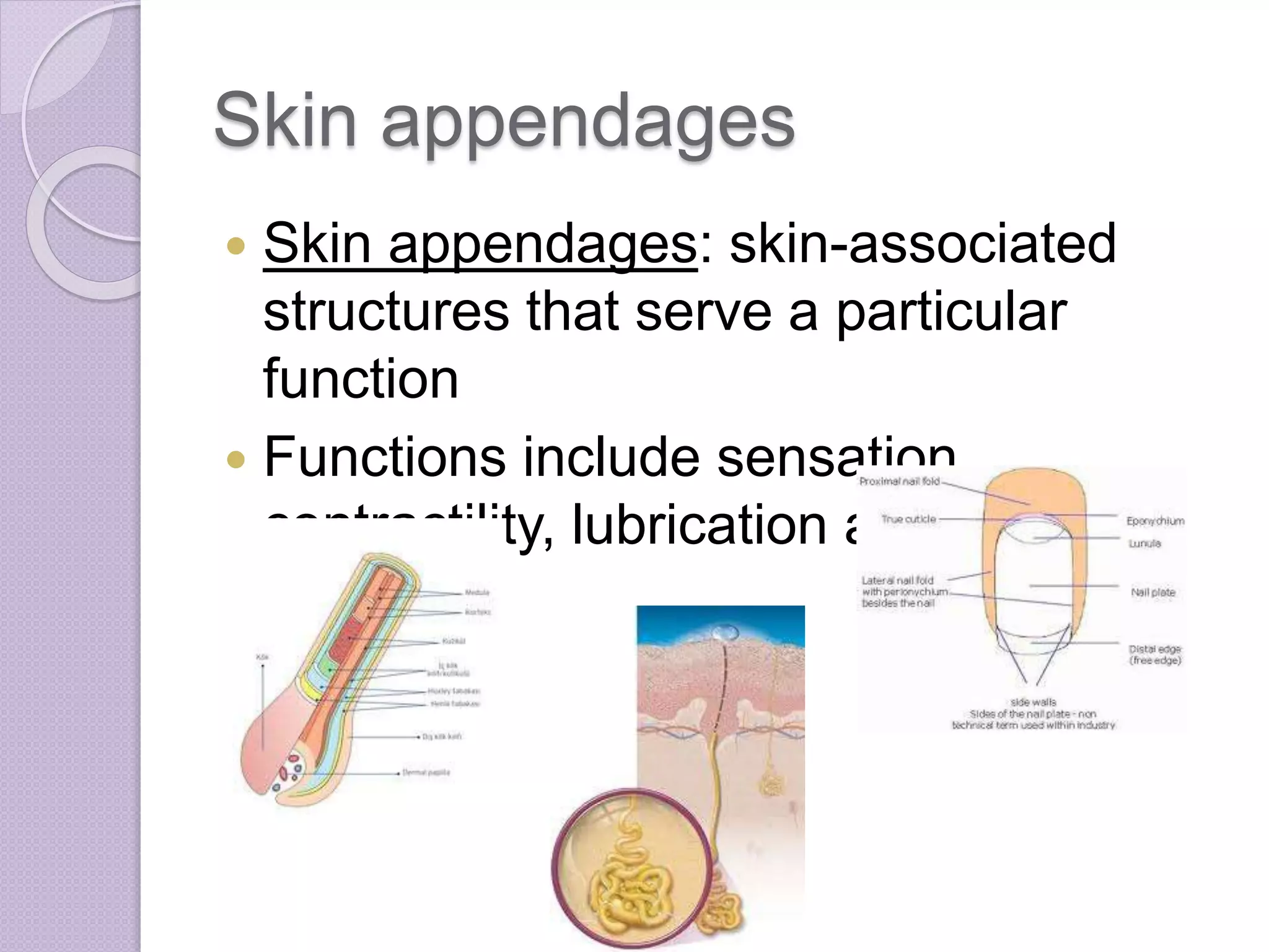
SlideShare a Scribd company logo
CHAPTER 4
SKIN AND BODY MEMBRANES
Body Membranes
 Body membranes cover surfaces, line
body cavities, and form protective
sheets around organs
 Fall into two major groups:
 1. epithelial membranes
include cutaneous, mucous, and serous
membranes
 2. connective tissue membranes
◦ Represented by synovial membranes
Body Membranes
 The functions of body membranes:
◦ 1. line or cover body surfaces
◦ 2. protect body surfaces
◦ 3. lubricate body surfaces
CLASSIFICATION OF
BODY MEMBRANES
Epithelial Membranes
 Epithelial
membranes
(covering and
lining
membranes)
include
cutaneous
membrane,
mucous
membranes, and
serous
Epithelial Membranes
 Calling these “epithelial” is inaccurate
 Do contain an epithelial sheet, but it is
always combined with an underlying
layer of connective tissue
 These are simple organs
Cutaneous Membranes
 Cutaneous Membrane: your skin
◦ Exposed to air
◦ Dry membrane
 The superficial epidermis
◦ keratinizing stratified squamous
epithelium
 The underlying dermis
◦ mostly dense connective tissue
Mucous Membranes
 Surface epithelium
◦ type depends on site
 Underlying loose
connective tissue
(lamina propia)
 Lines all body cavities
that open to the
exterior body surface
 Often adapted for
absorption and
secretion
Serous Membranes
 Surface simple squamous epithelium
 Underlying areolar connective tissue
 Lines open body cavities that are
closed to the exterior of the body
 Serous layers separated by serous
fluid
Serous Membranes
 Specific serous membranes
◦ Peritoneum
 Abdominal cavity
◦ Pleura
 Around the lungs
◦ Pericardium
 Around the heart
Connective Tissue
Membranes
 Synovial membrane
 Connective tissue only
 Lines fibrous capsules surrounding
joints
 Provide:
◦ Smooth surface
◦ Secrete lubricating fluid
Synovial Membrane
Connective Tissue
Membranes
 Synovial membrane
◦ Also line bursae and tendon sheaths
◦ Cushion organs moving against each
other during muscle activity
THE INTEGUMENTARY
SYSTEM (SKIN)
Integumentary System
 Skin (cutaneous membrane)
 Skin derivatives
◦ Sweat glands
◦ Oil glands
◦ Hairs
◦ Nails
Skin Functions
 Protects deeper tissues from:
◦ Mechanical damage
◦ Chemical damage
◦ Bacterial damage
◦ Thermal damage
◦ Ultraviolet radiation
◦ Desiccation
Skin Functions
 The uppermost layer of the skin is full
of keratin and is cornified, or
hardened, to help prevent water loss
from the body surface
Skin Functions
 Protects deeper tissues
 Aids in heat regulation
◦ Capillary network and sweat glands
 Aids in excretion of urea and uric acid
◦ Urea, salts, water
 Synthesizes Vitamin D
◦ Modified cholesterol molecules in the skin are
converted to Vitamin D by sunlight
 Cutaneous Sensory Receptors
◦ Tiny sensors detect touch, pressure,
temperature and pain
Skin Structure
 Epidermis – outer
layer
◦ Stratified
squamous
epithelium
◦ Often keratinized
 Dermis
◦ Dense connective
tissue
Skin Structure
Skin Structure
 Epidermis and dermis are firmly
connected
 A burn or friction may cause them to
separate
 Interstitial fluid accumulates, forms
blister
Skin Structure
 Deep to the dermis is the hypodermis
(subcutaneous tissue)
 Not part of the skin
 Anchors skin to underlying organs
 Composed mostly of adipose tissue
Skin Structure
EPIDERMIS
Epidermis
 Composed of up to five layers:
◦ Stratum basale
◦ Stratum spinosum
◦ Stratum granulosum
◦ Stratum lucidum
◦ Stratum corneum
Epidermis
 Most cells of the epidermis are
keratinocytes: keratin cells
Epidermis – Stratum basale
 Deepest cell layer in epidermis
 Lies closest to the dermis
 Receive the most nourishment,
through diffusion, from the dermis
 Cells are undergoing mitosis
 (also called the stratum germinativum)
 Pushed upward from this layer
Epidermis – Stratum basale
Epidermis – stratum spinsosum
and stratum granulosum
 Above the stratum
basale is the
stratum spinosum
and then the
stratum
granulosum
 Flatter, more
keratinized
Epidermis – Stratum lucidum
 Above the stratum granulosum is the
stratum lucidum
 Clear layer full of dead skin cells
 Not present in all skin regions
 Thick, hairless areas (palms, soles)
 High in keratin
 Far from blood supply
Epidermis – Stratum corneum
 Outermost layer
 Accounts for ¾ of epidermal thickness
 Shingle-like dead cells
 Completely filled with keratin
 Also called cornified or “horny cells”
Epidermis – stratum corneum
 The stratum corneum rubs and flakes
off slowly as dandruff
 The stratum corneum is replaced
quickly by rising cells from the stratum
basale
 We have an entirely new epidermis
every 25-45 days.
Epidermis - Melanin
 Melanin: pigment that ranges in color
from yellow to brown to black
 Melanin is produced by melanocytes
◦ Found mainly in the stratum basale
 Accumulates in membrane-bound
granules called melanosomes
Epidermis - Melanin
 The amount of
melanin in the
epidermis results
from genetics and
sunlight exposure
 Freckles and moles
are seen in areas
where melanin is
concentrated
DERMIS
Dermis
 Dermis – your hide
 Two layers:
 Papillary Layer (upper region)
 Reticular Layer (lower region)
Dermis – Papillary Layer
 Dermal Papillae
◦ Indent epidermis above
◦ Create fingerprints
 Capillary loops
◦ Nourish epidermis
 Pain and touch receptors
◦ Pain – free nerve endings
◦ Touch – Meissner’s corpuscles
Dermis – Reticular Layer
 Blood vessels
 Sweat and oil glands
 Deep pressure receptors – Pacinian
corpuscles
 Contain phagocytes that prevent
bacteria from reaching deeper tissues
Dermis
 Heavy in collagen and elastic fibers
 As age increases, these fibers
decrease as do fat cells and skin sags
 Abundantly supplied with blood
◦ Skin reds and warms with high body temp
 Rich nerve supply
SKIN COLOR
Skin Color
 Three pigments contribute to skin
color:
 1. the amount and kind of melanin
 2. the amount of carotene in the
stratum corneum and subcutaneous
tissue
 3. the amount of hemoglobin in the
dermal blood vessels
Skin Color
 People with a lot of melanin have
brown-toned skin
 People with less melanin have fair-
toned skin
Skin Color
 The hemoglobin in
the dermal blood
supply shows
through the
transparent cell
layers above
 This gives skin a
rosy glow
Skin Color
 Emotions also influence skin color,
and many alterations in skin color
signal disease:
 1. Redness (erythema) – reddened
skin
◦ Blushing, fever, hypertension,
inflammation, allergy
Skin Color
 2. Pallor (blanching)
◦ Emotional stress (fear, anger)
◦ Anemia, hypotension, impaired blood flow
 3. Jaundice (yellowing)
◦ Liver/Gallbladder disorders – excess of
bilirubin in the blood
Skin Color
 4. Bruises (black and blue coloring)
◦ Show where blood has escaped
circulation and has clotted in tissue
spaces
◦ Hematomas
◦ An unusual tendency to bruising may
signify a deficiency of vitamin C or
hemophilia
SKIN APPENDAGES
Skin appendages
 Skin appendages: skin-associated
structures that serve a particular
function
 Functions include sensation,
contractility, lubrication and heat loss
Skin Appendages
 Skin appendages
include cutaneous
glands, hair and hair
follicles, nails
 Arise from the
epidermis
 Play a role in
homeostasis
Cutaneous Glands
 Exocrine glands
◦ Release secretions to the skin surface via
ducts
 Two groups:
◦ Sebaceous glands
◦ Sweat glands
 Formed in the stratum basale and
push into the deeper layers of the
dermis
Sebaceous Glands
 Oil glands
 Found all over skin
except palms and soles
 Sebum: the product of
sebaceous glands
◦ Lubricate skin
◦ Kills bacteria
 Most with ducts empty
into hair follicles
 Increase production
during puberty
Sweat Glands
 Also called sudoriferous glands
 Widely distributed across the skin
 Two types:
 1. eccrine
◦ Open via duct to pore on skin
◦ Produce sweat
◦ Important in heat regulation
 2. apocrine
◦ Usually larger than eccrine glands
◦ Ducts empty into hair follicles
◦ Activated during pain and stress
Sweat Glands
Sweat and its Function
 Composition:
◦ Mostly water
◦ Some metabolic wastes (ammonia, urea,
etc.)
◦ Fatty acids and proteins (apocrine only)
 Function:
◦ Helps dissipate excess heat
◦ Excretes waste products
◦ Acidity decreases bacterial growth
 Odor is from associated bacteria
Hair and Hair Follicles
 Produced by hair
bulb
◦ Formed by well-
nourished stratum
basale cells in the
matrix (growth zone)
 Consists of hard,
keratinized
epithelial cells
 Melanocytes
provide pigment for
hair color
Hair and Hair Follicles
Hair Anatomy
 Central medulla
 Cortex surrounds
medulla
 Cuticle on outside
of cortex
◦ Most heavily
keratinized
Hair structures
 Hair follicle
◦ Dermal and epidermal sheath surround
hair root
 Arrector pili
◦ Smooth muscle
◦ Cause hair to stand up
◦ Goosebumps (piloerection)
 Sebaceous gland
◦ Lubricates hair
 Sweat gland
Hair function
 Warmth
◦ Limited in humans
 Protection
 Touch sense
 Non-verbal communication (eyebrows
and eyelashes)
Human Body Hair Types
 According to forensic scientists there
are six types of hair on the human
body:
 Head
 Eyebrow and eyelash
 Beard and moustache
 Body hair (Auxilairy)
 Pubic
 Armpit
Nails
 Scale-like modifications of the
epidermis
◦ Highly keratinized
 Stratum basale extends beneath the
nail bed
◦ Responsible for growth
 Lack of pigment makes them colorless
Nail Structures
 Free Edge
 Body
 Root of Nail
 Eponychium: proximal nail fold that
projects into the nail body
SKIN HOMEOSTATIC
IMBALANCES
Infections
 Athlete’s Foot
◦ Medically known as tinea pedis
◦ Caused by a fungal infection
◦ Thrives in warm, moist areas
◦ Contagious by contact
Infections
 Boils and Carbuncles
◦ Inflammation of hair follicles and
sebaceous glands
◦ Caused by bacterial infection
Infections
 Cold sores
◦ Caused by the Herpes Simplex Virus
◦ Can remain dormant for extended periods
of time
Infections
 Impetigo
◦ Caused by bacterial infection (staph)
◦ Highly contagious
◦ Common in elementary children
Allergies
 Contact dermatitis
◦ Exposure to substance causes an allergic
reaction
◦ Ex. poison ivy
Allergies
 Psoriasis
◦ Chronic, autoimmune condition
◦ Can be disfiguring
◦ Attacks are often triggered by trauma,
infection, stress or hormonal changes
BURNS
Burns
 Burn: tissue damage and cell death
caused by heat, electricity, UV
radiation (sunburns) or chemicals
Burns
 Immediate Associated Dangers:
 1. Dehydration
 2. Electrolyte Imbalance
 3. Circulatory Shock
Burns – Rule of Nines
 Way of determining the extent of
burns (how much of the body is
burned)
 Body is divided into 11 areas for quick
estimation
◦ Each area represents about 9% of the
body
◦ (last 1% is the area surrounding the
gentials)
Burns – Rule of Nines
Burns - Severity
 First-Degree
 Second – Degree
 Third-Degree
Burns - Severity
1st degree burns
 Only the epidermis is damaged
(superficial)
 Skin is red and swollen
 No blistering
 Minimal pain
 Usually heals in a week or less
 Cool, cover loosely, over the counter
pain medications
1st degree burns
2nd degree burns
 Epidermis and upper dermis are
damaged (partial thickness)
 Skin is red
 Blisters that sometimes break
 Severe pain
 Takes up to a month to heal
 Cool, cover loosely, over the counter
pain meds
 Be cautious of infections
2nd degree burns
3rd degree burns
 Destroy entire skin layer (full thickness)
 Skin is gray-white or black; may appear
waxy of charred
 Minimal pain at first due to nerve
damage
 Partial or complete skin grafts, biomask,
amnion
 Possible system shock and cardiac or
respiratory arrest
 Extended healing time
3rd degree burns
3rd degree burn treatment
Critical Burns
 Burns are considered critical if:
 Over 25% of the body has 2nd degree
burns
 Over 10% of the body has 3rd degree
burns
 There are third degree burns of the
face, hands or feet
Critical Burns
 Facial burns
◦ Particularly dangerous
◦ Possibility of burns in
respiratory passage
◦ Can cause swelling and
suffocation
 Joints
◦ Scar tissue can limit mobility
SKIN CANCER
Skin Cancer
 Cancer – abnormal cell mass
 Two types
 Benign
◦ Does not spread (encapsulated)
 Malignant
◦ Metastasized (moves) to other parts of the
body
 Most common type of cancer
Skin Cancer Types
 Basal Cell Carcinoma
◦ Least malignant
◦ Most common type
◦ Arises from stratum basale
Skin Cancer Types
 Squamous Cell Carcinoma
◦ Arises from stratum spinosum
◦ Metastasizes to lymph nodes
◦ Early removal allows for a good chance of
a cure
Skin Cancer Types
 Malignant Melanoma
◦ Most deadly type of skin cancer
◦ Cancer of melanocytes
◦ Metastasizes rapidly to lymph and blood
vessels
◦ Detection uses ABCD rule
ABCD Rules
ABCD Rules
 A = Asymmetry
◦ Two sides of pigmented mole do not
match
 B = Border Irregularity
◦ Borders of mole are not smooth
ABCD Rules
 C = Color
◦ Different colors in pigmented area
 D = Diameter
◦ Spot is larger than 6mm in diameter
ABCD Rules

More Related Content

What's hot

Chapter 7 - The Nervous System
Chapter 7 - The Nervous SystemChapter 7 - The Nervous System
Chapter 7 - The Nervous System
mpattani
 
Integumentary system by Dr. Sami
Integumentary system by Dr. SamiIntegumentary system by Dr. Sami
Integumentary system by Dr. Sami
Dr Adnan Sami
 
Epithelium, cells,tissues & histology
Epithelium, cells,tissues & histologyEpithelium, cells,tissues & histology
Epithelium, cells,tissues & histology
Dr. Waqas Nawaz
 
Temperature regulation by skin
Temperature regulation by skinTemperature regulation by skin
Temperature regulation by skin
Hashim Ali
 
The skin & integumentary system. ppt
The skin & integumentary system. pptThe skin & integumentary system. ppt
The skin & integumentary system. ppt
Bashant Kumar sah
 
Integumentary System
Integumentary SystemIntegumentary System
Integumentary System
clarot16
 
Anatomy and physiology Introduction Chapter 1 Notes
Anatomy and physiology Introduction Chapter 1 NotesAnatomy and physiology Introduction Chapter 1 Notes
Anatomy and physiology Introduction Chapter 1 Notes
mrhunterspage
 
Anatomy and Physiology Muscular System
Anatomy and Physiology Muscular SystemAnatomy and Physiology Muscular System
Anatomy and Physiology Muscular System
mrhunterspage
 
The integumentary system
The integumentary systemThe integumentary system
The integumentary system
Abhay Rajpoot
 
integumentary system
integumentary systemintegumentary system
integumentary system
Jay Patel
 
Integumentary System
Integumentary SystemIntegumentary System
Integumentary Systemwhitchur
 
Anatomy 1
 Anatomy 1 Anatomy 1
Tissues
TissuesTissues
SKIN - INTEGUMENTARY SYSTEM
SKIN - INTEGUMENTARY SYSTEMSKIN - INTEGUMENTARY SYSTEM
SKIN - INTEGUMENTARY SYSTEM
Dr Nilesh Kate
 
Anatomy and physiology Tissue Chapter
Anatomy and physiology Tissue ChapterAnatomy and physiology Tissue Chapter
Anatomy and physiology Tissue Chapter
mrhunterspage
 
Tissues
Tissues Tissues
Tissues
Sara Khan
 
Skeletal System Anatomy and Physiology
Skeletal System Anatomy and PhysiologySkeletal System Anatomy and Physiology
Skeletal System Anatomy and Physiology
Ronald Rubi
 
Endocrine System.ppt
Endocrine System.pptEndocrine System.ppt
Endocrine System.ppt
Vijay Salvekar
 
Chapter 1 Introduction to Anatomy and Physiology
 Chapter 1 Introduction to Anatomy and Physiology Chapter 1 Introduction to Anatomy and Physiology
Chapter 1 Introduction to Anatomy and Physiology
Yukti Sharma
 

What's hot (20)

Chapter 7 - The Nervous System
Chapter 7 - The Nervous SystemChapter 7 - The Nervous System
Chapter 7 - The Nervous System
 
Integumentary system by Dr. Sami
Integumentary system by Dr. SamiIntegumentary system by Dr. Sami
Integumentary system by Dr. Sami
 
Epithelium, cells,tissues & histology
Epithelium, cells,tissues & histologyEpithelium, cells,tissues & histology
Epithelium, cells,tissues & histology
 
Temperature regulation by skin
Temperature regulation by skinTemperature regulation by skin
Temperature regulation by skin
 
The skin & integumentary system. ppt
The skin & integumentary system. pptThe skin & integumentary system. ppt
The skin & integumentary system. ppt
 
Integumentary System
Integumentary SystemIntegumentary System
Integumentary System
 
Anatomy and physiology Introduction Chapter 1 Notes
Anatomy and physiology Introduction Chapter 1 NotesAnatomy and physiology Introduction Chapter 1 Notes
Anatomy and physiology Introduction Chapter 1 Notes
 
Anatomy and Physiology Muscular System
Anatomy and Physiology Muscular SystemAnatomy and Physiology Muscular System
Anatomy and Physiology Muscular System
 
The integumentary system
The integumentary systemThe integumentary system
The integumentary system
 
integumentary system
integumentary systemintegumentary system
integumentary system
 
Integumentary System
Integumentary SystemIntegumentary System
Integumentary System
 
Anatomy 1
 Anatomy 1 Anatomy 1
Anatomy 1
 
Tissues
TissuesTissues
Tissues
 
SKIN - INTEGUMENTARY SYSTEM
SKIN - INTEGUMENTARY SYSTEMSKIN - INTEGUMENTARY SYSTEM
SKIN - INTEGUMENTARY SYSTEM
 
Anatomy and physiology Tissue Chapter
Anatomy and physiology Tissue ChapterAnatomy and physiology Tissue Chapter
Anatomy and physiology Tissue Chapter
 
Tissues
Tissues Tissues
Tissues
 
Skin.ppt
Skin.pptSkin.ppt
Skin.ppt
 
Skeletal System Anatomy and Physiology
Skeletal System Anatomy and PhysiologySkeletal System Anatomy and Physiology
Skeletal System Anatomy and Physiology
 
Endocrine System.ppt
Endocrine System.pptEndocrine System.ppt
Endocrine System.ppt
 
Chapter 1 Introduction to Anatomy and Physiology
 Chapter 1 Introduction to Anatomy and Physiology Chapter 1 Introduction to Anatomy and Physiology
Chapter 1 Introduction to Anatomy and Physiology
 

Viewers also liked

Anatomy Of The Skin Lecture
Anatomy Of The Skin LectureAnatomy Of The Skin Lecture
Anatomy Of The Skin LectureLesley Castle
 
Ch. 4 Skin And Body Membranes
Ch. 4   Skin And Body MembranesCh. 4   Skin And Body Membranes
Ch. 4 Skin And Body MembranesWesley McCammon
 
The skeletal system (slide show)
The skeletal system (slide show)The skeletal system (slide show)
The skeletal system (slide show)
William Banaag
 
Skin Structure
Skin StructureSkin Structure
Skin Structure
saosmith
 
Chapter 3 cells and tissues
Chapter 3   cells and tissuesChapter 3   cells and tissues
Chapter 3 cells and tissues
mpattani
 
Body cavities
Body cavitiesBody cavities
Body cavities
Med Study
 
Skin Complications in Scleroderma
Skin Complications in SclerodermaSkin Complications in Scleroderma
Skin Complications in Scleroderma
Scleroderma Foundation of Greater Chicago
 
The Muscular System
The Muscular SystemThe Muscular System
The Muscular Systemitutor
 
Skeletal muscle mechanics
Skeletal muscle mechanicsSkeletal muscle mechanics
Skeletal muscle mechanics
Mubashir Iqbal
 
Muscle
MuscleMuscle
E integumentary system
E integumentary systemE integumentary system
E integumentary systemErica P
 
Muscles moving lower extremity.anat
Muscles moving lower extremity.anatMuscles moving lower extremity.anat
Muscles moving lower extremity.anat
guestc5fd8d
 
Lesson 1 skeletal system
Lesson 1   skeletal systemLesson 1   skeletal system
Lesson 1 skeletal systemDawn Chipchase
 
Seeley chapter 1
Seeley chapter 1Seeley chapter 1
Seeley chapter 1
tripp523
 
Dl141 chapt01 lecture
Dl141 chapt01 lectureDl141 chapt01 lecture
Dl141 chapt01 lecture
merrittl
 
Muscles study guide word doc
Muscles study guide word docMuscles study guide word doc
Muscles study guide word doc
Brittany Lynn
 
Chapter 6 - The Muscular System
Chapter 6 - The Muscular SystemChapter 6 - The Muscular System
Chapter 6 - The Muscular System
mpattani
 
Chicken wing dissection
Chicken wing dissectionChicken wing dissection
Chicken wing dissectionJenny Dixon
 

Viewers also liked (20)

Skin and body membranes
Skin and body membranesSkin and body membranes
Skin and body membranes
 
Anatomy Of The Skin Lecture
Anatomy Of The Skin LectureAnatomy Of The Skin Lecture
Anatomy Of The Skin Lecture
 
Ch. 4 Skin And Body Membranes
Ch. 4   Skin And Body MembranesCh. 4   Skin And Body Membranes
Ch. 4 Skin And Body Membranes
 
The skeletal system (slide show)
The skeletal system (slide show)The skeletal system (slide show)
The skeletal system (slide show)
 
Skin Structure
Skin StructureSkin Structure
Skin Structure
 
Chapter 3 cells and tissues
Chapter 3   cells and tissuesChapter 3   cells and tissues
Chapter 3 cells and tissues
 
Body cavities
Body cavitiesBody cavities
Body cavities
 
Skin Complications in Scleroderma
Skin Complications in SclerodermaSkin Complications in Scleroderma
Skin Complications in Scleroderma
 
The Muscular System
The Muscular SystemThe Muscular System
The Muscular System
 
Skeletal muscle mechanics
Skeletal muscle mechanicsSkeletal muscle mechanics
Skeletal muscle mechanics
 
Muscle
MuscleMuscle
Muscle
 
E integumentary system
E integumentary systemE integumentary system
E integumentary system
 
Muscles moving lower extremity.anat
Muscles moving lower extremity.anatMuscles moving lower extremity.anat
Muscles moving lower extremity.anat
 
Lesson 1 skeletal system
Lesson 1   skeletal systemLesson 1   skeletal system
Lesson 1 skeletal system
 
Body cavities
Body cavitiesBody cavities
Body cavities
 
Seeley chapter 1
Seeley chapter 1Seeley chapter 1
Seeley chapter 1
 
Dl141 chapt01 lecture
Dl141 chapt01 lectureDl141 chapt01 lecture
Dl141 chapt01 lecture
 
Muscles study guide word doc
Muscles study guide word docMuscles study guide word doc
Muscles study guide word doc
 
Chapter 6 - The Muscular System
Chapter 6 - The Muscular SystemChapter 6 - The Muscular System
Chapter 6 - The Muscular System
 
Chicken wing dissection
Chicken wing dissectionChicken wing dissection
Chicken wing dissection
 

Similar to Chapter 4 - Skin and Body Membranes

Skin An Essential Organ
Skin An Essential OrganSkin An Essential Organ
Skin An Essential Organ
Muhammadasif909
 
Lect 5 intergumentary
Lect 5   intergumentaryLect 5   intergumentary
Lect 5 intergumentarymissazyaziz
 
Skin anatomy & physiology for ayush students
Skin anatomy & physiology for ayush studentsSkin anatomy & physiology for ayush students
Skin anatomy & physiology for ayush students
Dr.arun Naragund
 
Integumentary system prep.pptx
Integumentary system prep.pptxIntegumentary system prep.pptx
Integumentary system prep.pptx
SamanthaEllaine
 
Janiola,structure-skincolor.pptx
Janiola,structure-skincolor.pptxJaniola,structure-skincolor.pptx
Janiola,structure-skincolor.pptx
NelJaniola
 
Skin
SkinSkin
Integumentary System Notes
Integumentary System NotesIntegumentary System Notes
Integumentary System Notesrachelkucera
 
5. The Integument and Related Structures
5. The Integument and Related Structures5. The Integument and Related Structures
5. The Integument and Related StructuresSUNY Ulster
 
Part 1 the integumentary system(kirin)
Part 1 the integumentary system(kirin)Part 1 the integumentary system(kirin)
Part 1 the integumentary system(kirin)
aiannanggansalim
 
Part 1 the integumentary system(ai-ann)
Part 1 the integumentary system(ai-ann)Part 1 the integumentary system(ai-ann)
Part 1 the integumentary system(ai-ann)aiannanggansalim
 
Integumentary system
Integumentary systemIntegumentary system
Integumentary system
Soneeshah
 
Pdf |Histology| - The skin -
Pdf |Histology| - The skin -Pdf |Histology| - The skin -
Pdf |Histology| - The skin -
Ameen ALzaidy
 
Anatomy of skin
Anatomy of skinAnatomy of skin
Anatomy of skin
A Y
 
The sense of touch, pain & temperature
The sense of touch, pain & temperatureThe sense of touch, pain & temperature
The sense of touch, pain & temperature
Hashim Ali
 
2. Integumentary system .ppt
2. Integumentary system .ppt2. Integumentary system .ppt
2. Integumentary system .ppt
TigabuAgmas1
 

Similar to Chapter 4 - Skin and Body Membranes (20)

Integument
IntegumentIntegument
Integument
 
Skin An Essential Organ
Skin An Essential OrganSkin An Essential Organ
Skin An Essential Organ
 
Lect 5 intergumentary
Lect 5   intergumentaryLect 5   intergumentary
Lect 5 intergumentary
 
Skin and body membranes
Skin and body membranesSkin and body membranes
Skin and body membranes
 
Skin anatomy & physiology for ayush students
Skin anatomy & physiology for ayush studentsSkin anatomy & physiology for ayush students
Skin anatomy & physiology for ayush students
 
Biosci
BiosciBiosci
Biosci
 
Integumentary system prep.pptx
Integumentary system prep.pptxIntegumentary system prep.pptx
Integumentary system prep.pptx
 
Janiola,structure-skincolor.pptx
Janiola,structure-skincolor.pptxJaniola,structure-skincolor.pptx
Janiola,structure-skincolor.pptx
 
Skin
SkinSkin
Skin
 
Integumentary System Notes
Integumentary System NotesIntegumentary System Notes
Integumentary System Notes
 
5. The Integument and Related Structures
5. The Integument and Related Structures5. The Integument and Related Structures
5. The Integument and Related Structures
 
Part 1 the integumentary system(kirin)
Part 1 the integumentary system(kirin)Part 1 the integumentary system(kirin)
Part 1 the integumentary system(kirin)
 
Part 1 the integumentary system(ai-ann)
Part 1 the integumentary system(ai-ann)Part 1 the integumentary system(ai-ann)
Part 1 the integumentary system(ai-ann)
 
Integumentary system
Integumentary systemIntegumentary system
Integumentary system
 
Pdf |Histology| - The skin -
Pdf |Histology| - The skin -Pdf |Histology| - The skin -
Pdf |Histology| - The skin -
 
Anatomy of skin
Anatomy of skinAnatomy of skin
Anatomy of skin
 
The sense of touch, pain & temperature
The sense of touch, pain & temperatureThe sense of touch, pain & temperature
The sense of touch, pain & temperature
 
2. Integumentary system .ppt
2. Integumentary system .ppt2. Integumentary system .ppt
2. Integumentary system .ppt
 
Integumentary System (unit VII).pptx
Integumentary System (unit VII).pptxIntegumentary System (unit VII).pptx
Integumentary System (unit VII).pptx
 
The Integumentary System
The Integumentary SystemThe Integumentary System
The Integumentary System
 

More from mpattani

Fingerprints
FingerprintsFingerprints
Fingerprints
mpattani
 
CSI and Evidence Collection
CSI and Evidence CollectionCSI and Evidence Collection
CSI and Evidence Collection
mpattani
 
JonBenet Ramsey
JonBenet RamseyJonBenet Ramsey
JonBenet Ramsey
mpattani
 
Forensic Science Chapter 1 Vocabulary
Forensic Science Chapter 1 VocabularyForensic Science Chapter 1 Vocabulary
Forensic Science Chapter 1 Vocabulary
mpattani
 
Perch dissection
Perch dissectionPerch dissection
Perch dissection
mpattani
 
Shark basics
Shark basicsShark basics
Shark basics
mpattani
 
Sea star dissection
Sea star dissectionSea star dissection
Sea star dissection
mpattani
 
Squid Dissection Prelab
Squid Dissection PrelabSquid Dissection Prelab
Squid Dissection Prelab
mpattani
 
Biology in Focus - Chapter 39
Biology in Focus - Chapter 39Biology in Focus - Chapter 39
Biology in Focus - Chapter 39
mpattani
 
Biology in Focus - Chapter 38
Biology in Focus - Chapter 38Biology in Focus - Chapter 38
Biology in Focus - Chapter 38
mpattani
 
Biology in Focus - Chapter 37
Biology in Focus - Chapter 37 Biology in Focus - Chapter 37
Biology in Focus - Chapter 37
mpattani
 
Biology in Focus - Chapter 36
Biology in Focus - Chapter 36Biology in Focus - Chapter 36
Biology in Focus - Chapter 36
mpattani
 
Reproductive System
Reproductive SystemReproductive System
Reproductive System
mpattani
 
Biology in Focus - Chapter 35
Biology in Focus - Chapter 35Biology in Focus - Chapter 35
Biology in Focus - Chapter 35
mpattani
 
Biology in Focus - Chapter 34
Biology in Focus - Chapter 34Biology in Focus - Chapter 34
Biology in Focus - Chapter 34
mpattani
 
Biology in Focus - Chapter 33
Biology in Focus - Chapter 33Biology in Focus - Chapter 33
Biology in Focus - Chapter 33
mpattani
 
Biology in Focus - Chapter 31
Biology in Focus - Chapter 31Biology in Focus - Chapter 31
Biology in Focus - Chapter 31
mpattani
 
Biology in Focus - Chapter 31
Biology in Focus - Chapter 31Biology in Focus - Chapter 31
Biology in Focus - Chapter 31
mpattani
 
Biology in Focus - Chapter 30
Biology in Focus - Chapter 30Biology in Focus - Chapter 30
Biology in Focus - Chapter 30
mpattani
 
Biology in Focus - Chapter 29
Biology in Focus - Chapter 29Biology in Focus - Chapter 29
Biology in Focus - Chapter 29
mpattani
 

More from mpattani (20)

Fingerprints
FingerprintsFingerprints
Fingerprints
 
CSI and Evidence Collection
CSI and Evidence CollectionCSI and Evidence Collection
CSI and Evidence Collection
 
JonBenet Ramsey
JonBenet RamseyJonBenet Ramsey
JonBenet Ramsey
 
Forensic Science Chapter 1 Vocabulary
Forensic Science Chapter 1 VocabularyForensic Science Chapter 1 Vocabulary
Forensic Science Chapter 1 Vocabulary
 
Perch dissection
Perch dissectionPerch dissection
Perch dissection
 
Shark basics
Shark basicsShark basics
Shark basics
 
Sea star dissection
Sea star dissectionSea star dissection
Sea star dissection
 
Squid Dissection Prelab
Squid Dissection PrelabSquid Dissection Prelab
Squid Dissection Prelab
 
Biology in Focus - Chapter 39
Biology in Focus - Chapter 39Biology in Focus - Chapter 39
Biology in Focus - Chapter 39
 
Biology in Focus - Chapter 38
Biology in Focus - Chapter 38Biology in Focus - Chapter 38
Biology in Focus - Chapter 38
 
Biology in Focus - Chapter 37
Biology in Focus - Chapter 37 Biology in Focus - Chapter 37
Biology in Focus - Chapter 37
 
Biology in Focus - Chapter 36
Biology in Focus - Chapter 36Biology in Focus - Chapter 36
Biology in Focus - Chapter 36
 
Reproductive System
Reproductive SystemReproductive System
Reproductive System
 
Biology in Focus - Chapter 35
Biology in Focus - Chapter 35Biology in Focus - Chapter 35
Biology in Focus - Chapter 35
 
Biology in Focus - Chapter 34
Biology in Focus - Chapter 34Biology in Focus - Chapter 34
Biology in Focus - Chapter 34
 
Biology in Focus - Chapter 33
Biology in Focus - Chapter 33Biology in Focus - Chapter 33
Biology in Focus - Chapter 33
 
Biology in Focus - Chapter 31
Biology in Focus - Chapter 31Biology in Focus - Chapter 31
Biology in Focus - Chapter 31
 
Biology in Focus - Chapter 31
Biology in Focus - Chapter 31Biology in Focus - Chapter 31
Biology in Focus - Chapter 31
 
Biology in Focus - Chapter 30
Biology in Focus - Chapter 30Biology in Focus - Chapter 30
Biology in Focus - Chapter 30
 
Biology in Focus - Chapter 29
Biology in Focus - Chapter 29Biology in Focus - Chapter 29
Biology in Focus - Chapter 29
 

Recently uploaded

plant biotechnology Lecture note ppt.pptx
plant biotechnology Lecture note ppt.pptxplant biotechnology Lecture note ppt.pptx
plant biotechnology Lecture note ppt.pptx
yusufzako14
 
platelets_clotting_biogenesis.clot retractionpptx
platelets_clotting_biogenesis.clot retractionpptxplatelets_clotting_biogenesis.clot retractionpptx
platelets_clotting_biogenesis.clot retractionpptx
muralinath2
 
insect taxonomy importance systematics and classification
insect taxonomy importance systematics and classificationinsect taxonomy importance systematics and classification
insect taxonomy importance systematics and classification
anitaento25
 
GBSN- Microbiology (Lab 3) Gram Staining
GBSN- Microbiology (Lab 3) Gram StainingGBSN- Microbiology (Lab 3) Gram Staining
GBSN- Microbiology (Lab 3) Gram Staining
Areesha Ahmad
 
filosofia boliviana introducción jsjdjd.pptx
filosofia boliviana introducción jsjdjd.pptxfilosofia boliviana introducción jsjdjd.pptx
filosofia boliviana introducción jsjdjd.pptx
IvanMallco1
 
In silico drugs analogue design: novobiocin analogues.pptx
In silico drugs analogue design: novobiocin analogues.pptxIn silico drugs analogue design: novobiocin analogues.pptx
In silico drugs analogue design: novobiocin analogues.pptx
AlaminAfendy1
 
Orion Air Quality Monitoring Systems - CWS
Orion Air Quality Monitoring Systems - CWSOrion Air Quality Monitoring Systems - CWS
Orion Air Quality Monitoring Systems - CWS
Columbia Weather Systems
 
Hemostasis_importance& clinical significance.pptx
Hemostasis_importance& clinical significance.pptxHemostasis_importance& clinical significance.pptx
Hemostasis_importance& clinical significance.pptx
muralinath2
 
Large scale production of streptomycin.pptx
Large scale production of streptomycin.pptxLarge scale production of streptomycin.pptx
Large scale production of streptomycin.pptx
Cherry
 
Body fluids_tonicity_dehydration_hypovolemia_hypervolemia.pptx
Body fluids_tonicity_dehydration_hypovolemia_hypervolemia.pptxBody fluids_tonicity_dehydration_hypovolemia_hypervolemia.pptx
Body fluids_tonicity_dehydration_hypovolemia_hypervolemia.pptx
muralinath2
 
Multi-source connectivity as the driver of solar wind variability in the heli...
Multi-source connectivity as the driver of solar wind variability in the heli...Multi-source connectivity as the driver of solar wind variability in the heli...
Multi-source connectivity as the driver of solar wind variability in the heli...
Sérgio Sacani
 
Citrus Greening Disease and its Management
Citrus Greening Disease and its ManagementCitrus Greening Disease and its Management
Citrus Greening Disease and its Management
subedisuryaofficial
 
Unveiling the Energy Potential of Marshmallow Deposits.pdf
Unveiling the Energy Potential of Marshmallow Deposits.pdfUnveiling the Energy Potential of Marshmallow Deposits.pdf
Unveiling the Energy Potential of Marshmallow Deposits.pdf
Erdal Coalmaker
 
general properties of oerganologametal.ppt
general properties of oerganologametal.pptgeneral properties of oerganologametal.ppt
general properties of oerganologametal.ppt
IqrimaNabilatulhusni
 
The ASGCT Annual Meeting was packed with exciting progress in the field advan...
The ASGCT Annual Meeting was packed with exciting progress in the field advan...The ASGCT Annual Meeting was packed with exciting progress in the field advan...
The ASGCT Annual Meeting was packed with exciting progress in the field advan...
Health Advances
 
Hemoglobin metabolism_pathophysiology.pptx
Hemoglobin metabolism_pathophysiology.pptxHemoglobin metabolism_pathophysiology.pptx
Hemoglobin metabolism_pathophysiology.pptx
muralinath2
 
Cancer cell metabolism: special Reference to Lactate Pathway
Cancer cell metabolism: special Reference to Lactate PathwayCancer cell metabolism: special Reference to Lactate Pathway
Cancer cell metabolism: special Reference to Lactate Pathway
AADYARAJPANDEY1
 
extra-chromosomal-inheritance[1].pptx.pdfpdf
extra-chromosomal-inheritance[1].pptx.pdfpdfextra-chromosomal-inheritance[1].pptx.pdfpdf
extra-chromosomal-inheritance[1].pptx.pdfpdf
DiyaBiswas10
 
Comparative structure of adrenal gland in vertebrates
Comparative structure of adrenal gland in vertebratesComparative structure of adrenal gland in vertebrates
Comparative structure of adrenal gland in vertebrates
sachin783648
 
NuGOweek 2024 Ghent - programme - final version
NuGOweek 2024 Ghent - programme - final versionNuGOweek 2024 Ghent - programme - final version
NuGOweek 2024 Ghent - programme - final version
pablovgd
 

Recently uploaded (20)

plant biotechnology Lecture note ppt.pptx
plant biotechnology Lecture note ppt.pptxplant biotechnology Lecture note ppt.pptx
plant biotechnology Lecture note ppt.pptx
 
platelets_clotting_biogenesis.clot retractionpptx
platelets_clotting_biogenesis.clot retractionpptxplatelets_clotting_biogenesis.clot retractionpptx
platelets_clotting_biogenesis.clot retractionpptx
 
insect taxonomy importance systematics and classification
insect taxonomy importance systematics and classificationinsect taxonomy importance systematics and classification
insect taxonomy importance systematics and classification
 
GBSN- Microbiology (Lab 3) Gram Staining
GBSN- Microbiology (Lab 3) Gram StainingGBSN- Microbiology (Lab 3) Gram Staining
GBSN- Microbiology (Lab 3) Gram Staining
 
filosofia boliviana introducción jsjdjd.pptx
filosofia boliviana introducción jsjdjd.pptxfilosofia boliviana introducción jsjdjd.pptx
filosofia boliviana introducción jsjdjd.pptx
 
In silico drugs analogue design: novobiocin analogues.pptx
In silico drugs analogue design: novobiocin analogues.pptxIn silico drugs analogue design: novobiocin analogues.pptx
In silico drugs analogue design: novobiocin analogues.pptx
 
Orion Air Quality Monitoring Systems - CWS
Orion Air Quality Monitoring Systems - CWSOrion Air Quality Monitoring Systems - CWS
Orion Air Quality Monitoring Systems - CWS
 
Hemostasis_importance& clinical significance.pptx
Hemostasis_importance& clinical significance.pptxHemostasis_importance& clinical significance.pptx
Hemostasis_importance& clinical significance.pptx
 
Large scale production of streptomycin.pptx
Large scale production of streptomycin.pptxLarge scale production of streptomycin.pptx
Large scale production of streptomycin.pptx
 
Body fluids_tonicity_dehydration_hypovolemia_hypervolemia.pptx
Body fluids_tonicity_dehydration_hypovolemia_hypervolemia.pptxBody fluids_tonicity_dehydration_hypovolemia_hypervolemia.pptx
Body fluids_tonicity_dehydration_hypovolemia_hypervolemia.pptx
 
Multi-source connectivity as the driver of solar wind variability in the heli...
Multi-source connectivity as the driver of solar wind variability in the heli...Multi-source connectivity as the driver of solar wind variability in the heli...
Multi-source connectivity as the driver of solar wind variability in the heli...
 
Citrus Greening Disease and its Management
Citrus Greening Disease and its ManagementCitrus Greening Disease and its Management
Citrus Greening Disease and its Management
 
Unveiling the Energy Potential of Marshmallow Deposits.pdf
Unveiling the Energy Potential of Marshmallow Deposits.pdfUnveiling the Energy Potential of Marshmallow Deposits.pdf
Unveiling the Energy Potential of Marshmallow Deposits.pdf
 
general properties of oerganologametal.ppt
general properties of oerganologametal.pptgeneral properties of oerganologametal.ppt
general properties of oerganologametal.ppt
 
The ASGCT Annual Meeting was packed with exciting progress in the field advan...
The ASGCT Annual Meeting was packed with exciting progress in the field advan...The ASGCT Annual Meeting was packed with exciting progress in the field advan...
The ASGCT Annual Meeting was packed with exciting progress in the field advan...
 
Hemoglobin metabolism_pathophysiology.pptx
Hemoglobin metabolism_pathophysiology.pptxHemoglobin metabolism_pathophysiology.pptx
Hemoglobin metabolism_pathophysiology.pptx
 
Cancer cell metabolism: special Reference to Lactate Pathway
Cancer cell metabolism: special Reference to Lactate PathwayCancer cell metabolism: special Reference to Lactate Pathway
Cancer cell metabolism: special Reference to Lactate Pathway
 
extra-chromosomal-inheritance[1].pptx.pdfpdf
extra-chromosomal-inheritance[1].pptx.pdfpdfextra-chromosomal-inheritance[1].pptx.pdfpdf
extra-chromosomal-inheritance[1].pptx.pdfpdf
 
Comparative structure of adrenal gland in vertebrates
Comparative structure of adrenal gland in vertebratesComparative structure of adrenal gland in vertebrates
Comparative structure of adrenal gland in vertebrates
 
NuGOweek 2024 Ghent - programme - final version
NuGOweek 2024 Ghent - programme - final versionNuGOweek 2024 Ghent - programme - final version
NuGOweek 2024 Ghent - programme - final version
 

Chapter 4 - Skin and Body Membranes

  • 1. CHAPTER 4 SKIN AND BODY MEMBRANES
  • 2. Body Membranes  Body membranes cover surfaces, line body cavities, and form protective sheets around organs  Fall into two major groups:  1. epithelial membranes include cutaneous, mucous, and serous membranes  2. connective tissue membranes ◦ Represented by synovial membranes
  • 3. Body Membranes  The functions of body membranes: ◦ 1. line or cover body surfaces ◦ 2. protect body surfaces ◦ 3. lubricate body surfaces
  • 5. Epithelial Membranes  Epithelial membranes (covering and lining membranes) include cutaneous membrane, mucous membranes, and serous
  • 6. Epithelial Membranes  Calling these “epithelial” is inaccurate  Do contain an epithelial sheet, but it is always combined with an underlying layer of connective tissue  These are simple organs
  • 7. Cutaneous Membranes  Cutaneous Membrane: your skin ◦ Exposed to air ◦ Dry membrane  The superficial epidermis ◦ keratinizing stratified squamous epithelium  The underlying dermis ◦ mostly dense connective tissue
  • 8. Mucous Membranes  Surface epithelium ◦ type depends on site  Underlying loose connective tissue (lamina propia)  Lines all body cavities that open to the exterior body surface  Often adapted for absorption and secretion
  • 9. Serous Membranes  Surface simple squamous epithelium  Underlying areolar connective tissue  Lines open body cavities that are closed to the exterior of the body  Serous layers separated by serous fluid
  • 10. Serous Membranes  Specific serous membranes ◦ Peritoneum  Abdominal cavity ◦ Pleura  Around the lungs ◦ Pericardium  Around the heart
  • 11. Connective Tissue Membranes  Synovial membrane  Connective tissue only  Lines fibrous capsules surrounding joints  Provide: ◦ Smooth surface ◦ Secrete lubricating fluid
  • 13. Connective Tissue Membranes  Synovial membrane ◦ Also line bursae and tendon sheaths ◦ Cushion organs moving against each other during muscle activity
  • 15. Integumentary System  Skin (cutaneous membrane)  Skin derivatives ◦ Sweat glands ◦ Oil glands ◦ Hairs ◦ Nails
  • 16. Skin Functions  Protects deeper tissues from: ◦ Mechanical damage ◦ Chemical damage ◦ Bacterial damage ◦ Thermal damage ◦ Ultraviolet radiation ◦ Desiccation
  • 17. Skin Functions  The uppermost layer of the skin is full of keratin and is cornified, or hardened, to help prevent water loss from the body surface
  • 18. Skin Functions  Protects deeper tissues  Aids in heat regulation ◦ Capillary network and sweat glands  Aids in excretion of urea and uric acid ◦ Urea, salts, water  Synthesizes Vitamin D ◦ Modified cholesterol molecules in the skin are converted to Vitamin D by sunlight  Cutaneous Sensory Receptors ◦ Tiny sensors detect touch, pressure, temperature and pain
  • 19. Skin Structure  Epidermis – outer layer ◦ Stratified squamous epithelium ◦ Often keratinized  Dermis ◦ Dense connective tissue
  • 21. Skin Structure  Epidermis and dermis are firmly connected  A burn or friction may cause them to separate  Interstitial fluid accumulates, forms blister
  • 22. Skin Structure  Deep to the dermis is the hypodermis (subcutaneous tissue)  Not part of the skin  Anchors skin to underlying organs  Composed mostly of adipose tissue
  • 25. Epidermis  Composed of up to five layers: ◦ Stratum basale ◦ Stratum spinosum ◦ Stratum granulosum ◦ Stratum lucidum ◦ Stratum corneum
  • 26. Epidermis  Most cells of the epidermis are keratinocytes: keratin cells
  • 27. Epidermis – Stratum basale  Deepest cell layer in epidermis  Lies closest to the dermis  Receive the most nourishment, through diffusion, from the dermis  Cells are undergoing mitosis  (also called the stratum germinativum)  Pushed upward from this layer
  • 29. Epidermis – stratum spinsosum and stratum granulosum  Above the stratum basale is the stratum spinosum and then the stratum granulosum  Flatter, more keratinized
  • 30. Epidermis – Stratum lucidum  Above the stratum granulosum is the stratum lucidum  Clear layer full of dead skin cells  Not present in all skin regions  Thick, hairless areas (palms, soles)  High in keratin  Far from blood supply
  • 31. Epidermis – Stratum corneum  Outermost layer  Accounts for ¾ of epidermal thickness  Shingle-like dead cells  Completely filled with keratin  Also called cornified or “horny cells”
  • 32. Epidermis – stratum corneum  The stratum corneum rubs and flakes off slowly as dandruff  The stratum corneum is replaced quickly by rising cells from the stratum basale  We have an entirely new epidermis every 25-45 days.
  • 33. Epidermis - Melanin  Melanin: pigment that ranges in color from yellow to brown to black  Melanin is produced by melanocytes ◦ Found mainly in the stratum basale  Accumulates in membrane-bound granules called melanosomes
  • 34. Epidermis - Melanin  The amount of melanin in the epidermis results from genetics and sunlight exposure  Freckles and moles are seen in areas where melanin is concentrated
  • 36. Dermis  Dermis – your hide  Two layers:  Papillary Layer (upper region)  Reticular Layer (lower region)
  • 37. Dermis – Papillary Layer  Dermal Papillae ◦ Indent epidermis above ◦ Create fingerprints  Capillary loops ◦ Nourish epidermis  Pain and touch receptors ◦ Pain – free nerve endings ◦ Touch – Meissner’s corpuscles
  • 38. Dermis – Reticular Layer  Blood vessels  Sweat and oil glands  Deep pressure receptors – Pacinian corpuscles  Contain phagocytes that prevent bacteria from reaching deeper tissues
  • 39. Dermis  Heavy in collagen and elastic fibers  As age increases, these fibers decrease as do fat cells and skin sags  Abundantly supplied with blood ◦ Skin reds and warms with high body temp  Rich nerve supply
  • 41. Skin Color  Three pigments contribute to skin color:  1. the amount and kind of melanin  2. the amount of carotene in the stratum corneum and subcutaneous tissue  3. the amount of hemoglobin in the dermal blood vessels
  • 42. Skin Color  People with a lot of melanin have brown-toned skin  People with less melanin have fair- toned skin
  • 43. Skin Color  The hemoglobin in the dermal blood supply shows through the transparent cell layers above  This gives skin a rosy glow
  • 44. Skin Color  Emotions also influence skin color, and many alterations in skin color signal disease:  1. Redness (erythema) – reddened skin ◦ Blushing, fever, hypertension, inflammation, allergy
  • 45. Skin Color  2. Pallor (blanching) ◦ Emotional stress (fear, anger) ◦ Anemia, hypotension, impaired blood flow  3. Jaundice (yellowing) ◦ Liver/Gallbladder disorders – excess of bilirubin in the blood
  • 46. Skin Color  4. Bruises (black and blue coloring) ◦ Show where blood has escaped circulation and has clotted in tissue spaces ◦ Hematomas ◦ An unusual tendency to bruising may signify a deficiency of vitamin C or hemophilia
  • 48. Skin appendages  Skin appendages: skin-associated structures that serve a particular function  Functions include sensation, contractility, lubrication and heat loss
  • 49. Skin Appendages  Skin appendages include cutaneous glands, hair and hair follicles, nails  Arise from the epidermis  Play a role in homeostasis
  • 50. Cutaneous Glands  Exocrine glands ◦ Release secretions to the skin surface via ducts  Two groups: ◦ Sebaceous glands ◦ Sweat glands  Formed in the stratum basale and push into the deeper layers of the dermis
  • 51. Sebaceous Glands  Oil glands  Found all over skin except palms and soles  Sebum: the product of sebaceous glands ◦ Lubricate skin ◦ Kills bacteria  Most with ducts empty into hair follicles  Increase production during puberty
  • 52. Sweat Glands  Also called sudoriferous glands  Widely distributed across the skin  Two types:  1. eccrine ◦ Open via duct to pore on skin ◦ Produce sweat ◦ Important in heat regulation  2. apocrine ◦ Usually larger than eccrine glands ◦ Ducts empty into hair follicles ◦ Activated during pain and stress
  • 54. Sweat and its Function  Composition: ◦ Mostly water ◦ Some metabolic wastes (ammonia, urea, etc.) ◦ Fatty acids and proteins (apocrine only)  Function: ◦ Helps dissipate excess heat ◦ Excretes waste products ◦ Acidity decreases bacterial growth  Odor is from associated bacteria
  • 55. Hair and Hair Follicles  Produced by hair bulb ◦ Formed by well- nourished stratum basale cells in the matrix (growth zone)  Consists of hard, keratinized epithelial cells  Melanocytes provide pigment for hair color
  • 56. Hair and Hair Follicles
  • 57. Hair Anatomy  Central medulla  Cortex surrounds medulla  Cuticle on outside of cortex ◦ Most heavily keratinized
  • 58. Hair structures  Hair follicle ◦ Dermal and epidermal sheath surround hair root  Arrector pili ◦ Smooth muscle ◦ Cause hair to stand up ◦ Goosebumps (piloerection)  Sebaceous gland ◦ Lubricates hair  Sweat gland
  • 59. Hair function  Warmth ◦ Limited in humans  Protection  Touch sense  Non-verbal communication (eyebrows and eyelashes)
  • 60. Human Body Hair Types  According to forensic scientists there are six types of hair on the human body:  Head  Eyebrow and eyelash  Beard and moustache  Body hair (Auxilairy)  Pubic  Armpit
  • 61. Nails  Scale-like modifications of the epidermis ◦ Highly keratinized  Stratum basale extends beneath the nail bed ◦ Responsible for growth  Lack of pigment makes them colorless
  • 62. Nail Structures  Free Edge  Body  Root of Nail  Eponychium: proximal nail fold that projects into the nail body
  • 64. Infections  Athlete’s Foot ◦ Medically known as tinea pedis ◦ Caused by a fungal infection ◦ Thrives in warm, moist areas ◦ Contagious by contact
  • 65. Infections  Boils and Carbuncles ◦ Inflammation of hair follicles and sebaceous glands ◦ Caused by bacterial infection
  • 66. Infections  Cold sores ◦ Caused by the Herpes Simplex Virus ◦ Can remain dormant for extended periods of time
  • 67. Infections  Impetigo ◦ Caused by bacterial infection (staph) ◦ Highly contagious ◦ Common in elementary children
  • 68. Allergies  Contact dermatitis ◦ Exposure to substance causes an allergic reaction ◦ Ex. poison ivy
  • 69. Allergies  Psoriasis ◦ Chronic, autoimmune condition ◦ Can be disfiguring ◦ Attacks are often triggered by trauma, infection, stress or hormonal changes
  • 70. BURNS
  • 71. Burns  Burn: tissue damage and cell death caused by heat, electricity, UV radiation (sunburns) or chemicals
  • 72. Burns  Immediate Associated Dangers:  1. Dehydration  2. Electrolyte Imbalance  3. Circulatory Shock
  • 73. Burns – Rule of Nines  Way of determining the extent of burns (how much of the body is burned)  Body is divided into 11 areas for quick estimation ◦ Each area represents about 9% of the body ◦ (last 1% is the area surrounding the gentials)
  • 74. Burns – Rule of Nines
  • 75. Burns - Severity  First-Degree  Second – Degree  Third-Degree
  • 77. 1st degree burns  Only the epidermis is damaged (superficial)  Skin is red and swollen  No blistering  Minimal pain  Usually heals in a week or less  Cool, cover loosely, over the counter pain medications
  • 79. 2nd degree burns  Epidermis and upper dermis are damaged (partial thickness)  Skin is red  Blisters that sometimes break  Severe pain  Takes up to a month to heal  Cool, cover loosely, over the counter pain meds  Be cautious of infections
  • 81. 3rd degree burns  Destroy entire skin layer (full thickness)  Skin is gray-white or black; may appear waxy of charred  Minimal pain at first due to nerve damage  Partial or complete skin grafts, biomask, amnion  Possible system shock and cardiac or respiratory arrest  Extended healing time
  • 83. 3rd degree burn treatment
  • 84. Critical Burns  Burns are considered critical if:  Over 25% of the body has 2nd degree burns  Over 10% of the body has 3rd degree burns  There are third degree burns of the face, hands or feet
  • 85. Critical Burns  Facial burns ◦ Particularly dangerous ◦ Possibility of burns in respiratory passage ◦ Can cause swelling and suffocation  Joints ◦ Scar tissue can limit mobility
  • 87. Skin Cancer  Cancer – abnormal cell mass  Two types  Benign ◦ Does not spread (encapsulated)  Malignant ◦ Metastasized (moves) to other parts of the body  Most common type of cancer
  • 88. Skin Cancer Types  Basal Cell Carcinoma ◦ Least malignant ◦ Most common type ◦ Arises from stratum basale
  • 89. Skin Cancer Types  Squamous Cell Carcinoma ◦ Arises from stratum spinosum ◦ Metastasizes to lymph nodes ◦ Early removal allows for a good chance of a cure
  • 90. Skin Cancer Types  Malignant Melanoma ◦ Most deadly type of skin cancer ◦ Cancer of melanocytes ◦ Metastasizes rapidly to lymph and blood vessels ◦ Detection uses ABCD rule
  • 92. ABCD Rules  A = Asymmetry ◦ Two sides of pigmented mole do not match  B = Border Irregularity ◦ Borders of mole are not smooth
  • 93. ABCD Rules  C = Color ◦ Different colors in pigmented area  D = Diameter ◦ Spot is larger than 6mm in diameter