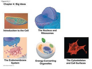
The document provides an overview of cell structures and organelles, including:
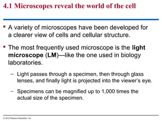
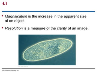
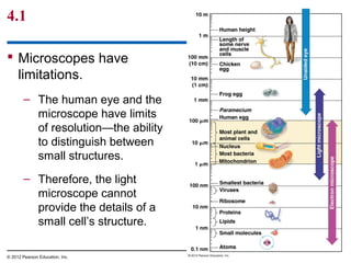
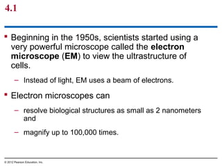
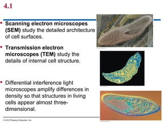
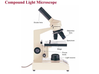
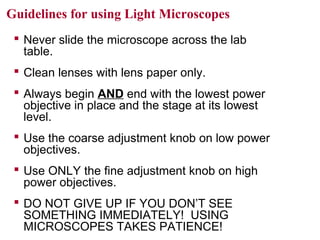
- Light microscopes and electron microscopes allow observation of cells at different magnifications.
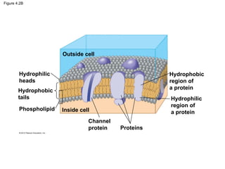
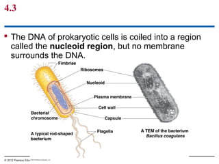
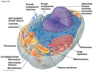
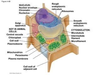
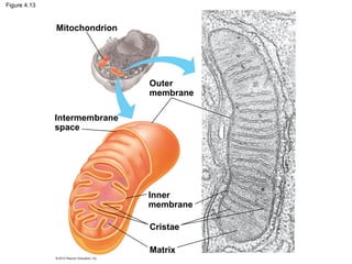
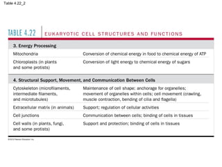
- Eukaryotic cells have internal membranes that create organelles, while prokaryotic cells lack these.
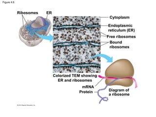
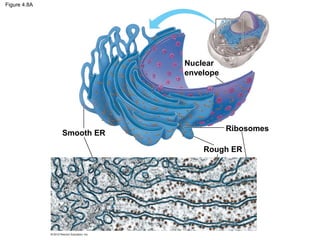
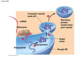
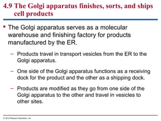
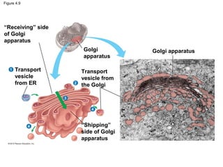
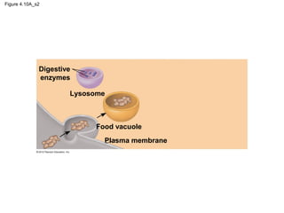
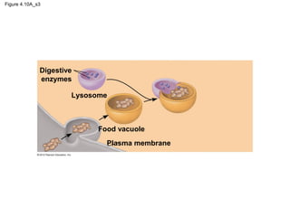
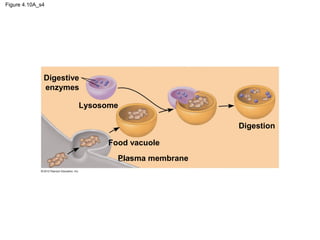
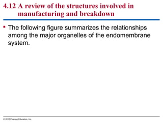
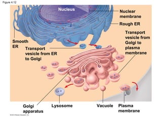
- The endomembrane system connects organelles like the ER, Golgi apparatus and plasma membrane to synthesize, modify and transport cell products.
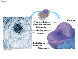
- The nucleus contains DNA and directs protein synthesis, while ribosomes build proteins using instructions from the nucleus.


![Chapter 6 cell energy [compatibility mode]](https://cdn.slidesharecdn.com/ss_thumbnails/chapter6-cellenergycompatibilitymode-141214133046-conversion-gate01-thumbnail.jpg?width=640&height=640&fit=bounds)


![Chapter 4 cell & tissues (1) [compatibility mode]](https://cdn.slidesharecdn.com/ss_thumbnails/chapter4-celltissues1compatibilitymode-141214131430-conversion-gate01-thumbnail.jpg?width=640&height=640&fit=bounds)


![Chapter 5 the working cells [compatibility mode]](https://cdn.slidesharecdn.com/ss_thumbnails/chapter5-theworkingcellscompatibilitymode-141214132834-conversion-gate01-thumbnail.jpg?width=640&height=640&fit=bounds)


![Chapter 8: Genetics [compatibility mode]](https://cdn.slidesharecdn.com/ss_thumbnails/chapter8-geneticscompatibilitymode-141214140247-conversion-gate02-thumbnail.jpg?width=640&height=640&fit=bounds)


![THEORY cell_biology__notes_print_1[1].pptx](https://cdn.slidesharecdn.com/ss_thumbnails/cellbiologynotesprint11-251210090642-34c62fe5-thumbnail.jpg?width=640&height=640&fit=bounds)