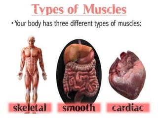
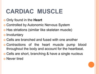
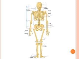
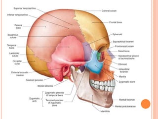
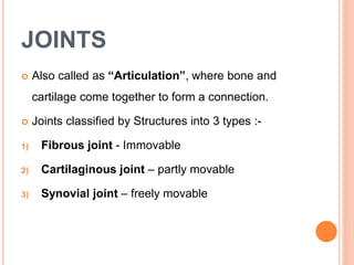
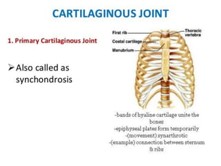
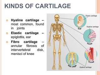
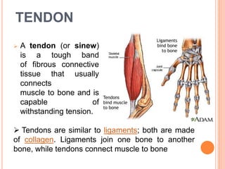
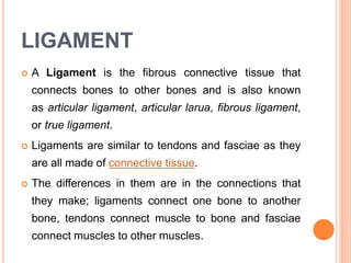
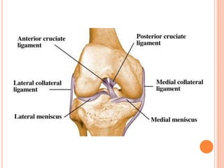
The musculoskeletal system consists of bones, muscles, cartilage, tendons and ligaments and works together to allow for movement and provide support to the body. It has three main functions - supporting the body, allowing for motion, and protecting vital organs. There are three main types of muscles - skeletal, smooth and cardiac. Skeletal muscle is voluntary and attached to bones via tendons. Smooth muscle is involuntary and found in internal organs. Cardiac muscle is only located in the heart. The skeletal system provides points of attachment for muscles, supports the body, protects organs, stores minerals, and makes blood cells. It consists of long, short, flat, irregular and sesamoid bones. Joints connect bones together and include fibrous


![MUSCLES
Our skeletal system has more than 650 muscles,
most of them disposed in pair to provide movement.
The muscles make up about 40 % of the body
mass.
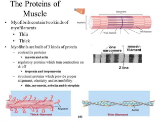
Muscular cells are called muscle fibers.
Every fibers contain thousand of myofibrils.
Inside each mayofibril there are many
myofilaments that are made of 2 proteins :
1] Actin (Thin)
2] Myosin (Thick)](https://image.slidesharecdn.com/musculoskeletonsystem-170404035622/85/Musculoskeleton-system-4-320.jpg)


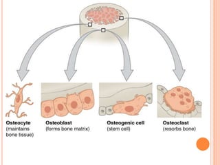
![BONE CELLS
[1] Osteogenic cells
- Develop into Osteoblasts
[2] Osteoblasts cells
- growing portion of bone, including periosteum
and endosteum
[3] Osteocytes cells
- maintain mineral concentation of matrix
[4] Osteoclasts cells
- Bone resorption](https://image.slidesharecdn.com/musculoskeletonsystem-170404035622/85/Musculoskeleton-system-32-320.jpg)
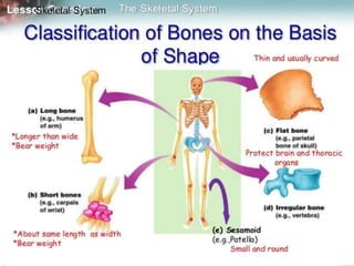
![CLASSIFICATION OF BONES
[1] Long Bones
Long bones have a
long shaft and two
distinct ends
All bones of the limbs
{except patella, wrist &
ankle}
The bones of the
fingers
They are usually
somewhat curved for
strength](https://image.slidesharecdn.com/musculoskeletonsystem-170404035622/85/Musculoskeleton-system-34-320.jpg)
![[2] SHORT BONES
Short bones are roughly
cubelike
Cube-shaped & have
approximately equal length
& width
Providing support and
stability with little
movement
Found in wrist & ankle
bones](https://image.slidesharecdn.com/musculoskeletonsystem-170404035622/85/Musculoskeleton-system-35-320.jpg)
![[3] FLAT BONES
Flat bones are thin,
flattened and usually
curved
Provide considerable
mechanical protection &
extensive surfaces for
muscle attachments
Ex. Scapula, Sternum,
Ribs, cranial bones](https://image.slidesharecdn.com/musculoskeletonsystem-170404035622/85/Musculoskeleton-system-36-320.jpg)
![[4] IRREGULAR BONES
Irregular bones don’t fit
into the previous
categories
Complicated shapes
Consist of cancellous
bone with a thin outer
layer of compact bone
Ex. Vertebrae, Mandible,
Hip, Sacrum](https://image.slidesharecdn.com/musculoskeletonsystem-170404035622/85/Musculoskeleton-system-37-320.jpg)
![[5] SESAMOID BONES
Short or irregular
bones, imbedded in a
tendon
It passes over a joint
which serves to
protect the tendon
Ex. Patellae
(kneecaps)](https://image.slidesharecdn.com/musculoskeletonsystem-170404035622/85/Musculoskeleton-system-38-320.jpg)


![[1] Bones that surround the spinal cord are classified as
________ bones.
A. Short
B. Sesamoid
C. Flat
D. Irregular
E. Long](https://image.slidesharecdn.com/musculoskeletonsystem-170404035622/85/Musculoskeleton-system-56-320.jpg)
![ [2] What is the difference between compact bone
and spongy bone?
A. They have different bone marrow.
B. They are made of different materials.
C. They have different sizes of bone cells.
D. They have different arrangement of bone cells.](https://image.slidesharecdn.com/musculoskeletonsystem-170404035622/85/Musculoskeleton-system-58-320.jpg)
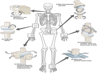
![[3] A joint between radius and carpal bone is known
as ..
A. Hinge joint
B. Saddle joint
C. Ball and socket joint
D. Condyloid joint
E. Plane joint](https://image.slidesharecdn.com/musculoskeletonsystem-170404035622/85/Musculoskeleton-system-60-320.jpg)
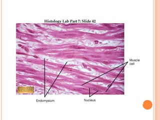
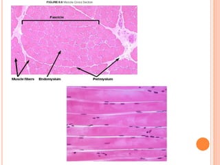
![[4] The tissue that surrounds a muscle cell (myofiber)
is known as……
A. Epimysium
B. Perimysium
C. Endomysium
D. Exomysium
E. Superficial mysium](https://image.slidesharecdn.com/musculoskeletonsystem-170404035622/85/Musculoskeleton-system-62-320.jpg)