Downloaded 396 times
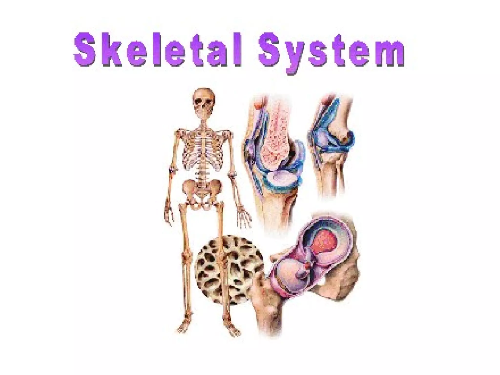
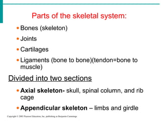

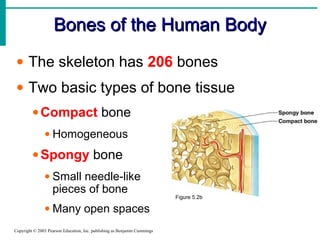
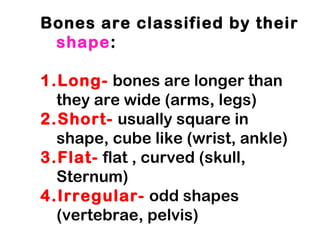
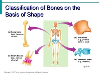
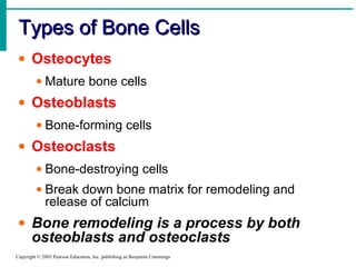
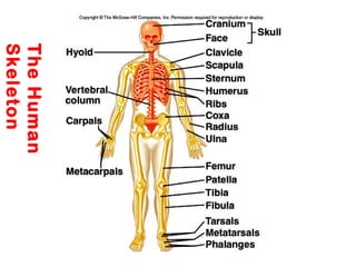
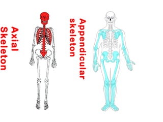
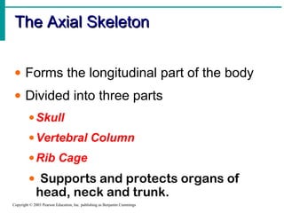

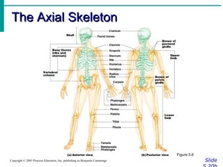
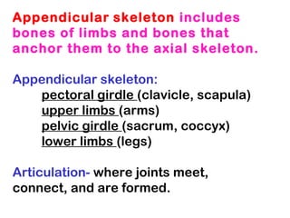
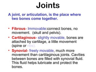
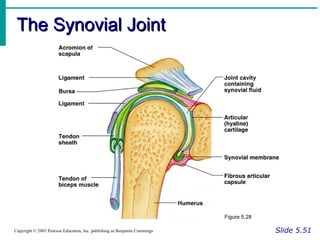
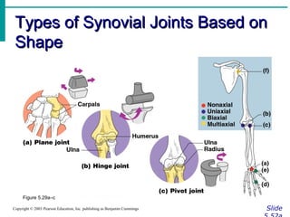
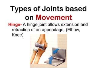
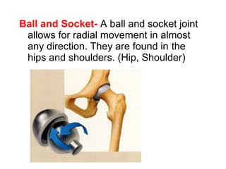
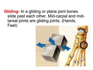
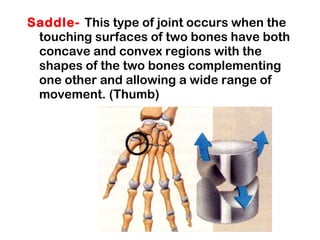

The skeletal system is divided into two sections - the axial skeleton which includes the skull, vertebral column, and rib cage, and the appendicular skeleton which includes the limbs and girdles attaching them to the axial skeleton. Bones provide structure, protection, movement, mineral storage, and blood cell formation. There are 206 bones in the human body which come in four shapes - long, short, flat, and irregular. Bones are made of two types of tissue and contain three main cell types. Joints connect and allow movement between bones. The main types of joints are fibrous, cartilaginous, and synovial, with synovial joints providing the most movement.


![481319198-Physical-Education-Skeletal-system-pptx[1].pptx](https://cdn.slidesharecdn.com/ss_thumbnails/481319198-physical-education-skeletal-system-pptx1-251017025828-969967ed-thumbnail.jpg?width=640&height=640&fit=bounds)