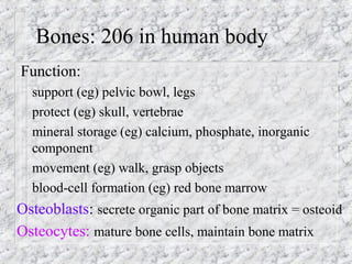
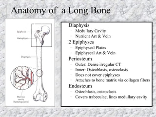
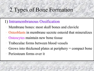
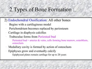
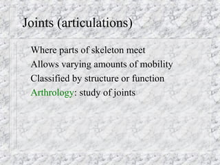
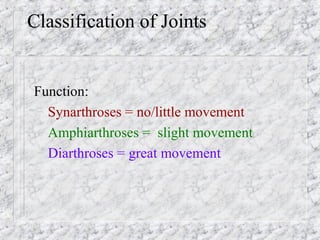
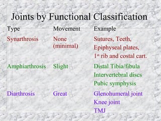
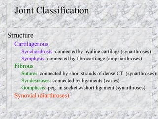
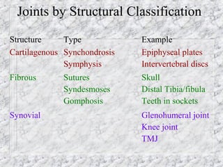
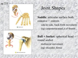
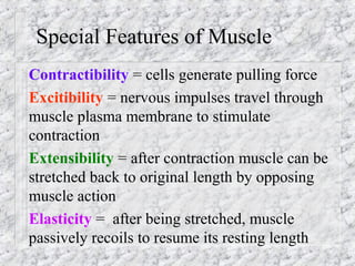
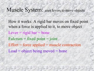
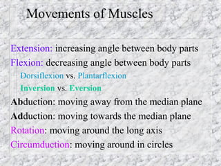
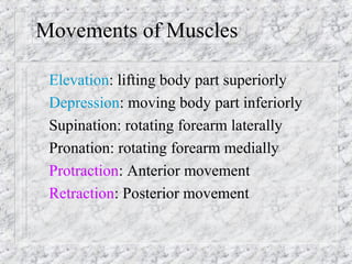
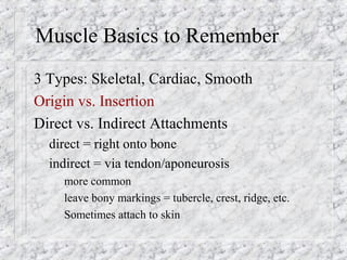
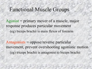
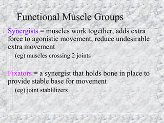
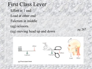
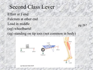
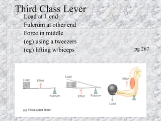
Bones, Joints and Muscles
Bones: 206 in human body
Function:
– support (eg) pelvic bowl, legs
– protect (eg) skull, vertebrae
– mineral storage (eg) calcium, phosphate, inorganic
component
– movement (eg) walk, grasp objects
– blood-cell formation (eg) red bone marrow
Osteoblasts: secrete organic part of bone matrix = osteoid
Osteocytes: mature bone cells, maintain bone matrix
Some Reminders about Bones
Bone = bone tissue (type of CT)
A Bone = an organ
Compact vs. Spongy Bone
Composition: Hydroxyapatite, protoplasm, collagen,
blood vessels, marrow
Skeleton = bones, cartilage (avascular, no nerves, 80%
H2O), joints, ligaments
Shapes of Bones
– Long, Flat, Irregular, Short
Before 8 weeks, embryo is all cartilage
Structure of Bone
Anatomy of a Long Bone
Diaphysis
– Medullary Cavity
– Nutrient Art & Vein
2 Epiphyses
– Epiphyseal Plates
– Epiphyseal Art & Vein
Periosteum
– Outer: Dense irregular CT
– Inner: Osteoblasts, osteoclasts
– Does not cover epiphyses
– Attaches to bone matrix via collagen fibers
Endosteum
– Osteoblasts, osteoclasts
– Covers trabeculae, lines medullary cavity
2 Types of Bone Formation
1) Intramembranous Ossification
– Membrane bones: most skull bones and clavicle
– Osteoblasts in membrane secrete osteoid that mineralizes
– Osteocytes maintain new bone tissue
– Trabeculae forms between blood vessels
– Grows into thickened plates at periphery = compact bone
– Periosteum forms over it
2 Types of Bone Formation :
2) Endochondral Ossification: All other bones
– Begins with a cartilaginous model
– Perichondrium becomes replaced by periosteum
– Cartilage in diaphysis calcifies
– Trabeculae forms from Periosteal bud
Periosteal bud = arteries & veins, cells forming bone marrow, osteoblasts,
osteoclasts
– Medullary cavity is formed by action of osteoclasts
– Epiphyses grow and eventually calcify
Epiphyseal plates remain cartilage for up to 20 years
Bone Growth & Remodeling
GROWTH
Appositional Growth = widening of bone
– Bone tissue added on surface by osteoblasts of periosteum
– Medullary cavity maintained by osteoclasts
Lengthening of Bone
– Epiphyseal plates enlarge by chondroblasts
– Matrix calcifies (chondrocytes die and disintegrate)
– Bone tissue replaces cartilage on diaphysis side
REMODELING
Due to mechanical stresses on bones, their tissue needs to be replaced
– Osteoclasts-take up bone ( = breakdown)
release Ca2++
, PO4 to body fluids from bone
– Osteoblasts-lay down bone
secrete osteoid to form new bone
Ideally osteoclasts and osteoblasts work at the same rate!
Joints (articulations)
Where parts of skeleton meet
Allows varying amounts of mobility
Classified by structure or function
Arthrology: study of joints
Classification of Joints
Function:
– Synarthroses = no/little movement
– Amphiarthroses = slight movement
– Diarthroses = great movement
Joints by Functional Classification
Type Movement Example
Synarthrosis None
(minimal)
Sutures, Teeth,
Epiphyseal plates,
1st
rib and costal cart.
Amphiarthrosis Slight Distal Tibia/fibula
Intervertebral discs
Pubic symphysis
Diarthrosis Great Glenohumeral joint
Knee joint
TMJ
Joint Classification
Structure
– Cartilagenous
Synchondrosis: connected by hyaline cartilage (synarthroses)
Symphysis: connected by fibrocartilage (amphiarthroses)
– Fibrous
Sutures: connected by short strands of dense CT (synarthroses)
Syndesmoses: connected by ligaments (varies)
Gomphosis: peg in socket w/short ligament (synarthroses)
– Synovial (diarthroses)
Joints by Structural Classification
Structure Type Example
Cartilagenous Synchondrosis
Symphysis
Epiphyseal plates
Intervertebral discs
Fibrous Sutures
Syndesmoses
Gomphosis
Skull
Distal Tibia/fibula
Teeth in sockets
Synovial Glenohumeral joint
Knee joint
TMJ
Components of SYNOVIAL JOINTS:
(Structural Joint Classification continued)
Articular cartilage: hyaline; covers ends of both bones
articulating
Synovial (joint) cavity: space holding synovial fluid
Articular capsule: Made of 2 layers
– Fibrous: external, dense CT for strength
– Synovial membrane: internal, produces synovial fluid
Synovial fluid: viscous; lubricates and nourishes;
contained in capsule and articular cartilages
Reinforcing ligaments: extracapsular/intracapsular
Nerves + vessels: Highly innervated, Highly vascular
Meniscus (some): fibrocartilage; improves the fit of 2 bones
to increase stability
Synovial Joint
pg 215
Bursae & Tendon Sheaths
Bursae: flat, fibrous sac
w/synovial membrane
lining
Tendon Sheaths:
elongated bursae that
wraps around tendons
3 Factors in Joint
Stability:
– Muscle Tone
– Ligaments
– Fit of Articular Surface
pg 219
Joint Shapes
Hinge: cylindrical end of 1
bone fits into trough shape of
other
– angular movement-1 plane (eg)
elbow, ankle, interphalangal
Plane: articular surface in flat
plane
– Short gliding movement
– (eg) intertarsal, articular processes
of vertebrae
pg 224
Joint Shapes
Condyloid: egg-shape articular
surface + oval concavity
– side-to-side, back+forth movement
– (eg) metacarpophalangeal (knuckle)
Pivot: round end fits into ring of
bone + ligament
– rotation on long axis
– (eg) prox. radius/ulna, atlas/dens
pg 225
Joint Shapes
Saddle: articular surface both
concave + convex
– side-to-side, back-forth movement
– (eg) carpometacarpal jt of thumb
–
Ball + Socket: spherical head +
round socket
– multiaxial movement
– (eg) shoulder, femur
pg 225
!Muscles!
Function: 1) movement
2) maintain posture
3) joint stability
4) generate heat
!Muscles!
Special Features of Muscle
Contractibility = cells generate pulling force
Excitibility = nervous impulses travel through
muscle plasma membrane to stimulate
contraction
Extensibility = after contraction muscle can be
stretched back to original length by opposing
muscle action
Elasticity = after being stretched, muscle
passively recoils to resume its resting length
Muscle System: uses levers to move objects
How it works: A rigid bar moves on fixed point
when a force is applied to it, to move object
Lever = rigid bar = bone
Fulcrum = fixed point = joint
Effort = force applied = muscle contraction
Load = object being moved = bone
Movements of Muscles
Extension: increasing angle between body parts
Flexion: decreasing angle between body parts
– Dorsiflexion vs. Plantarflexion
– Inversion vs. Eversion
Abduction: moving away from the median plane
Adduction: moving towards the median plane
Rotation: moving around the long axis
Circumduction: moving around in circles
Elevation: lifting body part superiorly
Depression: moving body part inferiorly
Supination: rotating forearm laterally
Pronation: rotating forearm medially
Protraction: Anterior movement
Retraction: Posterior movement
Movements of Muscles
Muscle Basics to Remember
3 Types: Skeletal, Cardiac, Smooth
Origin vs. Insertion
Direct vs. Indirect Attachments
– direct = right onto bone
– indirect = via tendon/aponeurosis
more common
leave bony markings = tubercle, crest, ridge, etc.
Sometimes attach to skin
Functional Muscle Groups
Agonist = primary mover of a muscle, major
response produces particular movement
– (eg) biceps brachii is main flexor of forearm
Antagonists = oppose/reverse particular
movement, prevent overshooting agonistic motion
– (eg) triceps brachii is antagonist to biceps brachii
Functional Muscle Groups
Synergists = muscles work together, adds extra
force to agonistic movement, reduce undesirable
extra movement
– (eg) muscles crossing 2 joints
Fixators = a synergist that holds bone in place to
provide stable base for movement
– (eg) joint stablilizers
Naming Muscles
Location: (eg) brachialis = arm
Shape: (eg) deltoid = triangle
Relative Size: (eg) minimus, maximus, longus
Direction of Fascicles: (eg) oblique, rectus
Location of Attachment: (eg) brachioradialis
Number of Origins: (eg) biceps, quadriceps
Action: (eg) flexor, adductor, extensor
Arrangement of Muscle Fibers
Parallel: long axis of fascicles parallel to axis of
muscle; straplike (eg) biceps, sternocleidomastoid
Convergent: O = broad, I = narrow, via tendon; fan
or triangle shaped (eg) pectoralis major
Circular: fascicles arranged in concentric circles;
sphincter (eg) around mouth
Arrangement of Muscle Fibers
Pennate: fascicles short + attached obliquely to
tendon running length of muscle; featherlike
– Unipennate = fascicles insert on only 1 side
(eg) flexor pollicis longus
– Bipennate = fascicles insert both sides
(eg) rectus femoris
– Multipennate = many bundles inserting together
(eg) deltoid
Arrangements of Muscle Fascicles
pg 269
STOP
More on Levers on the following pages
First Class Lever
Effort at 1 end
Load at other end
Fulcrum in middle
(eg) scissors
(eg) moving head up and down
pg 267
Second Class Lever
Effort at 1 end
Fulcrum at other end
Load in middle
(eg) wheelbarrel
(eg) standing on tip toes (not common in body)
pg 267
Third Class Lever
Load at 1 end
Fulcrum at other end
Force in middle
(eg) using a tweezers
(eg) lifting w/biceps pg 267
Mechanical Advantage
When the load is close to
the fulcrum, effort is
applied far from fulcrum
Small effort over large
distance = move large load
over short distance
(eg) Using a jack on a car
pg 266
Mechanical Disadvantage
When the load is farther
from the fulcrum than the
effort, the effort applied
must be greater than the
load being moved
Load moved quickly over
large distance
(eg) using a shovel
pg 266

Bones, joints, muscles

  • 1.
  • 2.
    Bones: 206 inhuman body Function: – support (eg) pelvic bowl, legs – protect (eg) skull, vertebrae – mineral storage (eg) calcium, phosphate, inorganic component – movement (eg) walk, grasp objects – blood-cell formation (eg) red bone marrow Osteoblasts: secrete organic part of bone matrix = osteoid Osteocytes: mature bone cells, maintain bone matrix
  • 3.
    Some Reminders aboutBones Bone = bone tissue (type of CT) A Bone = an organ Compact vs. Spongy Bone Composition: Hydroxyapatite, protoplasm, collagen, blood vessels, marrow Skeleton = bones, cartilage (avascular, no nerves, 80% H2O), joints, ligaments Shapes of Bones – Long, Flat, Irregular, Short Before 8 weeks, embryo is all cartilage
  • 4.
  • 5.
    Anatomy of aLong Bone Diaphysis – Medullary Cavity – Nutrient Art & Vein 2 Epiphyses – Epiphyseal Plates – Epiphyseal Art & Vein Periosteum – Outer: Dense irregular CT – Inner: Osteoblasts, osteoclasts – Does not cover epiphyses – Attaches to bone matrix via collagen fibers Endosteum – Osteoblasts, osteoclasts – Covers trabeculae, lines medullary cavity
  • 6.
    2 Types ofBone Formation 1) Intramembranous Ossification – Membrane bones: most skull bones and clavicle – Osteoblasts in membrane secrete osteoid that mineralizes – Osteocytes maintain new bone tissue – Trabeculae forms between blood vessels – Grows into thickened plates at periphery = compact bone – Periosteum forms over it
  • 7.
    2 Types ofBone Formation : 2) Endochondral Ossification: All other bones – Begins with a cartilaginous model – Perichondrium becomes replaced by periosteum – Cartilage in diaphysis calcifies – Trabeculae forms from Periosteal bud Periosteal bud = arteries & veins, cells forming bone marrow, osteoblasts, osteoclasts – Medullary cavity is formed by action of osteoclasts – Epiphyses grow and eventually calcify Epiphyseal plates remain cartilage for up to 20 years
  • 8.
    Bone Growth &Remodeling GROWTH Appositional Growth = widening of bone – Bone tissue added on surface by osteoblasts of periosteum – Medullary cavity maintained by osteoclasts Lengthening of Bone – Epiphyseal plates enlarge by chondroblasts – Matrix calcifies (chondrocytes die and disintegrate) – Bone tissue replaces cartilage on diaphysis side REMODELING Due to mechanical stresses on bones, their tissue needs to be replaced – Osteoclasts-take up bone ( = breakdown) release Ca2++ , PO4 to body fluids from bone – Osteoblasts-lay down bone secrete osteoid to form new bone Ideally osteoclasts and osteoblasts work at the same rate!
  • 9.
    Joints (articulations) Where partsof skeleton meet Allows varying amounts of mobility Classified by structure or function Arthrology: study of joints
  • 10.
    Classification of Joints Function: –Synarthroses = no/little movement – Amphiarthroses = slight movement – Diarthroses = great movement
  • 11.
    Joints by FunctionalClassification Type Movement Example Synarthrosis None (minimal) Sutures, Teeth, Epiphyseal plates, 1st rib and costal cart. Amphiarthrosis Slight Distal Tibia/fibula Intervertebral discs Pubic symphysis Diarthrosis Great Glenohumeral joint Knee joint TMJ
  • 12.
    Joint Classification Structure – Cartilagenous Synchondrosis:connected by hyaline cartilage (synarthroses) Symphysis: connected by fibrocartilage (amphiarthroses) – Fibrous Sutures: connected by short strands of dense CT (synarthroses) Syndesmoses: connected by ligaments (varies) Gomphosis: peg in socket w/short ligament (synarthroses) – Synovial (diarthroses)
  • 13.
    Joints by StructuralClassification Structure Type Example Cartilagenous Synchondrosis Symphysis Epiphyseal plates Intervertebral discs Fibrous Sutures Syndesmoses Gomphosis Skull Distal Tibia/fibula Teeth in sockets Synovial Glenohumeral joint Knee joint TMJ
  • 14.
    Components of SYNOVIALJOINTS: (Structural Joint Classification continued) Articular cartilage: hyaline; covers ends of both bones articulating Synovial (joint) cavity: space holding synovial fluid Articular capsule: Made of 2 layers – Fibrous: external, dense CT for strength – Synovial membrane: internal, produces synovial fluid Synovial fluid: viscous; lubricates and nourishes; contained in capsule and articular cartilages Reinforcing ligaments: extracapsular/intracapsular Nerves + vessels: Highly innervated, Highly vascular Meniscus (some): fibrocartilage; improves the fit of 2 bones to increase stability
  • 15.
  • 16.
    Bursae & TendonSheaths Bursae: flat, fibrous sac w/synovial membrane lining Tendon Sheaths: elongated bursae that wraps around tendons 3 Factors in Joint Stability: – Muscle Tone – Ligaments – Fit of Articular Surface pg 219
  • 17.
    Joint Shapes Hinge: cylindricalend of 1 bone fits into trough shape of other – angular movement-1 plane (eg) elbow, ankle, interphalangal Plane: articular surface in flat plane – Short gliding movement – (eg) intertarsal, articular processes of vertebrae pg 224
  • 18.
    Joint Shapes Condyloid: egg-shapearticular surface + oval concavity – side-to-side, back+forth movement – (eg) metacarpophalangeal (knuckle) Pivot: round end fits into ring of bone + ligament – rotation on long axis – (eg) prox. radius/ulna, atlas/dens pg 225
  • 19.
    Joint Shapes Saddle: articularsurface both concave + convex – side-to-side, back-forth movement – (eg) carpometacarpal jt of thumb – Ball + Socket: spherical head + round socket – multiaxial movement – (eg) shoulder, femur pg 225
  • 20.
    !Muscles! Function: 1) movement 2)maintain posture 3) joint stability 4) generate heat !Muscles!
  • 21.
    Special Features ofMuscle Contractibility = cells generate pulling force Excitibility = nervous impulses travel through muscle plasma membrane to stimulate contraction Extensibility = after contraction muscle can be stretched back to original length by opposing muscle action Elasticity = after being stretched, muscle passively recoils to resume its resting length
  • 22.
    Muscle System: useslevers to move objects How it works: A rigid bar moves on fixed point when a force is applied to it, to move object Lever = rigid bar = bone Fulcrum = fixed point = joint Effort = force applied = muscle contraction Load = object being moved = bone
  • 23.
    Movements of Muscles Extension:increasing angle between body parts Flexion: decreasing angle between body parts – Dorsiflexion vs. Plantarflexion – Inversion vs. Eversion Abduction: moving away from the median plane Adduction: moving towards the median plane Rotation: moving around the long axis Circumduction: moving around in circles
  • 24.
    Elevation: lifting bodypart superiorly Depression: moving body part inferiorly Supination: rotating forearm laterally Pronation: rotating forearm medially Protraction: Anterior movement Retraction: Posterior movement Movements of Muscles
  • 25.
    Muscle Basics toRemember 3 Types: Skeletal, Cardiac, Smooth Origin vs. Insertion Direct vs. Indirect Attachments – direct = right onto bone – indirect = via tendon/aponeurosis more common leave bony markings = tubercle, crest, ridge, etc. Sometimes attach to skin
  • 26.
    Functional Muscle Groups Agonist= primary mover of a muscle, major response produces particular movement – (eg) biceps brachii is main flexor of forearm Antagonists = oppose/reverse particular movement, prevent overshooting agonistic motion – (eg) triceps brachii is antagonist to biceps brachii
  • 27.
    Functional Muscle Groups Synergists= muscles work together, adds extra force to agonistic movement, reduce undesirable extra movement – (eg) muscles crossing 2 joints Fixators = a synergist that holds bone in place to provide stable base for movement – (eg) joint stablilizers
  • 28.
    Naming Muscles Location: (eg)brachialis = arm Shape: (eg) deltoid = triangle Relative Size: (eg) minimus, maximus, longus Direction of Fascicles: (eg) oblique, rectus Location of Attachment: (eg) brachioradialis Number of Origins: (eg) biceps, quadriceps Action: (eg) flexor, adductor, extensor
  • 29.
    Arrangement of MuscleFibers Parallel: long axis of fascicles parallel to axis of muscle; straplike (eg) biceps, sternocleidomastoid Convergent: O = broad, I = narrow, via tendon; fan or triangle shaped (eg) pectoralis major Circular: fascicles arranged in concentric circles; sphincter (eg) around mouth
  • 30.
    Arrangement of MuscleFibers Pennate: fascicles short + attached obliquely to tendon running length of muscle; featherlike – Unipennate = fascicles insert on only 1 side (eg) flexor pollicis longus – Bipennate = fascicles insert both sides (eg) rectus femoris – Multipennate = many bundles inserting together (eg) deltoid
  • 31.
    Arrangements of MuscleFascicles pg 269
  • 32.
    STOP More on Leverson the following pages
  • 33.
    First Class Lever Effortat 1 end Load at other end Fulcrum in middle (eg) scissors (eg) moving head up and down pg 267
  • 34.
    Second Class Lever Effortat 1 end Fulcrum at other end Load in middle (eg) wheelbarrel (eg) standing on tip toes (not common in body) pg 267
  • 35.
    Third Class Lever Loadat 1 end Fulcrum at other end Force in middle (eg) using a tweezers (eg) lifting w/biceps pg 267
  • 36.
    Mechanical Advantage When theload is close to the fulcrum, effort is applied far from fulcrum Small effort over large distance = move large load over short distance (eg) Using a jack on a car pg 266
  • 37.
    Mechanical Disadvantage When theload is farther from the fulcrum than the effort, the effort applied must be greater than the load being moved Load moved quickly over large distance (eg) using a shovel pg 266