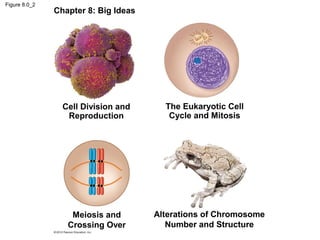
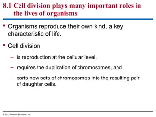
1) The chapter discusses the cellular basis of reproduction and inheritance. It covers topics like cell division, the cell cycle, meiosis, and alterations in chromosome structure.
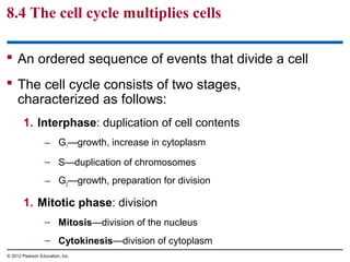
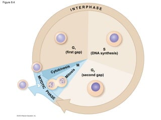
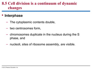
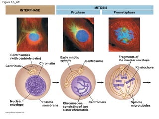
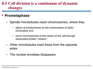
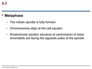
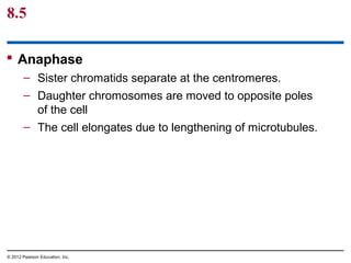
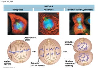
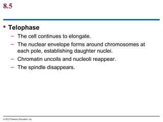
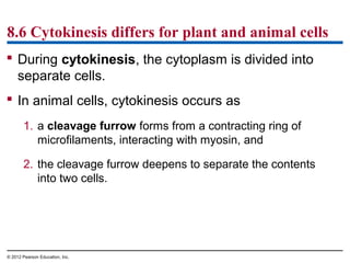
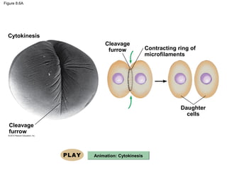
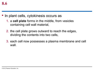
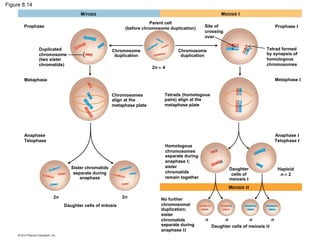
2) The key stages of the cell cycle are described, including interphase and the phases of mitosis (prophase, metaphase, anaphase, telophase). Cytokinesis is the final step that divides the cytoplasm.
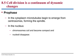
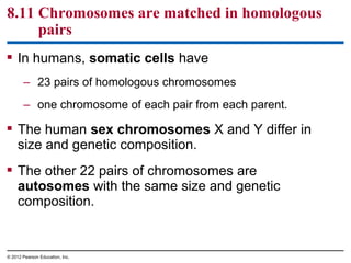
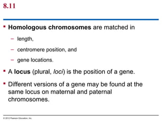
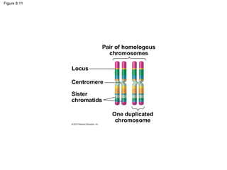
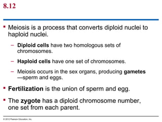
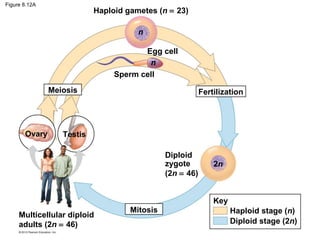
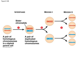
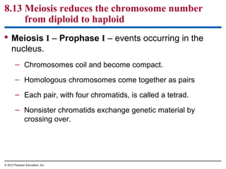
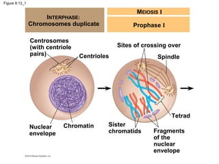
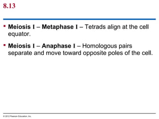
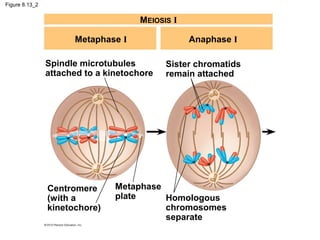
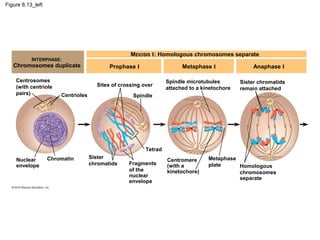
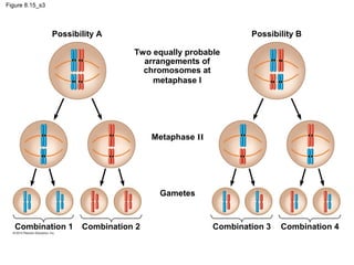
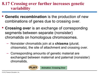
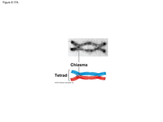
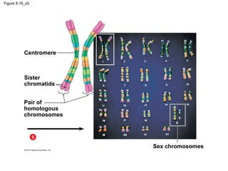
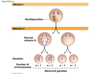
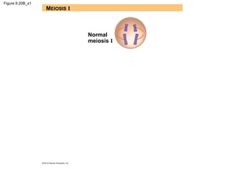
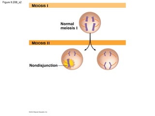
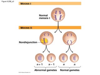
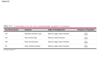
3) Meiosis is introduced as reducing the number of chromosomes by half to form gametes, while mitosis replicates chromosomes to form body cells. Homologous chromosomes pair and may exchange genetic material.


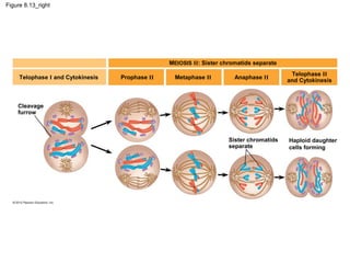


![Chapter 6 cell energy [compatibility mode]](https://cdn.slidesharecdn.com/ss_thumbnails/chapter6-cellenergycompatibilitymode-141214133046-conversion-gate01-thumbnail.jpg?width=640&height=640&fit=bounds)


![Chapter 8: Genetics [compatibility mode]](https://cdn.slidesharecdn.com/ss_thumbnails/chapter8-geneticscompatibilitymode-141214140247-conversion-gate02-thumbnail.jpg?width=640&height=640&fit=bounds)


![Mitosis p [compatibility mode]](https://cdn.slidesharecdn.com/ss_thumbnails/mitosispcompatibilitymode-111120223310-phpapp01-thumbnail.jpg?width=640&height=640&fit=bounds)


![Getting Started with Apache Spark: Big Data Made Simple [Free Meetup]](https://cdn.slidesharecdn.com/ss_thumbnails/apachesparkgettingstarted-260203175547-8361bcc3-thumbnail.jpg?width=640&height=640&fit=bounds)


