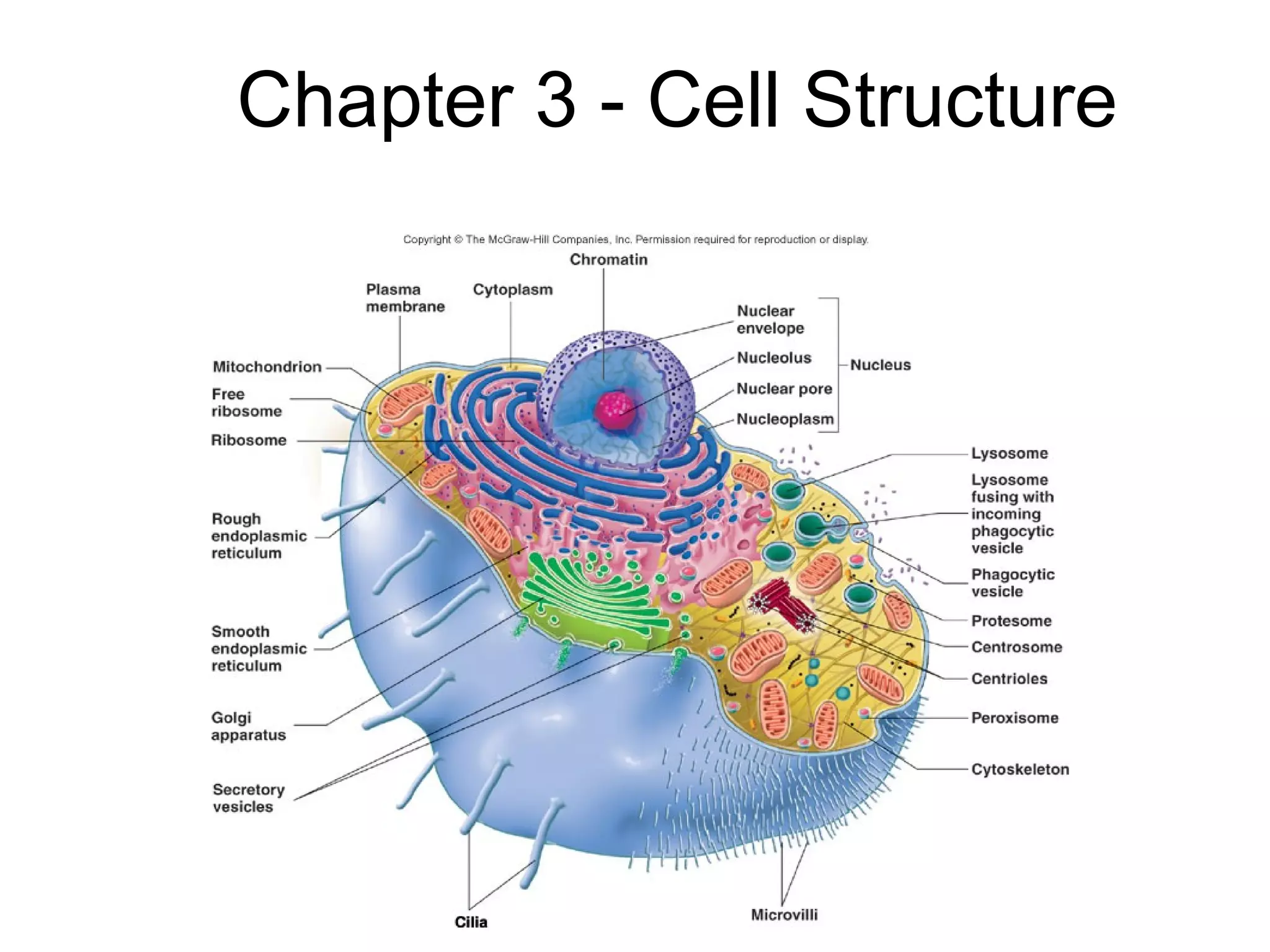
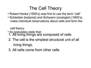
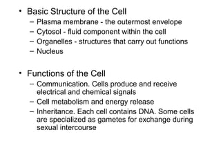
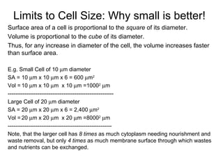
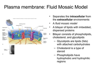
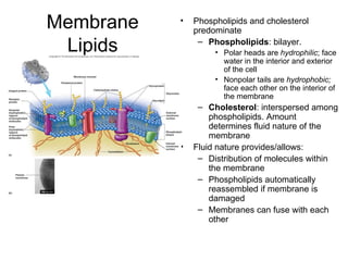
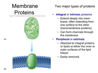
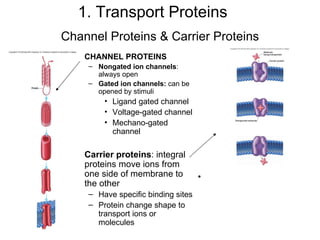
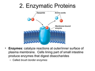
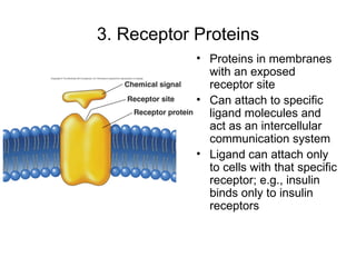
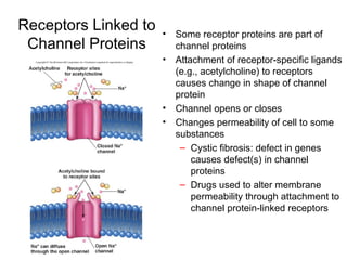
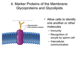
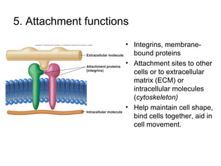
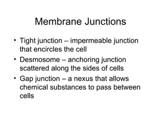
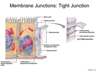
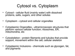
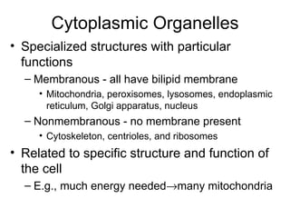
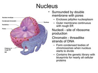
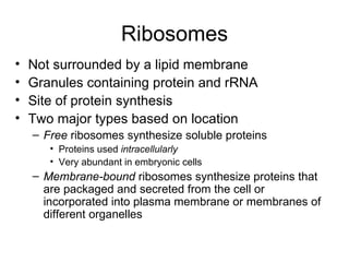
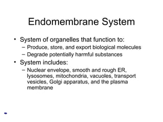
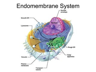
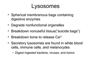
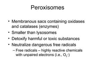
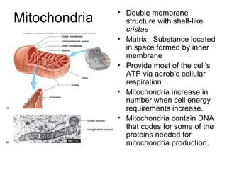
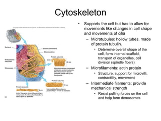
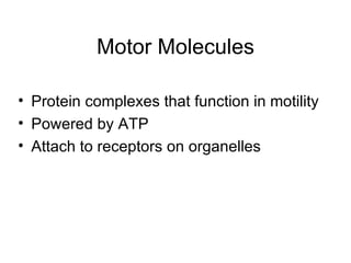
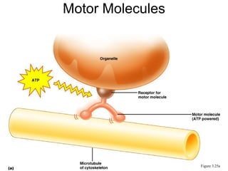
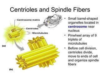
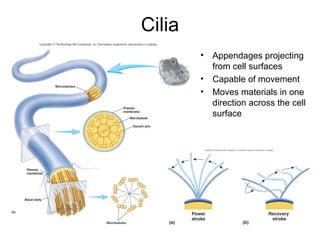
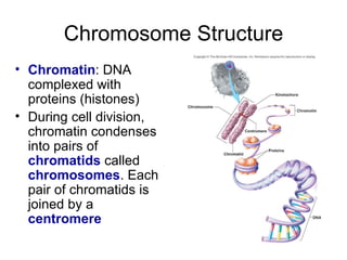
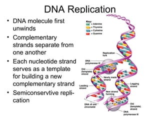
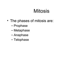
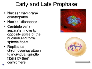
Chapter 3 - Cell Structure
The Cell Theory 1. All living things are composed of cells  2. The cell is the simplest structural unit of all  living things. 3. All cells come from other cells Robert Hooke (1500’s) was first to use the term “cell” Schleiden (botanist) and Schwann (zoologist) (1800’s)  make individual observations about cells and form the  cell theory. Its postulates state that:
Basic Structure of the Cell   Plasma membrane - the outermost envelope Cytosol - fluid component within the cell Organelles - structures that carry out functions  Nucleus Functions of the Cell Communication. Cells produce and receive electrical and chemical signals Cell metabolism and energy release Inheritance. Each cell contains DNA. Some cells are specialized as gametes for exchange during sexual intercourse
Limits to Cell Size: Why small is better! Surface area of a cell is proportional to the  square  of its diameter. Volume is proportional to the  cube  of its diameter. Thus, for any increase in diameter of the cell, the volume increases faster than surface area. E.g. Small Cell of 10   m diameter SA = 10   m x 10   m x 6 = 600   m 2 Vol =   10   m x 10   m  x 10   m =1000 2    m  --------------------------------------------------------- Large Cell of 20   m diameter SA = 20   m x 20   m x 6 = 2,400   m 2 Vol =   20   m x 20   m  x 20   m =8000 2    m -------------------------------------------------------- Note, that the larger cell has  8 times  as much cytoplasm needing nourishment and waste removal, but only  4 times  as much membrane surface through which wastes and nutrients can be exchanged.
Plasma membrane: Fluid Mosaic Model Separates the  intracellular  from the  extracellular  environments A  fluid mosaic model A bilayer of lipids with imbedded, dispersed proteins Bilayer consists of phospholipids, cholesterol, and glycolipids Glycolipids are lipids (fats) with attached carbohydrates Cholesterol is a type of steroid Phospholipids have hydrophobic and hydrophilic regions
Membrane Lipids Phospholipids and cholesterol predominate Phospholipids : bilayer.  Polar heads are  hydrophilic ; face water in the interior and exterior of the cell  Nonpolar tails are  hydrophobic;  face each other on the interior of the membrane Cholesterol : interspersed among phospholipids. Amount determines fluid nature of the membrane Fluid nature provides/allows:  Distribution of molecules within the membrane Phospholipids automatically reassembled if membrane is damaged Membranes can fuse with each other
Membrane Proteins   Integral  or  intrinsic   proteins Extend deeply into mem- brane, often   extending from one surface to the other (transmembrane proteins) Can form channels through the membrane Peripheral  or  extrinsic Attached to integral proteins or lipids at either the inner or outer surfaces of the lipid bilayer Easily removed Two major types of proteins
Functions of Membrane Proteins Transport - by channel or carrier proteins Enzymatic activity Receptors for signal transduction Cell-cell recognition Attachment to other cells, extracellular matrix, or cytoskeleton
Channel Proteins & Carrier Proteins CHANNEL PROTEINS Nongated ion channels : always open Gated ion channels:   can be opened by stimuli Ligand gated channel  Voltage-gated channel Mechano-gated channel Carrier proteins : integral proteins move ions from one side of membrane to the other Have specific binding sites Protein change shape to transport ions or molecules 1. Transport Proteins
2. Enzymatic Proteins Enzymes : catalyze reactions at outer/inner surface of plasma membrane.  Cells lining part of small intestine produce enzymes that digest disaccharides Called  brush border enzymes
3. Receptor Proteins Proteins in membranes with an exposed receptor site Can attach to specific ligand molecules and act as an intercellular communication system Ligand can attach only to cells with that specific receptor; e.g., insulin binds only to insulin receptors
Receptors Linked to Channel Proteins Some receptor proteins are part of channel proteins Attachment of receptor-specific ligands (e.g., acetylcholine) to receptors causes change in shape of channel protein Channel opens or closes Changes permeability of cell to some substances Cystic fibrosis: defect in genes causes defect(s) in channel proteins Drugs used to alter membrane permeability through attachment to channel protein-linked receptors
4. Marker Proteins of the Membrane: Glycoproteins and Glycolipids Allow cells to identify   one another or other molecules  Immunity Recognition of oocyte by sperm cell Intercellular communication
5. Attachment functions Integrins, membrane-bound proteins  Attachment sites to other cells or to extracellular matrix (ECM) or intracellular molecules ( cytoskeleton) Help maintain cell shape, bind cells together, aid in cell movement.
Membrane Junctions Tight junction – impermeable junction that encircles the cell  Desmosome – anchoring junction scattered along the sides of cells Gap junction – a nexus that allows chemical substances to pass between cells
Membrane Junctions: Tight Junction Figure 3.5a
Membrane Junctions: Desmosome Figure 3.5b
Membrane Junctions: Gap Junction Figure 3.5c
Cytosol vs. Cytoplasm Cytosol - cellular fluid (mainly water) with dissolved proteins, salts, sugars, and other solutes Cytoplasm - cytosol and cellular organelles  Cytoplasmic Organelles - ultramicroscopic structures that perform various cellular function; ribosomes, ER, mitochondria, etc Cytoskeleton - protein filaments and tubules that provide support, movement within the cell; cellular skeleton Cytoplasmic Inclusions - chemicals such as glycogen, fat, and pigments
Cytoplasmic Organelles Specialized structures with particular functions Membranous - all have bilipid membrane Mitochondria, peroxisomes, lysosomes, endoplasmic reticulum, Golgi apparatus, nucleus Nonmembranous - no membrane present Cytoskeleton, centrioles, and ribosomes Related to specific structure and function of the cell E.g., much energy needed  many mitochondria
Nucleus Surrounded by double membrane with pores Encloses jellylike nucleoplasm  Outer membrane continuous with rough ER  Nucleoli - site of ribosome production  Chromatin - threadlike strands of DNA Form condensed bodies of chromosomes when nucleus starts to divide Contains the genetic library with blueprints for nearly all cellular proteins
Ribosomes Not surrounded by a lipid membrane Granules containing protein and rRNA Site of protein synthesis Two major types based on location Free  ribosomes synthesize soluble proteins Proteins used  intracellularly Very abundant in embryonic cells Membrane-bound  ribosomes synthesize proteins that are packaged and secreted from the cell or incorporated into plasma membrane or membranes of different organelles
Endomembrane System System of organelles that function to:  Produce, store, and export biological molecules Degrade potentially harmful substances System includes: Nuclear envelope, smooth and rough ER, lysosomes, mitochondria, vacuoles, transport vesicles, Golgi apparatus, and the plasma membrane
Endomembrane System
Endoplasmic Reticulum
Endoplasmic Reticulum Interconnected tubes and parallel membranes enclosing cisternae; latter are spaces isolated from rest of cytoplasm 2 Types - rough ER and smooth ER Rough ER  - external surface studded with ribosomes Manufactures all secreted proteins Synthesizes integral membrane proteins   Smooth ER  - tubules arranged in a looping network; NO attached ribosomes.  Catalyzes the following reactions in various organs of the body. In the liver – lipid and cholesterol metabolism, breakdown of glycogen and, along with the kidneys, detoxification of drugs  In the testes – synthesis of steroid-based (fat) hormones  In the intestinal cells – absorption, synthesis, and transport of fats In skeletal and cardiac muscle – storage and release of calcium
Golgi Apparatus Stacked and flattened membranous sacs Functions in modification, packaging, distribution of proteins and lipids for secretion or internal use Transport vesicles from ER fuse with Golgi body After modification secretory vesicles leave the Golgi and move to designated parts of the cell
Role of the Golgi Apparatus Figure 3.21
Lysosomes Spherical membranous bags containing digestive enzymes Degrade nonfunctional organelles Breakdown nonuseful tissue(“suicide bags”) Breakdown bone to release Ca 2+ Secretory lysosomes are found in white blood cells, immune cells, and melanocytes Digest ingested bacteria, viruses, and toxins
Peroxisomes Membranous sacs containing oxidases and catalases (enzymes) Smaller than lysosomes Detoxify harmful or toxic substances Neutralize dangerous free radicals Free radicals – highly reactive chemicals with unpaired electrons (i.e., O 2 – )
Mitochondria Double membrane  structure with shelf-like  cristae Matrix:  Substance located in space formed by inner membrane Provide most of the cell’s ATP via aerobic cellular respiration Mitochondria increase in number when cell energy requirements increase.  Mitochondria contain DNA that codes for some of the proteins needed for mitochondria production.
Cytoskeleton Supports the cell but has to allow for movements like changes in cell shape and movements of cilia Microtubules: hollow tubes, made of protein tubulin. Determine overall shape of the cell, form internal scaffold, transport of organelles, cell division (spindle fibers) Microfilaments: actin protein Structure, support for microvilli, contractility, movement Intermediate filaments: provide mechanical strength Resist pulling forces on the cell and help form demosomes
Motor Molecules Protein complexes that function in motility Powered by ATP Attach to receptors on organelles
Motor Molecules Figure 3.25a
Centrioles and Spindle Fibers Small barrel-shaped organelles located in  centrosome  near nucleus Pinwheel array of 9 triplets of microtubules Before cell division, centrioles divide, move to ends of cell and organize spindle fibers
Cilia Appendages projecting from cell surfaces Capable of movement Moves materials in one direction across the cell surface
Flagella Similar to cilia but longer Usually only one per cell Move the cell itself in wave-like fashion Example:  sperm cell
Microvilli Extension of plasma membrane Increase the cell surface area Normally many on each cell One tenth to one twentieth size of cilia Do not move
Cell Division Essential for body growth and tissue repair Mitosis – nuclear division Cytokinesis – division of the cytoplasm
Cell Life Cycle Interphase :  phase between cell divisions Replication of DNA Ongoing normal cell activities Mitosis :  series of events that leads to the production of two cells by division of a mother cell into two daughter cells. Cells are genetically identical. Prophase Metaphase Anaphase Telophase Cytokinesis :  division of cell cytoplasm
Chromosome Structure Chromatin : DNA complexed with proteins (histones) During cell division, chromatin condenses into pairs of  chromatids  called  chromosomes . Each pair of chromatids is joined by a  centromere
DNA Replication DNA molecule first unwinds Complementary strands separate from one another Each nucleotide strand serves as a template for building a new complementary strand Semiconservtive repli-cation
Mitosis The phases of mitosis are: Prophase Metaphase Anaphase Telophase
Early and Late Prophase Nuclear membrane disintegrates Nucleoli disappear Centriole pairs separate, move to opposite poles of the nucleus and form spindle fibers Replicated chromosomes attach to individual spindle fibers by their centromere
Metaphase Metaphase plate Spindle Figure 3.32.4 Replicated chromosomes aligned along equator.
Anaphase Centromeres of the chromosomes split Motor proteins in kinetochores pull chromosomes toward poles
Telophase and Cytokinesis New sets of chromosomes extend into chromatin New nuclear membrane is formed from the rough ER Nucleoli reappear Generally cytokinesis (splitting of the cell into two) completes cell division
Cellular Aspects of Aging Cellular clock. After a certain amount of time or certain number of cell divisions, cells die. Death genes. Turn on late in life, or sometimes prematurely causing cells to deteriorate and die.  Apoptosis  - ‘programmed cell death’ DNA damage. Telomeres at ends of chromosomes TTAGGG.  During replication, nucleotides are lost.  Telomerase protects telomeres, enzymes seem to be lost with aging. Free radicals. DNA mutation caused by free radicals (atoms or molecules with an unpaired electron. Mitochondrial damage. Mitochondrial DNA may be more sensitive to free radicals.  Loss of energy, cell death.

Cell

  • 1.
    Chapter 3 -Cell Structure
  • 2.
    The Cell Theory1. All living things are composed of cells 2. The cell is the simplest structural unit of all living things. 3. All cells come from other cells Robert Hooke (1500’s) was first to use the term “cell” Schleiden (botanist) and Schwann (zoologist) (1800’s) make individual observations about cells and form the cell theory. Its postulates state that:
  • 3.
    Basic Structure ofthe Cell Plasma membrane - the outermost envelope Cytosol - fluid component within the cell Organelles - structures that carry out functions Nucleus Functions of the Cell Communication. Cells produce and receive electrical and chemical signals Cell metabolism and energy release Inheritance. Each cell contains DNA. Some cells are specialized as gametes for exchange during sexual intercourse
  • 4.
    Limits to CellSize: Why small is better! Surface area of a cell is proportional to the square of its diameter. Volume is proportional to the cube of its diameter. Thus, for any increase in diameter of the cell, the volume increases faster than surface area. E.g. Small Cell of 10  m diameter SA = 10  m x 10  m x 6 = 600  m 2 Vol = 10  m x 10  m x 10  m =1000 2  m --------------------------------------------------------- Large Cell of 20  m diameter SA = 20  m x 20  m x 6 = 2,400  m 2 Vol = 20  m x 20  m x 20  m =8000 2  m -------------------------------------------------------- Note, that the larger cell has 8 times as much cytoplasm needing nourishment and waste removal, but only 4 times as much membrane surface through which wastes and nutrients can be exchanged.
  • 5.
    Plasma membrane: FluidMosaic Model Separates the intracellular from the extracellular environments A fluid mosaic model A bilayer of lipids with imbedded, dispersed proteins Bilayer consists of phospholipids, cholesterol, and glycolipids Glycolipids are lipids (fats) with attached carbohydrates Cholesterol is a type of steroid Phospholipids have hydrophobic and hydrophilic regions
  • 6.
    Membrane Lipids Phospholipidsand cholesterol predominate Phospholipids : bilayer. Polar heads are hydrophilic ; face water in the interior and exterior of the cell Nonpolar tails are hydrophobic; face each other on the interior of the membrane Cholesterol : interspersed among phospholipids. Amount determines fluid nature of the membrane Fluid nature provides/allows: Distribution of molecules within the membrane Phospholipids automatically reassembled if membrane is damaged Membranes can fuse with each other
  • 7.
    Membrane Proteins Integral or intrinsic proteins Extend deeply into mem- brane, often extending from one surface to the other (transmembrane proteins) Can form channels through the membrane Peripheral or extrinsic Attached to integral proteins or lipids at either the inner or outer surfaces of the lipid bilayer Easily removed Two major types of proteins
  • 8.
    Functions of MembraneProteins Transport - by channel or carrier proteins Enzymatic activity Receptors for signal transduction Cell-cell recognition Attachment to other cells, extracellular matrix, or cytoskeleton
  • 9.
    Channel Proteins &Carrier Proteins CHANNEL PROTEINS Nongated ion channels : always open Gated ion channels: can be opened by stimuli Ligand gated channel Voltage-gated channel Mechano-gated channel Carrier proteins : integral proteins move ions from one side of membrane to the other Have specific binding sites Protein change shape to transport ions or molecules 1. Transport Proteins
  • 10.
    2. Enzymatic ProteinsEnzymes : catalyze reactions at outer/inner surface of plasma membrane. Cells lining part of small intestine produce enzymes that digest disaccharides Called brush border enzymes
  • 11.
    3. Receptor ProteinsProteins in membranes with an exposed receptor site Can attach to specific ligand molecules and act as an intercellular communication system Ligand can attach only to cells with that specific receptor; e.g., insulin binds only to insulin receptors
  • 12.
    Receptors Linked toChannel Proteins Some receptor proteins are part of channel proteins Attachment of receptor-specific ligands (e.g., acetylcholine) to receptors causes change in shape of channel protein Channel opens or closes Changes permeability of cell to some substances Cystic fibrosis: defect in genes causes defect(s) in channel proteins Drugs used to alter membrane permeability through attachment to channel protein-linked receptors
  • 13.
    4. Marker Proteinsof the Membrane: Glycoproteins and Glycolipids Allow cells to identify one another or other molecules Immunity Recognition of oocyte by sperm cell Intercellular communication
  • 14.
    5. Attachment functionsIntegrins, membrane-bound proteins Attachment sites to other cells or to extracellular matrix (ECM) or intracellular molecules ( cytoskeleton) Help maintain cell shape, bind cells together, aid in cell movement.
  • 15.
    Membrane Junctions Tightjunction – impermeable junction that encircles the cell Desmosome – anchoring junction scattered along the sides of cells Gap junction – a nexus that allows chemical substances to pass between cells
  • 16.
    Membrane Junctions: TightJunction Figure 3.5a
  • 17.
  • 18.
    Membrane Junctions: GapJunction Figure 3.5c
  • 19.
    Cytosol vs. CytoplasmCytosol - cellular fluid (mainly water) with dissolved proteins, salts, sugars, and other solutes Cytoplasm - cytosol and cellular organelles Cytoplasmic Organelles - ultramicroscopic structures that perform various cellular function; ribosomes, ER, mitochondria, etc Cytoskeleton - protein filaments and tubules that provide support, movement within the cell; cellular skeleton Cytoplasmic Inclusions - chemicals such as glycogen, fat, and pigments
  • 20.
    Cytoplasmic Organelles Specializedstructures with particular functions Membranous - all have bilipid membrane Mitochondria, peroxisomes, lysosomes, endoplasmic reticulum, Golgi apparatus, nucleus Nonmembranous - no membrane present Cytoskeleton, centrioles, and ribosomes Related to specific structure and function of the cell E.g., much energy needed  many mitochondria
  • 21.
    Nucleus Surrounded bydouble membrane with pores Encloses jellylike nucleoplasm Outer membrane continuous with rough ER Nucleoli - site of ribosome production Chromatin - threadlike strands of DNA Form condensed bodies of chromosomes when nucleus starts to divide Contains the genetic library with blueprints for nearly all cellular proteins
  • 22.
    Ribosomes Not surroundedby a lipid membrane Granules containing protein and rRNA Site of protein synthesis Two major types based on location Free ribosomes synthesize soluble proteins Proteins used intracellularly Very abundant in embryonic cells Membrane-bound ribosomes synthesize proteins that are packaged and secreted from the cell or incorporated into plasma membrane or membranes of different organelles
  • 23.
    Endomembrane System Systemof organelles that function to: Produce, store, and export biological molecules Degrade potentially harmful substances System includes: Nuclear envelope, smooth and rough ER, lysosomes, mitochondria, vacuoles, transport vesicles, Golgi apparatus, and the plasma membrane
  • 24.
  • 25.
  • 26.
    Endoplasmic Reticulum Interconnectedtubes and parallel membranes enclosing cisternae; latter are spaces isolated from rest of cytoplasm 2 Types - rough ER and smooth ER Rough ER - external surface studded with ribosomes Manufactures all secreted proteins Synthesizes integral membrane proteins Smooth ER - tubules arranged in a looping network; NO attached ribosomes. Catalyzes the following reactions in various organs of the body. In the liver – lipid and cholesterol metabolism, breakdown of glycogen and, along with the kidneys, detoxification of drugs In the testes – synthesis of steroid-based (fat) hormones In the intestinal cells – absorption, synthesis, and transport of fats In skeletal and cardiac muscle – storage and release of calcium
  • 27.
    Golgi Apparatus Stackedand flattened membranous sacs Functions in modification, packaging, distribution of proteins and lipids for secretion or internal use Transport vesicles from ER fuse with Golgi body After modification secretory vesicles leave the Golgi and move to designated parts of the cell
  • 28.
    Role of theGolgi Apparatus Figure 3.21
  • 29.
    Lysosomes Spherical membranousbags containing digestive enzymes Degrade nonfunctional organelles Breakdown nonuseful tissue(“suicide bags”) Breakdown bone to release Ca 2+ Secretory lysosomes are found in white blood cells, immune cells, and melanocytes Digest ingested bacteria, viruses, and toxins
  • 30.
    Peroxisomes Membranous sacscontaining oxidases and catalases (enzymes) Smaller than lysosomes Detoxify harmful or toxic substances Neutralize dangerous free radicals Free radicals – highly reactive chemicals with unpaired electrons (i.e., O 2 – )
  • 31.
    Mitochondria Double membrane structure with shelf-like cristae Matrix: Substance located in space formed by inner membrane Provide most of the cell’s ATP via aerobic cellular respiration Mitochondria increase in number when cell energy requirements increase. Mitochondria contain DNA that codes for some of the proteins needed for mitochondria production.
  • 32.
    Cytoskeleton Supports thecell but has to allow for movements like changes in cell shape and movements of cilia Microtubules: hollow tubes, made of protein tubulin. Determine overall shape of the cell, form internal scaffold, transport of organelles, cell division (spindle fibers) Microfilaments: actin protein Structure, support for microvilli, contractility, movement Intermediate filaments: provide mechanical strength Resist pulling forces on the cell and help form demosomes
  • 33.
    Motor Molecules Proteincomplexes that function in motility Powered by ATP Attach to receptors on organelles
  • 34.
  • 35.
    Centrioles and SpindleFibers Small barrel-shaped organelles located in centrosome near nucleus Pinwheel array of 9 triplets of microtubules Before cell division, centrioles divide, move to ends of cell and organize spindle fibers
  • 36.
    Cilia Appendages projectingfrom cell surfaces Capable of movement Moves materials in one direction across the cell surface
  • 37.
    Flagella Similar tocilia but longer Usually only one per cell Move the cell itself in wave-like fashion Example: sperm cell
  • 38.
    Microvilli Extension ofplasma membrane Increase the cell surface area Normally many on each cell One tenth to one twentieth size of cilia Do not move
  • 39.
    Cell Division Essentialfor body growth and tissue repair Mitosis – nuclear division Cytokinesis – division of the cytoplasm
  • 40.
    Cell Life CycleInterphase : phase between cell divisions Replication of DNA Ongoing normal cell activities Mitosis : series of events that leads to the production of two cells by division of a mother cell into two daughter cells. Cells are genetically identical. Prophase Metaphase Anaphase Telophase Cytokinesis : division of cell cytoplasm
  • 41.
    Chromosome Structure Chromatin: DNA complexed with proteins (histones) During cell division, chromatin condenses into pairs of chromatids called chromosomes . Each pair of chromatids is joined by a centromere
  • 42.
    DNA Replication DNAmolecule first unwinds Complementary strands separate from one another Each nucleotide strand serves as a template for building a new complementary strand Semiconservtive repli-cation
  • 43.
    Mitosis The phasesof mitosis are: Prophase Metaphase Anaphase Telophase
  • 44.
    Early and LateProphase Nuclear membrane disintegrates Nucleoli disappear Centriole pairs separate, move to opposite poles of the nucleus and form spindle fibers Replicated chromosomes attach to individual spindle fibers by their centromere
  • 45.
    Metaphase Metaphase plateSpindle Figure 3.32.4 Replicated chromosomes aligned along equator.
  • 46.
    Anaphase Centromeres ofthe chromosomes split Motor proteins in kinetochores pull chromosomes toward poles
  • 47.
    Telophase and CytokinesisNew sets of chromosomes extend into chromatin New nuclear membrane is formed from the rough ER Nucleoli reappear Generally cytokinesis (splitting of the cell into two) completes cell division
  • 48.
    Cellular Aspects ofAging Cellular clock. After a certain amount of time or certain number of cell divisions, cells die. Death genes. Turn on late in life, or sometimes prematurely causing cells to deteriorate and die. Apoptosis - ‘programmed cell death’ DNA damage. Telomeres at ends of chromosomes TTAGGG. During replication, nucleotides are lost. Telomerase protects telomeres, enzymes seem to be lost with aging. Free radicals. DNA mutation caused by free radicals (atoms or molecules with an unpaired electron. Mitochondrial damage. Mitochondrial DNA may be more sensitive to free radicals. Loss of energy, cell death.