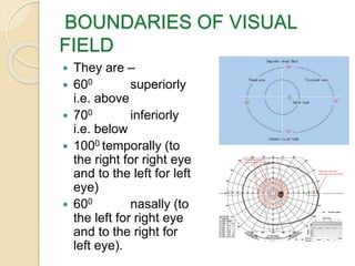
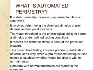
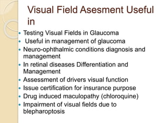
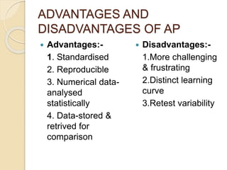
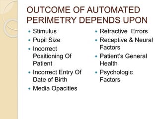
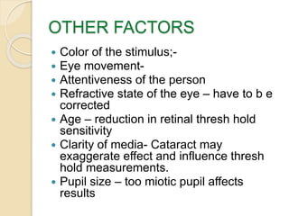
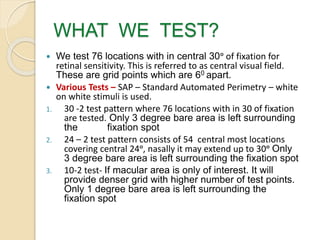
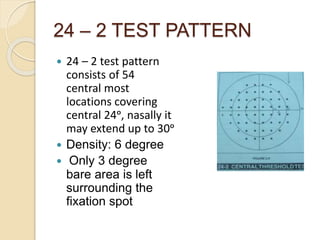
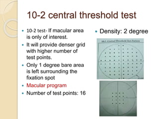
AUTOMATED PERIMETRY
DR. KISHOR BADHE
PROFESSOR & HOD
DEPT. OF OPHTHALMOLOGY
RURAL MEDICAL COLLEGE
PIMS, LONI
IMPORTANCE OF
PERIMETRY
 Evaluating the visual field is called as
perimetry.
 It is an important diagnostic tool in
glaucoma as well as in other conditions.
 Perimetry plays a crucial role in
diagnosing glaucoma and monitoring the
progression of the disease.
 Only perimetry can provide information
about the visual function which is crucial
for the patient.
WHAT IS VISUAL FIELD?
 It is island in the darkness of sea.
 When our eyes are directed straight forward,
we pay attention to the detail of the object at
which we are looking at.
 When doing this we are able to detect the
presence of other objects below, above and
to the sides of object of regard.
 Visual function is the ability to detect the
presence of any object in the field of vision
and to discern the details of an object when
we look directly at it.
HILL OF VISION
 It is also called as
the island of vision.
 It is three
dimensional graph of
visual sensitivity.
 The horizontal axis
represent locations
in the visual field.
 The altitude of the
hill (vertical direction)
at any location
represent the
sensitivity at that
location
 The acuity of vision
is sharpest at the top
of the hill
corresponding to
fovea and then
decreases to
periphery with nasal
slope steeper than
temporal
BOUNDARIES OF VISUAL
FIELD
 They are –
 600 superiorly
i.e. above
 700 inferiorly
i.e. below
 1000 temporally (to
the right for right eye
and to the left for left
eye)
 600 nasally (to
the left for right eye
and to the right for
left eye).
VISUAL CHARACTERISTICS IN
VISUAL FIELD
 Every point in visual
field has visual
characteristics of
visual function.
 Visual acuity is
greatest a point of
fixation.
 It is quantified in
terms of weakest
white spot or
stimulus visible in
each region of visual
field.
 The stimuli or white
spot can be adjusted
both in intensity and
size.
KINETIC PERIMETRY
 Stimuli is moved from a non seeing
area to seeing area along a set
meridian. Measurement is along X-Y
axis
 Aim is to find points in visual field of
equal retinal sensitivity.
 The perimeters are
 Lister Perimery
 Campimetry
 Goldman Perimatry
STATIC PERIMETRY
 Intensity of the stimuli at the same
predetermined spot is varied.
 Find out the theshhold at those
locations.
 More accurate than kinetic perimetry
 Gives 3 D picture of the hill of the
vision.
 Picks up field defects more accurately
KINETIC MANUAL
PERIMETRY
 Advantages:-
1.Tests full extent of
VF
2.Has a human
interface. Easy for
elderly & young
3.Better for
mapping shape of
defect
 Disadvantages:-
1.Requires highly
trained perimetrist
2.Not as
reproducible as
HVF
3.No numeric data
4.Numeric
comparison not
possible
WHAT IS AUTOMATED
PERIMETRY?
 It is static perimetry for measuring visual function out
side fovea.
 It involves determining the dimmest stimulus at pre-
determined test point locations.
 The visual threshold is the physiological ability to detect
a stimulus under defined testing conditions.
 It records the dimmest stimulus seen at the particular
location.
 This thresh hold testing involves precise quantification
of visual sensitivity, while supra threshold testing is used
mainly to establish whether visual function is with in
normal range.
 Compare with normal thresholds are stored in the
Visual Field Asesment Useful
in
 Testing Visual Fields in Glaucoma
 Useful in management of glaucoma
 Neuro-ophthalmic conditions diagnosis and
management
 In retinal diseases Differentiation and
Management
 Assessment of drivers visual function
 Issue certification for insurance purpose
 Drug induced maculopathy (chloroquine)
 Impairment of visual fields due to
blepharoptosis
ADVANTAGES AND
DISADVANTAGES OF AP
 Advantages:-
1. Standardised
2. Reproducible
3. Numerical data-
analysed
statistically
4. Data-stored &
retrived for
comparison
 Disadvantages:-
1.More challenging
& frustrating
2.Distinct learning
curve
3.Retest variability
OUTCOME OF AUTOMATED
PERIMETRY DEPENDS UPON
 Stimulus
 Pupil Size
 Incorrect
Positioning Of
Patient
 Incorrect Entry Of
Date of Birth
 Media Opacities
 Refractive Errors
 Receptive & Neural
Factors
 Patient’s General
Health
 Psychologic
Factors
IMPORTANT FACTORS
 Stimulus
 Stimulus Intensity
 Spot & Spot Size
 Background
Illumination
 Stimulus Duration
 Color of the
stimulus;
 Visual Threshold
 Fixation
 Movement;
 Stimulus – is spot of
light of predefined
size and intensity.
 The visibility of
stimulus depends
upon how far eye is
from screen and
background
illumination duration
and color of
stimulus and
background
INTENSITY OF STIMULUS
 Apostlib is an absolute measure of luminance
and is equal to 0.3183 candela m2 or 0.1
mililambert.
 The range of intensity of white light in perimeter
is in between 0.08 apostilbs and 10000 apostilbs
(asb) maximum brightness produced by
perimeter.
 The intensity expressed in log units. The
intensity can be varied over range of 5.1 log
units (51 decibels)
 The decibel value refers to retinal sensitivity
rather than stimulus intensity.
 0 db corresponds to maximum brightness
SENSITIVITY
 Lower the decibel value lower the
sensitivity; Higher the decibel value higher
the sensitivity
 At the point of fixation the eye is very
sensitive able to see the weakest stimulus.
Toward the periphery stronger illumination
is needed for the stimulus to be seen.
STIMULUS SIZE
 Stimulus Size - Goldman size III stimulus is
used. . It is 0.43 in diameter which is small
enough to have detailed examination and large
enough to do the field charting.
 It has the advantage that its visibility is less
affected by the refractive errors.
 It may scatter some light because of the size
but it does not matter for the other advantages.
 The large targets may be required in advanced
stage glaucoma
BACKGROUND ILLUMINATION
 It is important because visibility of stimulus
depends upon it.
 It determines the degree of light or dark adaptation
of the retina (photopic, Mesopic, or Scotopic).
 This influences whether or not dim stimuli are
seen.
 The background illumination is set at 31.5
apostilbs. This approximates the brightness of
photopic or day light vision which depends upon
retinal cone function rather than on rods.
 The advantage of testing photopic visual system is
that visibility depends upon object contrast rather
on absolute brightness as in rod vision.
STIMULUS DURATION
 The stimulus duration is 200
milliseconds (ms)
 It is shorter than the latency of the
voluntary eye movements (about 250
ms).
 The patient does not have the time to
see peripheral visual field and look
toward it.
 The visibility of stimulus increases with
duration.
 Visibility does not change after critical
rd
FIXATION
 The fixation of the patient is monitored either by
Heijl – Krakau Blind spot monitoring technique
or Gaze tracker.
 The positive response when stimuli is presented
in the blind spot indicate poor fixation the blind
spot is 60 in diameter so positive shift of 3-40 can
be detected.
 Gaze Tracker monitoring – measures gaze
tracking with precision of about 10 and record a
measurement each time the stimulus is
presented.
 At the bottom is line with up and down lines. The
down lines indicate it was unsuccessful in
measuring gaze direction during particular
stimulation
STIMULUS PRESENTATION
 The machine start with presenting
stimuli in each quadrant
 If stimulus is seen then it is dimmed by
3-4 decibels step by step till no longer
seen.
 If the stimuli is not seen initially then
the intensity is increased in steps until
the stimulus is seen.
 The testing is done in other test
locations until threshold sensitivities
are determined.
OTHER FACTORS
 Color of the stimulus;-
 Eye movement-
 Attentiveness of the person
 Refractive state of the eye – have to b e
corrected
 Age – reduction in retinal thresh hold
sensitivity
 Clarity of media- Cataract may
exaggerate effect and influence thresh
hold measurements.
 Pupil size – too miotic pupil affects
results
WHAT WE TEST?
 We test 76 locations with in central 30ᵒ of fixation for
retinal sensitivity. This is referred to as central visual field.
These are grid points which are 60 apart.
 Various Tests – SAP – Standard Automated Perimetry – white
on white stimuli is used.
1. 30 -2 test pattern where 76 locations with in 30 of fixation
are tested. Only 3 degree bare area is left surrounding
the fixation spot
2. 24 – 2 test pattern consists of 54 central most locations
covering central 24ᵒ, nasally it may extend up to 30ᵒ Only
3 degree bare area is left surrounding the fixation spot
3. 10-2 test- If macular area is only of interest. It will
provide denser grid with higher number of test points.
Only 1 degree bare area is left surrounding the
fixation spot
24 – 2 TEST PATTERN
 24 – 2 test pattern
consists of 54
central most
locations covering
central 24ᵒ, nasally it
may extend up to 30ᵒ
 Density: 6 degree
 Only 3 degree
bare area is left
surrounding the
fixation spot
10-2 central threshold test
 10-2 test- If macular area
is only of interest.
 It will provide denser grid
with higher number of
test points.
 Only 1 degree bare area
is left surrounding the
fixation spot
 Macular program
 Number of test points: 16
 Density: 2 degree
 Advantages of computer-
1. Random presentation of targets
2. Estimation of patient reliability
3. Reduced variability
4. Statistical evaluation of data at many
levels
5. Threshold strategies.
SWAP – Short Wavelength
Automated Perimetry
 Known as blue yellow perimetry -
Goldman V stimulus is presented on
the yellow background. The yellow
background reduces the
responsiveness of the red and green
cones so that the blue stimuli are
primarily seen by the blue cone
system.
 It is more sensitive than standard
perimetry.
 Can detect glaucomatous visual
field loss at earlier stages than white
on white perimetry. In this the
 It is used in detecting neuro-
ophthalmic diseases, ARMD,
Diabetic Macular Edema, and
Migraine.
BASIC COMPONENTS OF
PERIMETER
 Perimetric Unit –
bowl type of screen
 Control Unit –
Computer, dialogue
screen, key board or
light pen,. It monitors
instrumentation
function according to
perimetrist request.
Has printer & record
information in
computer
PROCEDURE - TECHNIQUE
 Tell the patient what
the test is for?
 Show how the
stimulus will look
like.
 Where it may appear
 How long the test will
last
 How to sit
 When to blink.
 How to pause test
 Have positive
attitude
 Normal distance is
30 cms
 Fully presbiopic
patients are provided
by +3.25 D
correction
 Refractive blur
corrected
 Avoid testing fields
with spectacles
 One eye tested at
one time
 Patch the other eye
TESTING STRATEGY
 Full threshold –
 Threshold
 Suprathreshold
SUPRA THRESHOLD
 Staircase method (4-2 bracketing
strategy)
 Used to detect threshold
 Intensity of stimulus is decreased in 4-
db step till stimulus is no longer seen
 Increasing the stimulus in 2-db step till
stimulus is seen again
 Threshold perimetry
 Threshold found at predetermined points
 Time consuming process
 Supra-threshold perimetry
 Intensity of stimulus shown at a spot
much higher than threshold at that spot
 Mainly for screening & Picks up gross
visual defects
 Fastpac:
 Decreases the test time by 40%
 3-db increment instead of 4-db
 Threshold crossed only once
THANKS

Automated perimetry

  • 1.
    AUTOMATED PERIMETRY DR. KISHORBADHE PROFESSOR & HOD DEPT. OF OPHTHALMOLOGY RURAL MEDICAL COLLEGE PIMS, LONI
  • 2.
    IMPORTANCE OF PERIMETRY  Evaluatingthe visual field is called as perimetry.  It is an important diagnostic tool in glaucoma as well as in other conditions.  Perimetry plays a crucial role in diagnosing glaucoma and monitoring the progression of the disease.  Only perimetry can provide information about the visual function which is crucial for the patient.
  • 3.
    WHAT IS VISUALFIELD?  It is island in the darkness of sea.  When our eyes are directed straight forward, we pay attention to the detail of the object at which we are looking at.  When doing this we are able to detect the presence of other objects below, above and to the sides of object of regard.  Visual function is the ability to detect the presence of any object in the field of vision and to discern the details of an object when we look directly at it.
  • 4.
    HILL OF VISION It is also called as the island of vision.  It is three dimensional graph of visual sensitivity.  The horizontal axis represent locations in the visual field.  The altitude of the hill (vertical direction) at any location represent the sensitivity at that location  The acuity of vision is sharpest at the top of the hill corresponding to fovea and then decreases to periphery with nasal slope steeper than temporal
  • 5.
    BOUNDARIES OF VISUAL FIELD They are –  600 superiorly i.e. above  700 inferiorly i.e. below  1000 temporally (to the right for right eye and to the left for left eye)  600 nasally (to the left for right eye and to the right for left eye).
  • 6.
    VISUAL CHARACTERISTICS IN VISUALFIELD  Every point in visual field has visual characteristics of visual function.  Visual acuity is greatest a point of fixation.  It is quantified in terms of weakest white spot or stimulus visible in each region of visual field.  The stimuli or white spot can be adjusted both in intensity and size.
  • 7.
    KINETIC PERIMETRY  Stimuliis moved from a non seeing area to seeing area along a set meridian. Measurement is along X-Y axis  Aim is to find points in visual field of equal retinal sensitivity.  The perimeters are  Lister Perimery  Campimetry  Goldman Perimatry
  • 8.
    STATIC PERIMETRY  Intensityof the stimuli at the same predetermined spot is varied.  Find out the theshhold at those locations.  More accurate than kinetic perimetry  Gives 3 D picture of the hill of the vision.  Picks up field defects more accurately
  • 9.
    KINETIC MANUAL PERIMETRY  Advantages:- 1.Testsfull extent of VF 2.Has a human interface. Easy for elderly & young 3.Better for mapping shape of defect  Disadvantages:- 1.Requires highly trained perimetrist 2.Not as reproducible as HVF 3.No numeric data 4.Numeric comparison not possible
  • 10.
    WHAT IS AUTOMATED PERIMETRY? It is static perimetry for measuring visual function out side fovea.  It involves determining the dimmest stimulus at pre- determined test point locations.  The visual threshold is the physiological ability to detect a stimulus under defined testing conditions.  It records the dimmest stimulus seen at the particular location.  This thresh hold testing involves precise quantification of visual sensitivity, while supra threshold testing is used mainly to establish whether visual function is with in normal range.  Compare with normal thresholds are stored in the
  • 11.
    Visual Field AsesmentUseful in  Testing Visual Fields in Glaucoma  Useful in management of glaucoma  Neuro-ophthalmic conditions diagnosis and management  In retinal diseases Differentiation and Management  Assessment of drivers visual function  Issue certification for insurance purpose  Drug induced maculopathy (chloroquine)  Impairment of visual fields due to blepharoptosis
  • 12.
    ADVANTAGES AND DISADVANTAGES OFAP  Advantages:- 1. Standardised 2. Reproducible 3. Numerical data- analysed statistically 4. Data-stored & retrived for comparison  Disadvantages:- 1.More challenging & frustrating 2.Distinct learning curve 3.Retest variability
  • 13.
    OUTCOME OF AUTOMATED PERIMETRYDEPENDS UPON  Stimulus  Pupil Size  Incorrect Positioning Of Patient  Incorrect Entry Of Date of Birth  Media Opacities  Refractive Errors  Receptive & Neural Factors  Patient’s General Health  Psychologic Factors
  • 14.
    IMPORTANT FACTORS  Stimulus Stimulus Intensity  Spot & Spot Size  Background Illumination  Stimulus Duration  Color of the stimulus;  Visual Threshold  Fixation  Movement;  Stimulus – is spot of light of predefined size and intensity.  The visibility of stimulus depends upon how far eye is from screen and background illumination duration and color of stimulus and background
  • 15.
    INTENSITY OF STIMULUS Apostlib is an absolute measure of luminance and is equal to 0.3183 candela m2 or 0.1 mililambert.  The range of intensity of white light in perimeter is in between 0.08 apostilbs and 10000 apostilbs (asb) maximum brightness produced by perimeter.  The intensity expressed in log units. The intensity can be varied over range of 5.1 log units (51 decibels)  The decibel value refers to retinal sensitivity rather than stimulus intensity.  0 db corresponds to maximum brightness
  • 16.
    SENSITIVITY  Lower thedecibel value lower the sensitivity; Higher the decibel value higher the sensitivity  At the point of fixation the eye is very sensitive able to see the weakest stimulus. Toward the periphery stronger illumination is needed for the stimulus to be seen.
  • 17.
    STIMULUS SIZE  StimulusSize - Goldman size III stimulus is used. . It is 0.43 in diameter which is small enough to have detailed examination and large enough to do the field charting.  It has the advantage that its visibility is less affected by the refractive errors.  It may scatter some light because of the size but it does not matter for the other advantages.  The large targets may be required in advanced stage glaucoma
  • 18.
    BACKGROUND ILLUMINATION  Itis important because visibility of stimulus depends upon it.  It determines the degree of light or dark adaptation of the retina (photopic, Mesopic, or Scotopic).  This influences whether or not dim stimuli are seen.  The background illumination is set at 31.5 apostilbs. This approximates the brightness of photopic or day light vision which depends upon retinal cone function rather than on rods.  The advantage of testing photopic visual system is that visibility depends upon object contrast rather on absolute brightness as in rod vision.
  • 19.
    STIMULUS DURATION  Thestimulus duration is 200 milliseconds (ms)  It is shorter than the latency of the voluntary eye movements (about 250 ms).  The patient does not have the time to see peripheral visual field and look toward it.  The visibility of stimulus increases with duration.  Visibility does not change after critical rd
  • 20.
    FIXATION  The fixationof the patient is monitored either by Heijl – Krakau Blind spot monitoring technique or Gaze tracker.  The positive response when stimuli is presented in the blind spot indicate poor fixation the blind spot is 60 in diameter so positive shift of 3-40 can be detected.  Gaze Tracker monitoring – measures gaze tracking with precision of about 10 and record a measurement each time the stimulus is presented.  At the bottom is line with up and down lines. The down lines indicate it was unsuccessful in measuring gaze direction during particular stimulation
  • 21.
    STIMULUS PRESENTATION  Themachine start with presenting stimuli in each quadrant  If stimulus is seen then it is dimmed by 3-4 decibels step by step till no longer seen.  If the stimuli is not seen initially then the intensity is increased in steps until the stimulus is seen.  The testing is done in other test locations until threshold sensitivities are determined.
  • 22.
    OTHER FACTORS  Colorof the stimulus;-  Eye movement-  Attentiveness of the person  Refractive state of the eye – have to b e corrected  Age – reduction in retinal thresh hold sensitivity  Clarity of media- Cataract may exaggerate effect and influence thresh hold measurements.  Pupil size – too miotic pupil affects results
  • 23.
    WHAT WE TEST? We test 76 locations with in central 30ᵒ of fixation for retinal sensitivity. This is referred to as central visual field. These are grid points which are 60 apart.  Various Tests – SAP – Standard Automated Perimetry – white on white stimuli is used. 1. 30 -2 test pattern where 76 locations with in 30 of fixation are tested. Only 3 degree bare area is left surrounding the fixation spot 2. 24 – 2 test pattern consists of 54 central most locations covering central 24ᵒ, nasally it may extend up to 30ᵒ Only 3 degree bare area is left surrounding the fixation spot 3. 10-2 test- If macular area is only of interest. It will provide denser grid with higher number of test points. Only 1 degree bare area is left surrounding the fixation spot
  • 24.
    24 – 2TEST PATTERN  24 – 2 test pattern consists of 54 central most locations covering central 24ᵒ, nasally it may extend up to 30ᵒ  Density: 6 degree  Only 3 degree bare area is left surrounding the fixation spot
  • 25.
    10-2 central thresholdtest  10-2 test- If macular area is only of interest.  It will provide denser grid with higher number of test points.  Only 1 degree bare area is left surrounding the fixation spot  Macular program  Number of test points: 16  Density: 2 degree
  • 26.
     Advantages ofcomputer- 1. Random presentation of targets 2. Estimation of patient reliability 3. Reduced variability 4. Statistical evaluation of data at many levels 5. Threshold strategies.
  • 27.
    SWAP – ShortWavelength Automated Perimetry  Known as blue yellow perimetry - Goldman V stimulus is presented on the yellow background. The yellow background reduces the responsiveness of the red and green cones so that the blue stimuli are primarily seen by the blue cone system.  It is more sensitive than standard perimetry.  Can detect glaucomatous visual field loss at earlier stages than white on white perimetry. In this the  It is used in detecting neuro- ophthalmic diseases, ARMD, Diabetic Macular Edema, and Migraine.
  • 28.
    BASIC COMPONENTS OF PERIMETER Perimetric Unit – bowl type of screen  Control Unit – Computer, dialogue screen, key board or light pen,. It monitors instrumentation function according to perimetrist request. Has printer & record information in computer
  • 29.
    PROCEDURE - TECHNIQUE Tell the patient what the test is for?  Show how the stimulus will look like.  Where it may appear  How long the test will last  How to sit  When to blink.  How to pause test  Have positive attitude  Normal distance is 30 cms  Fully presbiopic patients are provided by +3.25 D correction  Refractive blur corrected  Avoid testing fields with spectacles  One eye tested at one time  Patch the other eye
  • 30.
    TESTING STRATEGY  Fullthreshold –  Threshold  Suprathreshold
  • 31.
    SUPRA THRESHOLD  Staircasemethod (4-2 bracketing strategy)  Used to detect threshold  Intensity of stimulus is decreased in 4- db step till stimulus is no longer seen  Increasing the stimulus in 2-db step till stimulus is seen again
  • 32.
     Threshold perimetry Threshold found at predetermined points  Time consuming process  Supra-threshold perimetry  Intensity of stimulus shown at a spot much higher than threshold at that spot  Mainly for screening & Picks up gross visual defects  Fastpac:  Decreases the test time by 40%  3-db increment instead of 4-db  Threshold crossed only once
  • 33.

Editor's Notes

  • #4 What is Visual Field? It is island in the darkness of sea. When our eyes are directed straight forward, we pay attention to the detail of the object at which we are looking at. When doing this we are able to detect the presence of other objects below, above and to the sides of object of regard. Visual function is the ability to detect the presence of any object in the filed of vision and to discern the details of an object when we look directly at it.
  • #5 Hill of Vision – It is also called as the island of vision in the darkness of sea. . It is three dimensional structure of visual sensitivity. The horizontal axis represent locations in the visual field. The altitude of the hill (vertical direction) at any location represent the sensitivity at that location. The acuity of vision is sharpest at the top of the hill corresponding to fovea and then decreases to periphery with nasal slope steeper than temporal
  • #7 Visual Characteristics in Visual Filed -- Every point in the visual field has certain characteristics of visual function. The visual acuity is greatest at point of fixation. To find the visual acuity of other points ask the patient to fix at certain point and placing the chart at selected locations say at 100 temporally, 200 nasally or 300 superiorly or inferiorly and so on. The smallest letter which the patient is able to read at on the chart is the VA of that point. In practice it is quantified in terms of weakest white spot that can be seen in each region of the visual field. The stimuli in projection perimeters are the white spots that can be adjusted both in intensity and size.  
  • #11 What is automated perimetry?— It is static and most important clinical tool for measuring visual function out side fovea. It involves determining the dimmest stimulus at predetermined test point locations. The visual threshold is the physiological ability to detect a stimulus under defined testing conditions. The dimmest stimulus seen at the particular location is recorded. This thresh hold testing involves precise quantification of visual sensitivity, while supra threshold testing is used mainly to establish whether visual function is with in normal range. The normal thresholds are stored in the computer and against these the patient’s values are compared. The machines compare the patient’s sensitivity against this data.
  • #13 Advantages and Disadvantages of AP Advantages:- 1. Standardised 2. Reproducible 3. Numerical data- analysed statistically 4. Data-stored & retrived for comparison
  • #14 Outcome of Automated Perimetry depends upon: Stimulus Pupil Size Incorrect Positioning Of Patient Incorrect Entry Of Date Of Birth Media Opacities Refractive Errors Receptive & Neural Factors Patient’s General Health Psychologic Factors
  • #16 In the center portion of the visual field near the point of fixation the eye is very sensitive able to see the weakest stimulus. Toward the periphery stronger illumination is needed for the stimulus to be seen. The apostilb is the unit of luminance used in perimeter and is equal to 0.3183 candela m2 or 0.1 mililambert. Decibel is a measure of sensitivity of retina  Inversely proportional  It is a relative measure varies from machine to machine
  • #18 The mean retinal sensitivity increases with increase in size or luminosity relative to that of background. If the diameter of stimulus is increased it is visible as less intense larger stimulus. This is due to spatial summation.
  • #19 Background Illumination – it is important because a stimulus of certain intensity will be less visible against light background than against the dark one. It determines the degree of light or dark adaptation of the retina (photopic, Mesopic, or Scotopic). This influences whether or not dim stimuli are seen. The background illumination is set at 31.5 apostilbs. This approximates the brightness of photopic or day light vision. This is the vision which depends upon retinal cone function rather than on rods. The advantage of testing photopic visual system is that the visibility depends upon object contrast rather on absolute brightness as in rod vision.
  • #20 Stimulus Duration -- The stimulus duration is 200 milliseconds (ms). This is long enough for the visibility to be affected by small variations and durations. It is shorter than the latency of the voluntary eye movements (about 250 ms). Thus the patient does not have the time to see peripheral visual field and look toward it. It is easy to see a spot shown for longer time Duration – important for brief flash (1/100 second v/s 2/100 second) but the duration does not change the visibility of the stimulus after critical time – after 1/3 second or longer.
  • #21 Fixation -- The fixation of the patient is monitored either by Heijl – Krakau Blind spot monitoring technique or Gaze tracker. It provides an index of quality of patient fixation during an examination by periodically presenting stimuli in the blind spot. The positive response when stimuli is presented in the blind spot indicate poor fixation the blind spot is 60 in diameter so positive shift of 3-40 can be detected. Gaze Tracker monitoring – measures gaze tracking with precision of about 10 and record a measurement each time the stimulus is presented.
  • #23 Refractive errors- Myopia of 1 D does not affect how ever large errors need to be corrected and hyperopia alters thresh hold sensitivity
  • #26 Macular programme  Number of test points: 16  Density: 2 degree  Only 1 degree bare area is left surrounding the fixation spot
  • #28 SWAP – Short Wavelength Automated Perimetry – also known as blue yellow perimetry can detect glaucomatous visual field loss at earlier stages than white on white perimetry. In this the Goldman V stimulus is presented on the yellow background. The yellow background reduces the responsiveness of the red and green cones so that the blue stimuli are primarily seen by the blue cone system. It is more sensitive than standard perimetry. It is used in detecting neuro-ophthalmic diseases, ARMD, Diabetic Macular Edema, and Migraine.
  • #33 Sita standard:  Takes half time than full threshold method  Sita fast:  Takes half time than fast pac threshold method