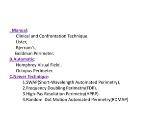
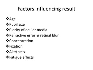
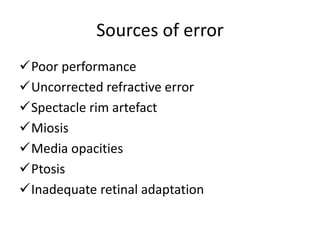
Visual Field Testing(Peripheral)
Mahir Faiyaz
B.Optom 3rd Year
7th Batch,ICO
Visual Field
• The visual field is the total area in which objects can be
seen in the peripheral vision while the eye is focused on a
central point
• The most peripheral portions of the visual field are least
sensitive ; objects must be large and bright to be visualized.
• Outside the borders of the visual field, no visual perception
occurs
Few Terminologies
Fixation
Central & Peripheral visual field
Isopter
Threshold
Depression
Scotoma
Fluctuation
Perimetry
Kinetic Perimetry
• In kinetic perimetry,stimulus is presented in
the non seeing periphery & moved towards
the point of fixation until the patient first
perceives it.
• The procedure is repeated with the use of
same stimulus along other meridians,usually
spaced every 15degrees.
• By joining these areas an isopter is formed.
Static Perimetry
• A stimulus is presented at a known location
for a known duration with varying luminance
to find local threshold.
• The stimulus is not moved as in kinetic
perimetry.
• This is a stationary target.
. Manual:
Clinical and Confrontation Technique.
Lister,
Bjerrum’s,
Goldman Perimeter.
B.Automatic:
Humphrey Visual Field .
Octopus Perimeter.
C.Newer Technique:
1.SWAP(Short-Wavelength Automated Perimetry).
2.Frequency Doubling Perimetry(FDP).
3.High-Pas Resolution Perimetry(HPRP).
4.Random Dot Motion Automated Perimetry(RDMAP)
CONFRONTATION TECHNIQUE
• Preparing the patient for the test:
• Examiner and patient should sit facing each
other about 1m apart.
• Test should be in good lighting which comes
from behind the patient and uniformly
illuminates the patient’s visual field.
• Plain background should be in front of the
patient , to allow detection of the stimulus
more reliably.
• Patient is instructed to look directly into the
examiner’s eye.
• When testing the right eye , the patient’s left
eye should be occluded.
• The visual field of the patient’s right eye will
correspond to the visual field of the
examiner’s left eye.
Indications
Field examination by neurologists on the bed
side.
By optometrists &ophthalmologists as a rapid
screening visual field test.
Only way to evaluate visual field in bedridden
patients
Patient with lack of attention & morbid
patient who cannot undergo longer test.
Disadvantages
Less sensitive
It gives qualatative assessment not
quantative.
Examiner dependent.
Apostilb & Decibel
Luminance can be indicated in units called as
apostilb(asb).
Apostilb is an absolute value & remain constant
from machine to machine.
Decibel scale is a logarithmic scale that is
reciprocally related to the intensity of light.
asb & dB values go in opposite directions.
In humphrey test,
-10asb=30dB
-100asb=20dB.
AUTOMATED PERIMETRY VARIABLES
• Stimulus Size:
• *5 Sizes(I-V),
• *Stimulus Size III is Commonly Used.
• *Distance from Eye 30 cm.
• Stimulus Duration:0.2 Sec
• Stimulus intensity: 0(max) to 50(min) dB
• Background illumination:31.5asb
Procedures
• Setting up the perimeter
– Enter patient data and examination data
– Typing error in the birth year results in a uniform artifact depression
– Calibrate the unit
– Apply the trial lenses before seating the patient
• Instructing the patient
– Maintain comfortable position. Do not move
– Always Look straight ahead at the fixation target
– Blink regularly to avoid discomfort
– Press the button only when the stimulus is seen as not all
stimuli are visible
• Instructing the patient
– If uncomfortable, may close eye for a moment. The test is
interrupted and only continues after eye opens
-To ask for assistance, keep the button pressed
• Setting up the patient
– The patient seated comfortably on a height-adjustable
chair with a back rest
– The patient is asked to press the button a few times and
occluder is applied making sure the patient can blink freely
• Refractive errors
– If patient does not have sharp image of the
stimulus, the d.l. sensitivity values are reduced
-For direct projection type perimeters (OCTOPUS
300) far correction is required
-For cupola perimeter near correction (42.5cm) is
needed
• Refraction and trial lenses
– Cylinder correction
• Screening:
2 Strategies:
– Single-level suprathreshold test
– 2-Level suprathreshold test
Single-level suprathreshold test
• Stimulus 2-6 dB higher than threshold or expected hill
of vision
• Result: seen (normal) or nor seen (defect)
 2-Level suprathreshold test
• 3 categories: Normal, relative defect, absolute defect
• Done as per single-level but abnormal spots are tested
with 0 dB attenuation: Threshold-Related Strategy
(Humphrey
Factors influencing result
Age
Pupil size
Clarity of ocular media
Refractive error & retinal blur
Concentration
Fixation
Alertness
Fatigue effects
THRESHOLD STRATEGY
1.Suprathreshold Perimetry
2.Threshold Perimetry-
a.Threshold:
Full Threshold-30-2(Total Point 76,Field 30)
24-2(Total Point 54.Field 24)
10-2(Total Point 68,Field 10)
Macular(Total Point 16,Field 5).
Full Field(120 Degree).
b.Fast pac
c.SITA fast
d.SITA Standard
Supra threshold Static Perimetry
Stimulus brighter than the anticipated normal
value for the corresponding retinal locations.
Mainly used for screening purposes.
Only provide information about depth &
contour of field defect.
Threshold Perimetry
Full threshold periemtry
• Measure the retinal threshold at 70-80 points
within the central 24-30degrees.
• Determines the threshold value at each point by
the bracketing technique
• Appropriate for a patients first test as it helps in
determining the future data points.
ADVANTAGES OVER MANUAL
PERIMETRY
• 1. STANDARDIZATION OF TEST CONDITION
• 2. ESTIMATION OF PATIENT RELIABILITY
• 3. COMPUTERIZED ANALYSIS OF FIELD
• 4. EASY FOLLOW-UP
Sources of error
Poor performance
Uncorrected refractive error
Spectacle rim artefact
Miosis
Media opacities
Ptosis
Inadequate retinal adaptation
Printout
Reproductibility Stage
Reliability Indices
Gray Scale
Total Deviation Plot
Pattern Deviation Plot
Global Indices
GHT
Gaze Tracker
Reliability indices
• False Positive (FP):
– Sound cue without light source or subthreshold
stimulus
– SITA: FP = response within the patient’s response time
• False Negative (FN):
– No response to a 9 dB brighter stimulus at a location
that had a measurable threshold earlier.
– “Clover leaf” pattern with fatigue
• Fixation Losses (FL):
– Heijl-Krakau method of periodic testing of the
blind spot
– Video monitor ± IR camera to detect pupil
movement
– Pseudofixation losses
Grey scale
• It provides only a semiquantitative
assessment of the visual field, with
interpolated values printed between tested
points, and are insufficient for full analysis.
• Gives the idea of retinal threshold at a glance.
– Darker means lower retinal sensitivity
– Alarming sign Beware!!
Total Deviation Plot
• Represents the deviation of the patient’s result from the
age matched controls.
• The upper numerical display illustrates the difference in
decibel and the lower display exhibits these differences in
grey symbols.
• Deviation from age matched controls even less than 4
decibel is shown.(differ. from Octopus)
PATTERN DEVIATION PLOT
Derived from the total deviation plot
Gives an idea about the resemblance of
patients visual field to the shape of the hill of
vision.
Localized defects
Global indices
 MD
 PSD
 SF
 CPSD
 VFI
GHT
• 5 sectors in the upper field are compared to
five mirror images in the lower
• If value in two sectors differ to an extent that
found in
– <0.5% of the normal population ( highly sensitive)
– <1% of normal population (outside normal limit)
– <3% of the normal population (Boderline)
– <5% of the normal population ( can be a normal
plot)
Criteria
Pattern deviation plot
- ≥3non edge points with p>5%
-one point with p>1%
-Cluster in arcuate area
CPSD or PSD
-depressed with p>5%
Abnormal GHT
Recent advances in automated
perimetry
• Goldman kinetic module
• High-pass resolution perimetry
• Short wavelenght sensitive perimetry
• Flicker perimetry
• Motion perimetry
• Rarebit perimetry
• Pupil perimetry
• Multifocal VEP
Thanks for
your
Attention

Peripheral visual field

  • 1.
    Visual Field Testing(Peripheral) MahirFaiyaz B.Optom 3rd Year 7th Batch,ICO
  • 2.
    Visual Field • Thevisual field is the total area in which objects can be seen in the peripheral vision while the eye is focused on a central point • The most peripheral portions of the visual field are least sensitive ; objects must be large and bright to be visualized. • Outside the borders of the visual field, no visual perception occurs
  • 3.
    Few Terminologies Fixation Central &Peripheral visual field Isopter Threshold Depression Scotoma Fluctuation Perimetry
  • 4.
    Kinetic Perimetry • Inkinetic perimetry,stimulus is presented in the non seeing periphery & moved towards the point of fixation until the patient first perceives it. • The procedure is repeated with the use of same stimulus along other meridians,usually spaced every 15degrees. • By joining these areas an isopter is formed.
  • 5.
    Static Perimetry • Astimulus is presented at a known location for a known duration with varying luminance to find local threshold. • The stimulus is not moved as in kinetic perimetry. • This is a stationary target.
  • 6.
    . Manual: Clinical andConfrontation Technique. Lister, Bjerrum’s, Goldman Perimeter. B.Automatic: Humphrey Visual Field . Octopus Perimeter. C.Newer Technique: 1.SWAP(Short-Wavelength Automated Perimetry). 2.Frequency Doubling Perimetry(FDP). 3.High-Pas Resolution Perimetry(HPRP). 4.Random Dot Motion Automated Perimetry(RDMAP)
  • 7.
    CONFRONTATION TECHNIQUE • Preparingthe patient for the test: • Examiner and patient should sit facing each other about 1m apart. • Test should be in good lighting which comes from behind the patient and uniformly illuminates the patient’s visual field. • Plain background should be in front of the patient , to allow detection of the stimulus more reliably.
  • 8.
    • Patient isinstructed to look directly into the examiner’s eye. • When testing the right eye , the patient’s left eye should be occluded. • The visual field of the patient’s right eye will correspond to the visual field of the examiner’s left eye.
  • 9.
    Indications Field examination byneurologists on the bed side. By optometrists &ophthalmologists as a rapid screening visual field test. Only way to evaluate visual field in bedridden patients Patient with lack of attention & morbid patient who cannot undergo longer test.
  • 10.
    Disadvantages Less sensitive It givesqualatative assessment not quantative. Examiner dependent.
  • 11.
    Apostilb & Decibel Luminancecan be indicated in units called as apostilb(asb). Apostilb is an absolute value & remain constant from machine to machine. Decibel scale is a logarithmic scale that is reciprocally related to the intensity of light. asb & dB values go in opposite directions. In humphrey test, -10asb=30dB -100asb=20dB.
  • 12.
    AUTOMATED PERIMETRY VARIABLES •Stimulus Size: • *5 Sizes(I-V), • *Stimulus Size III is Commonly Used. • *Distance from Eye 30 cm. • Stimulus Duration:0.2 Sec • Stimulus intensity: 0(max) to 50(min) dB • Background illumination:31.5asb
  • 13.
    Procedures • Setting upthe perimeter – Enter patient data and examination data – Typing error in the birth year results in a uniform artifact depression – Calibrate the unit – Apply the trial lenses before seating the patient • Instructing the patient – Maintain comfortable position. Do not move – Always Look straight ahead at the fixation target – Blink regularly to avoid discomfort – Press the button only when the stimulus is seen as not all stimuli are visible
  • 14.
    • Instructing thepatient – If uncomfortable, may close eye for a moment. The test is interrupted and only continues after eye opens -To ask for assistance, keep the button pressed • Setting up the patient – The patient seated comfortably on a height-adjustable chair with a back rest – The patient is asked to press the button a few times and occluder is applied making sure the patient can blink freely
  • 15.
    • Refractive errors –If patient does not have sharp image of the stimulus, the d.l. sensitivity values are reduced -For direct projection type perimeters (OCTOPUS 300) far correction is required -For cupola perimeter near correction (42.5cm) is needed • Refraction and trial lenses – Cylinder correction
  • 16.
    • Screening: 2 Strategies: –Single-level suprathreshold test – 2-Level suprathreshold test
  • 17.
    Single-level suprathreshold test •Stimulus 2-6 dB higher than threshold or expected hill of vision • Result: seen (normal) or nor seen (defect)  2-Level suprathreshold test • 3 categories: Normal, relative defect, absolute defect • Done as per single-level but abnormal spots are tested with 0 dB attenuation: Threshold-Related Strategy (Humphrey
  • 18.
    Factors influencing result Age Pupilsize Clarity of ocular media Refractive error & retinal blur Concentration Fixation Alertness Fatigue effects
  • 19.
    THRESHOLD STRATEGY 1.Suprathreshold Perimetry 2.ThresholdPerimetry- a.Threshold: Full Threshold-30-2(Total Point 76,Field 30) 24-2(Total Point 54.Field 24) 10-2(Total Point 68,Field 10) Macular(Total Point 16,Field 5). Full Field(120 Degree). b.Fast pac c.SITA fast d.SITA Standard
  • 20.
    Supra threshold StaticPerimetry Stimulus brighter than the anticipated normal value for the corresponding retinal locations. Mainly used for screening purposes. Only provide information about depth & contour of field defect.
  • 21.
    Threshold Perimetry Full thresholdperiemtry • Measure the retinal threshold at 70-80 points within the central 24-30degrees. • Determines the threshold value at each point by the bracketing technique • Appropriate for a patients first test as it helps in determining the future data points.
  • 23.
    ADVANTAGES OVER MANUAL PERIMETRY •1. STANDARDIZATION OF TEST CONDITION • 2. ESTIMATION OF PATIENT RELIABILITY • 3. COMPUTERIZED ANALYSIS OF FIELD • 4. EASY FOLLOW-UP
  • 24.
    Sources of error Poorperformance Uncorrected refractive error Spectacle rim artefact Miosis Media opacities Ptosis Inadequate retinal adaptation
  • 25.
    Printout Reproductibility Stage Reliability Indices GrayScale Total Deviation Plot Pattern Deviation Plot Global Indices GHT Gaze Tracker
  • 26.
    Reliability indices • FalsePositive (FP): – Sound cue without light source or subthreshold stimulus – SITA: FP = response within the patient’s response time • False Negative (FN): – No response to a 9 dB brighter stimulus at a location that had a measurable threshold earlier. – “Clover leaf” pattern with fatigue
  • 27.
    • Fixation Losses(FL): – Heijl-Krakau method of periodic testing of the blind spot – Video monitor ± IR camera to detect pupil movement – Pseudofixation losses
  • 28.
    Grey scale • Itprovides only a semiquantitative assessment of the visual field, with interpolated values printed between tested points, and are insufficient for full analysis. • Gives the idea of retinal threshold at a glance. – Darker means lower retinal sensitivity – Alarming sign Beware!!
  • 30.
    Total Deviation Plot •Represents the deviation of the patient’s result from the age matched controls. • The upper numerical display illustrates the difference in decibel and the lower display exhibits these differences in grey symbols. • Deviation from age matched controls even less than 4 decibel is shown.(differ. from Octopus)
  • 31.
    PATTERN DEVIATION PLOT Derivedfrom the total deviation plot Gives an idea about the resemblance of patients visual field to the shape of the hill of vision. Localized defects
  • 32.
    Global indices  MD PSD  SF  CPSD  VFI
  • 34.
    GHT • 5 sectorsin the upper field are compared to five mirror images in the lower • If value in two sectors differ to an extent that found in – <0.5% of the normal population ( highly sensitive) – <1% of normal population (outside normal limit) – <3% of the normal population (Boderline) – <5% of the normal population ( can be a normal plot)
  • 37.
    Criteria Pattern deviation plot -≥3non edge points with p>5% -one point with p>1% -Cluster in arcuate area CPSD or PSD -depressed with p>5% Abnormal GHT
  • 38.
    Recent advances inautomated perimetry • Goldman kinetic module • High-pass resolution perimetry • Short wavelenght sensitive perimetry • Flicker perimetry • Motion perimetry • Rarebit perimetry • Pupil perimetry • Multifocal VEP
  • 39.