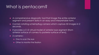
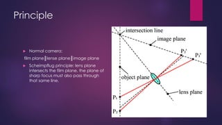
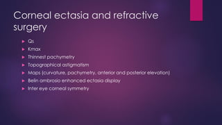
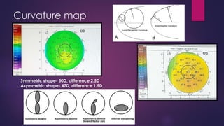
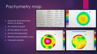
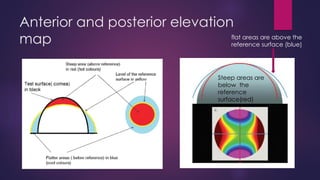
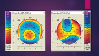
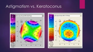
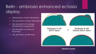
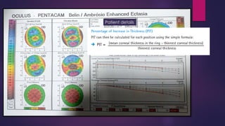
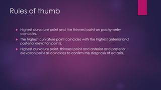
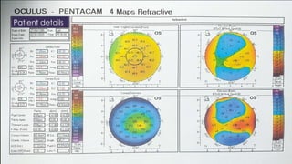
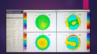
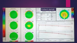
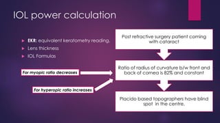
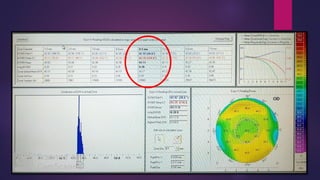
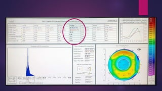
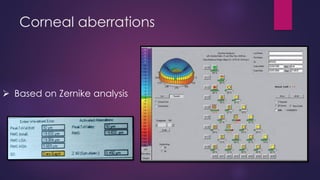
PENTACAM
SIMI AFROZ
Assistant Professor
Department of optometry
School of Medical and Allied Health Sciences
G D GOENKA UNIVERSITY
B.Optom (A.I.I.M.S.), M.Optom (Amity University)
What is pentacam?
 A comprehensive diagnostic tool that image the entire anterior
segment and present data in an easy and interpretable form.
 Contain rotating scheimpflug camera which capture 50 images in 2
seconds.
 It provides a 3 -D virtual model of anterior eye segment (from
anterior surface of cornea to posterior surface of lens).
 2 camera:-
 One to scan the eye
 Other to monitor the fixation
Principle
 Normal camera:
film plane║lense plane║image plane
 Scheimpflug principle: lens plane
intersects the film plane, the plane of
sharp focus must also pass through
that same line.
Applications
 Corneal ectasia
 Refractive surgery
 Corrected IOP
 Corneal aberrations
 IOL power calculation
 Densitometry
Corneal ectasia and refractive
surgery
 Qs
 Kmax
 Thinnest pachymetry
 Topographical astigmatism
 Maps (curvature, pachymetry, anterior and posterior elevation)
 Belin ambrosio enhanced ectasia display
 Inter eye corneal symmetry
Curvature map
Symmetric shape- 50D, difference 2.5D
Asymmetric shape- 47D, difference 1.5D
Pachymetry map
 Measure thickness from
limbus to limbus.
 At centre of pupil.
 At the apex of cone.
 At the thinnest point.
 Relative pachymetry map
 Corneal volume.
Anterior and posterior elevation
map
Steep areas are
below the
reference
surface(red)
flat areas are above the
reference surface (blue)
Astigmatism vs. Keratoconus
Belin - ambrosio enhanced ectasia
display
 Df(deviation of front elevation)
 Db( deviation of back elevation)
 Dp( deviation of average
pachemetry progression)
 Dt( deviation of minimum
thickness)
 Da( deviation of ART Max)
Normal cornea
Before & After Exclusion Software
Patient details
Rules of thumb
 Highest curvature point and the thinnest point on pachymetry
coincides.
 The highest curvature point coincides with the highest anterior and
posterior elevation points.
 Highest curvature point, thinnest point and anterior and posterior
elevation point all coincides to confirm the diagnosis of ectasia.
Patient details
Patient details
Patient details
Patient details
Patient details
IOL power calculation
 EKR: equivalent keratometry reading.
 Lens thickness
 IOL Formulas
Placido based topographers have blind
spot in the centre.
Ratio of radius of curvature b/w front and
back of cornea is 82% and constant
Post refractive surgery patient coming
with cataract
For myopic ratio decreases
For hyperopic ratio increases
Corneal aberrations
➢ Based on Zernike analysis
SIMI AFROZ
Assistant Professor (ADHOC)
Department of optometry
School of Allied Health Sciences and Research
SHARDA UNIVERSITY
B.Optom (A.I.I.M.S.), M.Optom (Amity University)
simi.afroz@sharda.ac.in

Pentacam

  • 1.
    PENTACAM SIMI AFROZ Assistant Professor Departmentof optometry School of Medical and Allied Health Sciences G D GOENKA UNIVERSITY B.Optom (A.I.I.M.S.), M.Optom (Amity University)
  • 2.
    What is pentacam? A comprehensive diagnostic tool that image the entire anterior segment and present data in an easy and interpretable form.  Contain rotating scheimpflug camera which capture 50 images in 2 seconds.  It provides a 3 -D virtual model of anterior eye segment (from anterior surface of cornea to posterior surface of lens).  2 camera:-  One to scan the eye  Other to monitor the fixation
  • 3.
    Principle  Normal camera: filmplane║lense plane║image plane  Scheimpflug principle: lens plane intersects the film plane, the plane of sharp focus must also pass through that same line.
  • 4.
    Applications  Corneal ectasia Refractive surgery  Corrected IOP  Corneal aberrations  IOL power calculation  Densitometry
  • 5.
    Corneal ectasia andrefractive surgery  Qs  Kmax  Thinnest pachymetry  Topographical astigmatism  Maps (curvature, pachymetry, anterior and posterior elevation)  Belin ambrosio enhanced ectasia display  Inter eye corneal symmetry
  • 6.
    Curvature map Symmetric shape-50D, difference 2.5D Asymmetric shape- 47D, difference 1.5D
  • 7.
    Pachymetry map  Measurethickness from limbus to limbus.  At centre of pupil.  At the apex of cone.  At the thinnest point.  Relative pachymetry map  Corneal volume.
  • 8.
    Anterior and posteriorelevation map Steep areas are below the reference surface(red) flat areas are above the reference surface (blue)
  • 10.
  • 11.
    Belin - ambrosioenhanced ectasia display  Df(deviation of front elevation)  Db( deviation of back elevation)  Dp( deviation of average pachemetry progression)  Dt( deviation of minimum thickness)  Da( deviation of ART Max) Normal cornea
  • 12.
    Before & AfterExclusion Software
  • 13.
  • 14.
    Rules of thumb Highest curvature point and the thinnest point on pachymetry coincides.  The highest curvature point coincides with the highest anterior and posterior elevation points.  Highest curvature point, thinnest point and anterior and posterior elevation point all coincides to confirm the diagnosis of ectasia.
  • 15.
  • 16.
  • 17.
  • 19.
  • 20.
  • 21.
    IOL power calculation EKR: equivalent keratometry reading.  Lens thickness  IOL Formulas Placido based topographers have blind spot in the centre. Ratio of radius of curvature b/w front and back of cornea is 82% and constant Post refractive surgery patient coming with cataract For myopic ratio decreases For hyperopic ratio increases
  • 24.
    Corneal aberrations ➢ Basedon Zernike analysis
  • 26.
    SIMI AFROZ Assistant Professor(ADHOC) Department of optometry School of Allied Health Sciences and Research SHARDA UNIVERSITY B.Optom (A.I.I.M.S.), M.Optom (Amity University) simi.afroz@sharda.ac.in