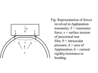
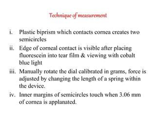
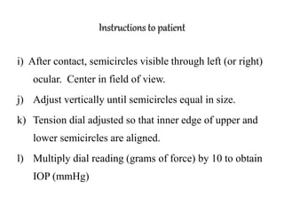
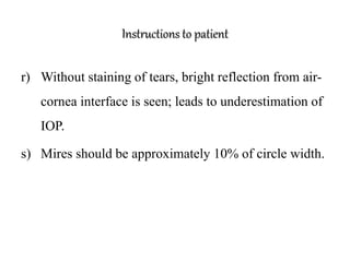
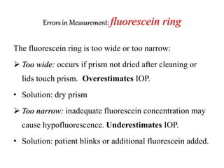
Goldman Applanation Tonometer
 In Goldman Applanation tonometry, slit-lamp is used to
illuminate the tonometer tip and fluorescein – stained
cornea. Magnification has very little importance in this
procedure.
 Applanation tonometery is the best available procedure to
record intraocular tension. It is least affected by scleral
rigidity, which is an inherent deterrent of indentation
tonometry (1) (PK Muk: 119)
Optical Principle of gat
Goldmann Applanation tonometer is based on the
Imbert–Fick principle, which states that for a dry
thin-walled sphere, the pressure (P) inside the sphere
equals the force (F) necessary to flatten its surface
divided by the area (A) of flattening (i.e. P = F/A). It
applies to surfaces which are perfectly spherical, dry,
flexible, elastic, and infinitely thin.
Optical Principle of gat
Theoretically, average corneal rigidity (taken as 520 μm
for GAT) and the capillary attraction of the tear
meniscus cancel each other out when the flattened area
has the 3.06 mm diameter contact surface of the
Goldmann prism, which is applied to the cornea using
the Goldmann tonometer with a measurable amount of
force from which the IOP is deduced.
Fig: Representation of forces
involved in Applanation
tonometry. F = tonometer
force; s = surface tension
of precorneal tear
film; P = intraocular
pressure; A = area of
Applanation; b = corneal
rigidity/resistance to
bending.
Technique of measurement
i. Plastic biprism which contacts cornea creates two
semicircles
ii. Edge of corneal contact is visible after placing
fluorescein into tear film & viewing with cobalt
blue light
iii. Manually rotate the dial calibrated in grams, force is
adjusted by changing the length of a spring within
the device.
iv. Inner margins of semicircles touch when 3.06 mm
of cornea is applanated.
Instructions to patient
a) Press head firmly against chin and forehead rest.
b) look straight ahead and fixate on a target (e.g.
examiners opposite ear)
c) breathe normally, do not hold your breath
d) Blink immediately prior to measurement to moisten
cornea.
Instructions to patient
e) Position patient’s head with forehead rest well above
eyebrows, allowing raising of eyebrows.
f) anesthetic & fluorescein (0.25%), drop applied
g) maximal illumination of biprism the lamp is moved
toward the eye until the tip of biprism contacts the
apex of the cornea
h) stop moving forward when limbus shines with light,
Instructions to patient
i) After contact, semicircles visible through left (or right)
ocular. Center in field of view.
j) Adjust vertically until semicircles equal in size.
k) Tension dial adjusted so that inner edge of upper and
lower semicircles are aligned.
l) Multiply dial reading (grams of force) by 10 to obtain
IOP (mmHg)
Instructions to patient
m) Read at median over which arcs glide to control for
excursions due to ocular pulsations.
n) If slit-lamp moved too far toward patient the
pressure arm will push against a spring which will
press against the eye with a low inoffensive force.
o) Mires (flattened area) too large, moving dial doesn’t
alter appearance.
Instructions to patient
p) Solution: Draw back until regular pulsation noted
and appearance of mires normalizes.
q) Blue central area represents applanated cornea,
green semicircles are fluorescein-stained tears, inner
border of ring is demarcation between flattened and
non-flattened cornea.
Instructions to patient
r) Without staining of tears, bright reflection from air-
cornea interface is seen; leads to underestimation of
IOP.
s) Mires should be approximately 10% of circle width.
Errors in Measurement: fluorescein ring
The fluorescein ring is too wide or too narrow:
 Too wide: occurs if prism not dried after cleaning or
lids touch prism. Overestimates IOP.
• Solution: dry prism
 Too narrow: inadequate fluorescein concentration may
cause hypofluorescence. Underestimates IOP.
• Solution: patient blinks or additional fluorescein added.
Errors in Measurement: Corneal astigmatism
• Corneal astigmatism: When regular astigmatism is present, an
elliptical contact with tonometer head occurs.
• This results in an under estimation of IOP in with-the-rule
astigmatism and
• an over estimation with against-the-rule astigmatism,
• with an error range of about -2.5 to +2.5 mmHg.
Errors in Measurement: Corneal curvature
Steeper corneas need to be indented more to produce the
standard area of contact, necessitating more force and
therefore indicating a higher IOP reading. It has been
suggested that over the range of corneal curvature of
40 to 49 diopters, the error in IOP reading is about
3mmHg
Errors in Measurement: Corneal oedema
Goldmann Applanation Tonometry underestimates the
IOP in eyes with moderate corneal edema. This
underestimation was attributed to the observation that
the epithelium of edematous corneas is easier to
indent than normal epithelium.
Errors in Measurement: Corneal thickness
Thin corneas tend to produce underestimation and thick
corneas produce overestimation of IOP. Clinical
implication of this fact in patients with thin corneas
may be wrongly diagnosed as normal tension glaucoma
and thick corneas wrongly as ocular hypertension
emphasizing importance of checking central corneal
thickness on a routine basis in glaucoma clinics.

Goldman Applanation Tonometer

  • 2.
    Goldman Applanation Tonometer In Goldman Applanation tonometry, slit-lamp is used to illuminate the tonometer tip and fluorescein – stained cornea. Magnification has very little importance in this procedure.  Applanation tonometery is the best available procedure to record intraocular tension. It is least affected by scleral rigidity, which is an inherent deterrent of indentation tonometry (1) (PK Muk: 119)
  • 3.
    Optical Principle ofgat Goldmann Applanation tonometer is based on the Imbert–Fick principle, which states that for a dry thin-walled sphere, the pressure (P) inside the sphere equals the force (F) necessary to flatten its surface divided by the area (A) of flattening (i.e. P = F/A). It applies to surfaces which are perfectly spherical, dry, flexible, elastic, and infinitely thin.
  • 4.
    Optical Principle ofgat Theoretically, average corneal rigidity (taken as 520 μm for GAT) and the capillary attraction of the tear meniscus cancel each other out when the flattened area has the 3.06 mm diameter contact surface of the Goldmann prism, which is applied to the cornea using the Goldmann tonometer with a measurable amount of force from which the IOP is deduced.
  • 5.
    Fig: Representation offorces involved in Applanation tonometry. F = tonometer force; s = surface tension of precorneal tear film; P = intraocular pressure; A = area of Applanation; b = corneal rigidity/resistance to bending.
  • 6.
    Technique of measurement i.Plastic biprism which contacts cornea creates two semicircles ii. Edge of corneal contact is visible after placing fluorescein into tear film & viewing with cobalt blue light iii. Manually rotate the dial calibrated in grams, force is adjusted by changing the length of a spring within the device. iv. Inner margins of semicircles touch when 3.06 mm of cornea is applanated.
  • 7.
    Instructions to patient a)Press head firmly against chin and forehead rest. b) look straight ahead and fixate on a target (e.g. examiners opposite ear) c) breathe normally, do not hold your breath d) Blink immediately prior to measurement to moisten cornea.
  • 8.
    Instructions to patient e)Position patient’s head with forehead rest well above eyebrows, allowing raising of eyebrows. f) anesthetic & fluorescein (0.25%), drop applied g) maximal illumination of biprism the lamp is moved toward the eye until the tip of biprism contacts the apex of the cornea h) stop moving forward when limbus shines with light,
  • 9.
    Instructions to patient i)After contact, semicircles visible through left (or right) ocular. Center in field of view. j) Adjust vertically until semicircles equal in size. k) Tension dial adjusted so that inner edge of upper and lower semicircles are aligned. l) Multiply dial reading (grams of force) by 10 to obtain IOP (mmHg)
  • 10.
    Instructions to patient m)Read at median over which arcs glide to control for excursions due to ocular pulsations. n) If slit-lamp moved too far toward patient the pressure arm will push against a spring which will press against the eye with a low inoffensive force. o) Mires (flattened area) too large, moving dial doesn’t alter appearance.
  • 11.
    Instructions to patient p)Solution: Draw back until regular pulsation noted and appearance of mires normalizes. q) Blue central area represents applanated cornea, green semicircles are fluorescein-stained tears, inner border of ring is demarcation between flattened and non-flattened cornea.
  • 12.
    Instructions to patient r)Without staining of tears, bright reflection from air- cornea interface is seen; leads to underestimation of IOP. s) Mires should be approximately 10% of circle width.
  • 13.
    Errors in Measurement:fluorescein ring The fluorescein ring is too wide or too narrow:  Too wide: occurs if prism not dried after cleaning or lids touch prism. Overestimates IOP. • Solution: dry prism  Too narrow: inadequate fluorescein concentration may cause hypofluorescence. Underestimates IOP. • Solution: patient blinks or additional fluorescein added.
  • 14.
    Errors in Measurement:Corneal astigmatism • Corneal astigmatism: When regular astigmatism is present, an elliptical contact with tonometer head occurs. • This results in an under estimation of IOP in with-the-rule astigmatism and • an over estimation with against-the-rule astigmatism, • with an error range of about -2.5 to +2.5 mmHg.
  • 15.
    Errors in Measurement:Corneal curvature Steeper corneas need to be indented more to produce the standard area of contact, necessitating more force and therefore indicating a higher IOP reading. It has been suggested that over the range of corneal curvature of 40 to 49 diopters, the error in IOP reading is about 3mmHg
  • 16.
    Errors in Measurement:Corneal oedema Goldmann Applanation Tonometry underestimates the IOP in eyes with moderate corneal edema. This underestimation was attributed to the observation that the epithelium of edematous corneas is easier to indent than normal epithelium.
  • 17.
    Errors in Measurement:Corneal thickness Thin corneas tend to produce underestimation and thick corneas produce overestimation of IOP. Clinical implication of this fact in patients with thin corneas may be wrongly diagnosed as normal tension glaucoma and thick corneas wrongly as ocular hypertension emphasizing importance of checking central corneal thickness on a routine basis in glaucoma clinics.