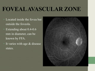
Downloaded 791 times

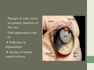


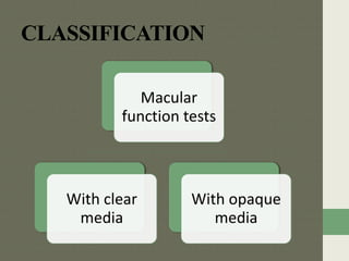
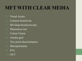


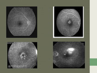


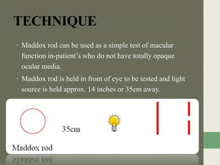

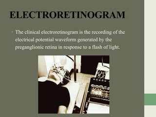
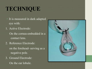


The document discusses the anatomy, embryology, and function tests of the macula lutea. It describes the macula lutea as a 5.5mm circular area at the posterior pole of the retina that subserves central vision. It notes the macula's delayed development until 8 months gestation and specialization of the fovea which contains the highest concentration of cones. The document outlines various macular function tests used to evaluate macular diseases, including visual acuity, Amsler grid, microperimetry, and electroretinography. It provides details on the anatomy and cell layers of the fovea centralis and techniques for assessing macular integrity with tests like the Maddox rod.


