Anatomy & Physiology Lecture Notes - Ch. 3 cells - part 3
•Download as PPT, PDF•
5 likes•3,516 views

website: http://www.am-medicine.com Facebook page : https://www.facebook.com/pages/Am-medicine/207726329406832 Facebook group: https://www.facebook.com/groups/1409138472653811/
Report
Share
Report
Share

Recommended
Anatomy & Physiology Lecture Notes - Ch. 3 cells - part 2
website: http://www.am-medicine.com
Facebook page : https://www.facebook.com/pages/Am-medicine/207726329406832
Facebook group: https://www.facebook.com/groups/1409138472653811/
Unit1: Organisation of Human Body
This document provides an overview of the organization of the human body from the subatomic to the organ system level. It begins by defining the different levels of organization, from subatomic to cellular to tissue to organ to system. Key points include that cells are the basic functional units that form tissues, tissues combine to form organs, and organ systems work together in an organized way. The document then discusses cell structure and function, including the cell membrane and different mechanisms of transport. It also covers the differentiation of cells into tissues, the main tissue types, and how organs are formed from tissues to perform specific functions. Finally, it classifies the main organ systems by their role in nutrition, reproduction, or interaction and notes that all systems work in
Anatomy & Physiology Lecture Notes - Ch. 3 cells - part 1
website: http://www.am-medicine.com
Facebook page : https://www.facebook.com/pages/Am-medicine/207726329406832
Facebook group: https://www.facebook.com/groups/1409138472653811/
Cell anatomy
The document summarizes key aspects of cell structure and function. It describes that cells have three main parts - the plasma membrane, cytoplasm, and nucleus. The plasma membrane encloses the cell and regulates what enters and exits. The cytoplasm contains organelles that carry out metabolic functions, and the nucleus houses genetic material and controls cellular activities. Specific organelles like mitochondria, ribosomes, and the endoplasmic reticulum are discussed in detail. The mechanisms of passive transport like diffusion and osmosis, as well as active transport processes, are also summarized.
Cell and cell organelles
This document summarizes the main components and functions of cells and cell organelles. It describes that cells can be either prokaryotic or eukaryotic. The key organelles discussed include the cell membrane, mitochondria, nucleus, ribosomes, endoplasmic reticulum, Golgi bodies, lysosomes, peroxisomes, cytosol and cytoskeleton. Each organelle is briefly described in terms of its structure and main functions in the cell.
Plasma Membrane
The plasma membrane acts as a gatekeeper that regulates what enters and exits the cell. It uses both passive and active transport. Passive transport relies on diffusion and moves substances along concentration gradients, while active transport requires energy and can move substances against concentration gradients using mechanisms like the sodium-potassium pump. The plasma membrane is a lipid bilayer with embedded proteins that gives cells structure and allows for selective permeability and transport of molecules in and out of the cell.
introduction to human physiology
The document discusses several key topics in human physiology:
1) It describes homeostasis as the maintenance of nearly constant internal conditions in the body. Various organ systems like the respiratory, gastrointestinal, and musculoskeletal systems help maintain homeostasis.
2) It discusses the structure and functions of cells, including their membranous structures, organelles, water and ion content, and proteins.
3) It examines the locomotion of cells through ameboid movement and ciliary movement, describing the mechanisms by which these types of cell movement occur.
Transport across cell membrane
cell biology topic transport across cell membrane. transport of important structures accross plasma mebrane of different types of cell in humans. structure and function of cell membane
Cell
Cell basic stuctural and functional unit , Prokaryotic cell , Eukaryotic cells , cytoplasm ,Nucleus , Ribosome , lysosomes , Endoplasmic Reticulum , Golgi apparatus , Mitochondria , cytosol , animal cell , plant cell , biology , Important
Points , discovery , definition , meaning , Robert Hooke
Cell and cell organlles
This document compares and contrasts animal and plant cells. It notes that both cell types have a cell membrane and plastids, while only plant cells have a cell wall. It then provides more detailed information about various cell organelles found in both animal and plant cells, including their structure, functions, and role in cellular processes like transport and energy production. Key organelles discussed include the nucleus, cytoplasm, endoplasmic reticulum, Golgi complex, lysosomes, mitochondria, and vacuoles.
mbbs ims msu
Cell structure and function can be summarized in 3 points:
1. All living things are made of cells, which are the basic functional units. Cells come from preexisting cells through cell division.
2. Cells can be either prokaryotic (lacking organelles) or eukaryotic (containing organelles). Eukaryotic cells, which include plants and animals, have internal structures like a nucleus bounded by a nuclear membrane.
3. A typical animal cell is enclosed by a cell membrane and contains a nucleus, cytoplasm, mitochondria, endoplasmic reticulum, Golgi apparatus, lysosomes, and ribosomes that allow the cell to carry out life functions like respiration, protein
Anatomy terminology
This document provides definitions and descriptions of key anatomical and physiological concepts. It defines anatomy as the study of structure and relationships between structures, and physiology as the study of how body structures function. It describes the structural hierarchy of the human body from chemicals to cells to tissues to organs to systems. Key concepts covered include homeostasis, feedback loops, disease, anatomical position and planes, and directional terms.
Transport Across Cell Membranes
The document discusses transport across cell membranes. It begins by describing the structure and function of cell membranes, including their semipermeable nature. It then explains various transport mechanisms like diffusion, osmosis, facilitated diffusion, active transport, and endocytosis/exocytosis that allow materials to move across membranes. Specific examples are given of how these transport mechanisms function in cells, lungs, and other organisms and systems to maintain homeostasis.
Anatomy and physiology Introduction Chapter 1 Notes
The document provides an introduction to human anatomy and physiology, outlining key concepts such as anatomical position, body cavities, homeostasis, and levels of organization. It defines anatomy and physiology and describes the basic functions of the human body including movement, growth, digestion, and excretion. The reading also explains homeostasis and feedback systems that help maintain stable internal body conditions.
Skeletal System
The skeletal system consists of bones, joints, cartilage, and ligaments that make up the endoskeleton. It has two divisions: the axial skeleton including the skull and spine, and the appendicular skeleton including the limbs and girdles. Bones provide structure, protect organs, allow movement, store minerals, and form blood cells. The skeleton contains over 200 bones including long, short, flat, and irregular bones. Bones are living tissues that undergo remodeling through bone cells like osteoblasts and osteoclasts. Joints allow movement and include hinge, ball-and-socket, and gliding joints. Common skeletal conditions include osteoporosis which weakens bones, and curvatures of the spine like scol
Cell
The document discusses cell structure and function. It covers the cell theory, basic structures of the cell including the plasma membrane and organelles, and functions of the cell like communication and metabolism. It describes limits to cell size and provides details on the fluid mosaic model of the plasma membrane. It also summarizes the structure and roles of various organelles and discusses cell division and the life cycle.
Nervous system
The nervous system has two main parts - the central nervous system (CNS) and the peripheral nervous system (PNS). The CNS is made up of the brain and spinal cord, and acts as the main control center. The brain controls all body functions and processes sensory information. The spinal cord relays messages between the brain and body and coordinates reflexes. The PNS connects the CNS to the rest of the body using cranial and spinal nerves. It links the CNS to sensory receptors and muscles throughout the body.
Biology, cytoplasm
The cytoplasm is a jelly-like material that makes up the region between the cell membrane and nucleus. It contains cytosol, organelles, and inclusions. Cytosol is a solution of water, proteins, sugars, and dissolved gases that provides the environment for biochemical reactions. Organelles such as mitochondria, lysosomes, and chloroplasts carry out specialized functions. Inclusions are non-living substances like glycogen granules, lipid droplets, and pigments that cells store or secrete. The cytoplasm is the site where most cell activities occur.
Introduction of tissue - Epithelial tissue
This document discusses the different types of tissues in the body, with a focus on epithelial tissue. It defines tissue as groups of cells organized to perform specific functions. The four basic tissue types are epithelial, connective, muscular and nervous tissue. Epithelial tissue forms coverings and linings and has closely packed cells held together by junctions. Epithelial tissue is classified based on cell shape and layer arrangement into simple and stratified types, including squamous, cuboidal, columnar, pseudostratified and transitional epithelium. The document provides detailed descriptions of each epithelial tissue type including their location and functions such as secretion, protection and absorption.
1 obj331 cellbiol
The document summarizes cellular structures and functions. It identifies the five chief cellular functions as movement, conductivity, metabolic absorption, secretion, and excretion. It then describes the structures and functions of key cellular organelles including the nucleus, ribosomes, endoplasmic reticulum, Golgi apparatus, lysosomes, and mitochondria. It also discusses plasma membrane structure and functions such as transport, protection, and cell communication.
Class 1 Human cell physiology
Cell physiology is the biological study of the activities that take place in a cell to keep it alive. The term physiology refers to normal functions in a living organism.
Anatomy and Physiology; Introduction to the human body
A&P terminology introduced, a brief history of the study of anatomy, body systems, life processes, homeostasis, positive and negative feedback systems, directional terms and regions of the body terminology are introduced
TISSUE.pptx
Tissue Definition
Tissues are groups of cells that have a similar structure and act together to perform a specific function. The word tissue comes from a form of an old French verb meaning “to weave”. There are four different types of tissues in animals: connective, muscle, nervous, and epithelial. In plants, tissues are divided into three types: vascular, ground, and epidermal. Groups of tissues make up organs in the body such as the brain and heart.
Types of Animal Tissues
Connective
Connective tissue connects or separates groups of other tissues. It is found in between all the other tissues and organs in the body. Connective tissue is made up of cells and ground substance, which is a gel that surrounds cells. Most connective tissue, except for lymph and blood, also contains fibers, which are long, narrow proteins. Fibers can be collagenous, which bind bones to tissues; elastic, which allow organs like the lungs to move; or reticular, which provide physical support to cells. Connective tissue also allows oxygen to diffuse from blood vessels into cells.
About 1 in 10 people are have a disorder involving connective tissue. Some connective tissue disorders include sarcomas, Marfan syndrome, lupus, and scurvy, which is a Vitamin C deficiency that leads to fragile connective tissue.
Muscle
Muscle tissue comprises all the muscles in the body, and the specialized nature of the tissue is what allows muscles to contract. There are three types of muscle tissue: skeletal muscle, cardiac muscle, and smooth muscle. Skeletal muscle anchors tendons to bones and allows the body to move. Cardiac muscle is found in the heart and contracts to pump blood. Smooth muscle is found in the intestines, where it helps move food through the digestive tract, and it is also found in other organs like blood vessels, the uterus, and the bladder. Skeletal and cardiac muscles are striated; this means that they contain sarcomeres (a unit of muscle tissue) that are arranged in a uniform pattern. Smooth muscle does not have sarcomeres.
Duchenne muscular dystrophy is an example of a muscle tissue disorder. It is an inherited disorder that causes muscles to atrophy over time. The muscles shorten as they atrophy, which can cause scoliosis and immobile joints. Individuals with the disorder are usually male because the gene responsible for it is found on the X chromosome (of which males have only one).
Nervous
Nervous tissue is found in the brain, spinal cord, and peripheral nerves, which are all parts of the nervous system. It is made up of neurons, which are nerve cells, and neuroglia, which are cells that help nerve impulses travel. Nervous tissue is grouped into four types: gray matter and white matter in the brain, and nerves and ganglia in the peripheral nervous system. The main difference between gray and white matter is that axons of the neurons in gray matter are unmyelinated, while white matter is myelinated. Myelin is a white, fatty substance that insulates neurons and
Anatomy 1st lecture
This document provides an introduction to basic human anatomy. It defines anatomy as the study of body structures and discusses different types of anatomical studies such as microscopic, gross, and functional anatomy. The document explains that the human body is organized from cells to tissues to organs to systems. It then describes the main regions of the body - the head/neck, back/trunk, and upper and lower limbs. The anatomical position and different planes of the body are defined to provide a standard reference for describing anatomy.
Introduction to physiology lecture
The document summarizes key aspects of cell physiology:
- Cells are the basic units of structure and function in the body, with over 100 trillion cells that come in a variety of shapes and sizes.
- All cells share certain characteristics like mechanisms for obtaining and using energy from nutrients.
- The basic structures of cells include a plasma membrane, cytoplasm containing organelles like the nucleus, mitochondria and ribosomes, and the nucleus which houses genetic material.
- The plasma membrane is selectively permeable and controls what enters and exits the cell. It contains proteins, lipids and carbohydrates.
- The cytoplasm and organelles work together to carry out specialized functions and transport materials within the cell.
- The nucleus contains
Introduction to Human Anatomy
This document provides an introduction to anatomy and physiology. It defines anatomy as the study of body parts and their relationships, while physiology is the study of body functions. Anatomy can be examined on a microscopic or developmental level, while physiology analyzes systems and their functions. The document then outlines the structural hierarchy of the body from chemicals to organ systems. It describes several key organ systems and homeostasis, concluding with important anatomical terminology.
Cell Structure and Function
The document provides an overview of cell structure and function. It defines the cell and outlines the cell theory. The major components of plant cells are described, including organelles like the nucleus, chloroplasts, mitochondria, and cell membrane. It also discusses the cytoskeleton and movement of substances across the cell membrane through passive diffusion, facilitated transport, osmosis, and active transport processes like endocytosis and exocytosis. The overall purpose is to introduce learners to key aspects of cell biology.
Brain
The brain is a three-pound organ responsible for all mental functions and control of vital activities. It contains over 100 billion neurons at birth. The document describes the development of the brain before and at birth, including the separation of the medulla, cerebellum, and cerebrum by the fourth month and the appearance of an indent on the cerebrum by the sixth month. It then provides details about several parts of the brain in response to a series of questions.
001 Anatomy Physiology Introduction
Topics Covered
1. Intro to Anat Physio
2. Anat Physio Terminologies
3. Cell Structure and Functions
4. Cell Organelles
5. Tissue Organization
Epithelial Tissue
Connective Tissue
Muscular Tissue
Nervous Tissue
6. Tissue Embryology
7. Tissue Injury and Inflammation
CELL ANATOMY
All organisms are composed of one or more cells, which are the basic unit of life. A typical animal cell contains organelles such as a nucleus, cytoplasm, mitochondria, endoplasmic reticulum, Golgi apparatus, lysosomes, and microtubules. The nucleus houses the cell's DNA and directs cell activities, while organelles such as mitochondria generate energy and the endoplasmic reticulum and Golgi apparatus modify and transport proteins within the cell. Cells arise only through division of preexisting cells, demonstrating the cell theory that cells are the fundamental unit of life.
More Related Content
What's hot
Cell
Cell basic stuctural and functional unit , Prokaryotic cell , Eukaryotic cells , cytoplasm ,Nucleus , Ribosome , lysosomes , Endoplasmic Reticulum , Golgi apparatus , Mitochondria , cytosol , animal cell , plant cell , biology , Important
Points , discovery , definition , meaning , Robert Hooke
Cell and cell organlles
This document compares and contrasts animal and plant cells. It notes that both cell types have a cell membrane and plastids, while only plant cells have a cell wall. It then provides more detailed information about various cell organelles found in both animal and plant cells, including their structure, functions, and role in cellular processes like transport and energy production. Key organelles discussed include the nucleus, cytoplasm, endoplasmic reticulum, Golgi complex, lysosomes, mitochondria, and vacuoles.
mbbs ims msu
Cell structure and function can be summarized in 3 points:
1. All living things are made of cells, which are the basic functional units. Cells come from preexisting cells through cell division.
2. Cells can be either prokaryotic (lacking organelles) or eukaryotic (containing organelles). Eukaryotic cells, which include plants and animals, have internal structures like a nucleus bounded by a nuclear membrane.
3. A typical animal cell is enclosed by a cell membrane and contains a nucleus, cytoplasm, mitochondria, endoplasmic reticulum, Golgi apparatus, lysosomes, and ribosomes that allow the cell to carry out life functions like respiration, protein
Anatomy terminology
This document provides definitions and descriptions of key anatomical and physiological concepts. It defines anatomy as the study of structure and relationships between structures, and physiology as the study of how body structures function. It describes the structural hierarchy of the human body from chemicals to cells to tissues to organs to systems. Key concepts covered include homeostasis, feedback loops, disease, anatomical position and planes, and directional terms.
Transport Across Cell Membranes
The document discusses transport across cell membranes. It begins by describing the structure and function of cell membranes, including their semipermeable nature. It then explains various transport mechanisms like diffusion, osmosis, facilitated diffusion, active transport, and endocytosis/exocytosis that allow materials to move across membranes. Specific examples are given of how these transport mechanisms function in cells, lungs, and other organisms and systems to maintain homeostasis.
Anatomy and physiology Introduction Chapter 1 Notes
The document provides an introduction to human anatomy and physiology, outlining key concepts such as anatomical position, body cavities, homeostasis, and levels of organization. It defines anatomy and physiology and describes the basic functions of the human body including movement, growth, digestion, and excretion. The reading also explains homeostasis and feedback systems that help maintain stable internal body conditions.
Skeletal System
The skeletal system consists of bones, joints, cartilage, and ligaments that make up the endoskeleton. It has two divisions: the axial skeleton including the skull and spine, and the appendicular skeleton including the limbs and girdles. Bones provide structure, protect organs, allow movement, store minerals, and form blood cells. The skeleton contains over 200 bones including long, short, flat, and irregular bones. Bones are living tissues that undergo remodeling through bone cells like osteoblasts and osteoclasts. Joints allow movement and include hinge, ball-and-socket, and gliding joints. Common skeletal conditions include osteoporosis which weakens bones, and curvatures of the spine like scol
Cell
The document discusses cell structure and function. It covers the cell theory, basic structures of the cell including the plasma membrane and organelles, and functions of the cell like communication and metabolism. It describes limits to cell size and provides details on the fluid mosaic model of the plasma membrane. It also summarizes the structure and roles of various organelles and discusses cell division and the life cycle.
Nervous system
The nervous system has two main parts - the central nervous system (CNS) and the peripheral nervous system (PNS). The CNS is made up of the brain and spinal cord, and acts as the main control center. The brain controls all body functions and processes sensory information. The spinal cord relays messages between the brain and body and coordinates reflexes. The PNS connects the CNS to the rest of the body using cranial and spinal nerves. It links the CNS to sensory receptors and muscles throughout the body.
Biology, cytoplasm
The cytoplasm is a jelly-like material that makes up the region between the cell membrane and nucleus. It contains cytosol, organelles, and inclusions. Cytosol is a solution of water, proteins, sugars, and dissolved gases that provides the environment for biochemical reactions. Organelles such as mitochondria, lysosomes, and chloroplasts carry out specialized functions. Inclusions are non-living substances like glycogen granules, lipid droplets, and pigments that cells store or secrete. The cytoplasm is the site where most cell activities occur.
Introduction of tissue - Epithelial tissue
This document discusses the different types of tissues in the body, with a focus on epithelial tissue. It defines tissue as groups of cells organized to perform specific functions. The four basic tissue types are epithelial, connective, muscular and nervous tissue. Epithelial tissue forms coverings and linings and has closely packed cells held together by junctions. Epithelial tissue is classified based on cell shape and layer arrangement into simple and stratified types, including squamous, cuboidal, columnar, pseudostratified and transitional epithelium. The document provides detailed descriptions of each epithelial tissue type including their location and functions such as secretion, protection and absorption.
1 obj331 cellbiol
The document summarizes cellular structures and functions. It identifies the five chief cellular functions as movement, conductivity, metabolic absorption, secretion, and excretion. It then describes the structures and functions of key cellular organelles including the nucleus, ribosomes, endoplasmic reticulum, Golgi apparatus, lysosomes, and mitochondria. It also discusses plasma membrane structure and functions such as transport, protection, and cell communication.
Class 1 Human cell physiology
Cell physiology is the biological study of the activities that take place in a cell to keep it alive. The term physiology refers to normal functions in a living organism.
Anatomy and Physiology; Introduction to the human body
A&P terminology introduced, a brief history of the study of anatomy, body systems, life processes, homeostasis, positive and negative feedback systems, directional terms and regions of the body terminology are introduced
TISSUE.pptx
Tissue Definition
Tissues are groups of cells that have a similar structure and act together to perform a specific function. The word tissue comes from a form of an old French verb meaning “to weave”. There are four different types of tissues in animals: connective, muscle, nervous, and epithelial. In plants, tissues are divided into three types: vascular, ground, and epidermal. Groups of tissues make up organs in the body such as the brain and heart.
Types of Animal Tissues
Connective
Connective tissue connects or separates groups of other tissues. It is found in between all the other tissues and organs in the body. Connective tissue is made up of cells and ground substance, which is a gel that surrounds cells. Most connective tissue, except for lymph and blood, also contains fibers, which are long, narrow proteins. Fibers can be collagenous, which bind bones to tissues; elastic, which allow organs like the lungs to move; or reticular, which provide physical support to cells. Connective tissue also allows oxygen to diffuse from blood vessels into cells.
About 1 in 10 people are have a disorder involving connective tissue. Some connective tissue disorders include sarcomas, Marfan syndrome, lupus, and scurvy, which is a Vitamin C deficiency that leads to fragile connective tissue.
Muscle
Muscle tissue comprises all the muscles in the body, and the specialized nature of the tissue is what allows muscles to contract. There are three types of muscle tissue: skeletal muscle, cardiac muscle, and smooth muscle. Skeletal muscle anchors tendons to bones and allows the body to move. Cardiac muscle is found in the heart and contracts to pump blood. Smooth muscle is found in the intestines, where it helps move food through the digestive tract, and it is also found in other organs like blood vessels, the uterus, and the bladder. Skeletal and cardiac muscles are striated; this means that they contain sarcomeres (a unit of muscle tissue) that are arranged in a uniform pattern. Smooth muscle does not have sarcomeres.
Duchenne muscular dystrophy is an example of a muscle tissue disorder. It is an inherited disorder that causes muscles to atrophy over time. The muscles shorten as they atrophy, which can cause scoliosis and immobile joints. Individuals with the disorder are usually male because the gene responsible for it is found on the X chromosome (of which males have only one).
Nervous
Nervous tissue is found in the brain, spinal cord, and peripheral nerves, which are all parts of the nervous system. It is made up of neurons, which are nerve cells, and neuroglia, which are cells that help nerve impulses travel. Nervous tissue is grouped into four types: gray matter and white matter in the brain, and nerves and ganglia in the peripheral nervous system. The main difference between gray and white matter is that axons of the neurons in gray matter are unmyelinated, while white matter is myelinated. Myelin is a white, fatty substance that insulates neurons and
Anatomy 1st lecture
This document provides an introduction to basic human anatomy. It defines anatomy as the study of body structures and discusses different types of anatomical studies such as microscopic, gross, and functional anatomy. The document explains that the human body is organized from cells to tissues to organs to systems. It then describes the main regions of the body - the head/neck, back/trunk, and upper and lower limbs. The anatomical position and different planes of the body are defined to provide a standard reference for describing anatomy.
Introduction to physiology lecture
The document summarizes key aspects of cell physiology:
- Cells are the basic units of structure and function in the body, with over 100 trillion cells that come in a variety of shapes and sizes.
- All cells share certain characteristics like mechanisms for obtaining and using energy from nutrients.
- The basic structures of cells include a plasma membrane, cytoplasm containing organelles like the nucleus, mitochondria and ribosomes, and the nucleus which houses genetic material.
- The plasma membrane is selectively permeable and controls what enters and exits the cell. It contains proteins, lipids and carbohydrates.
- The cytoplasm and organelles work together to carry out specialized functions and transport materials within the cell.
- The nucleus contains
Introduction to Human Anatomy
This document provides an introduction to anatomy and physiology. It defines anatomy as the study of body parts and their relationships, while physiology is the study of body functions. Anatomy can be examined on a microscopic or developmental level, while physiology analyzes systems and their functions. The document then outlines the structural hierarchy of the body from chemicals to organ systems. It describes several key organ systems and homeostasis, concluding with important anatomical terminology.
Cell Structure and Function
The document provides an overview of cell structure and function. It defines the cell and outlines the cell theory. The major components of plant cells are described, including organelles like the nucleus, chloroplasts, mitochondria, and cell membrane. It also discusses the cytoskeleton and movement of substances across the cell membrane through passive diffusion, facilitated transport, osmosis, and active transport processes like endocytosis and exocytosis. The overall purpose is to introduce learners to key aspects of cell biology.
Brain
The brain is a three-pound organ responsible for all mental functions and control of vital activities. It contains over 100 billion neurons at birth. The document describes the development of the brain before and at birth, including the separation of the medulla, cerebellum, and cerebrum by the fourth month and the appearance of an indent on the cerebrum by the sixth month. It then provides details about several parts of the brain in response to a series of questions.
What's hot (20)
Anatomy and physiology Introduction Chapter 1 Notes
Anatomy and physiology Introduction Chapter 1 Notes
Anatomy and Physiology; Introduction to the human body
Anatomy and Physiology; Introduction to the human body
Viewers also liked
001 Anatomy Physiology Introduction
Topics Covered
1. Intro to Anat Physio
2. Anat Physio Terminologies
3. Cell Structure and Functions
4. Cell Organelles
5. Tissue Organization
Epithelial Tissue
Connective Tissue
Muscular Tissue
Nervous Tissue
6. Tissue Embryology
7. Tissue Injury and Inflammation
CELL ANATOMY
All organisms are composed of one or more cells, which are the basic unit of life. A typical animal cell contains organelles such as a nucleus, cytoplasm, mitochondria, endoplasmic reticulum, Golgi apparatus, lysosomes, and microtubules. The nucleus houses the cell's DNA and directs cell activities, while organelles such as mitochondria generate energy and the endoplasmic reticulum and Golgi apparatus modify and transport proteins within the cell. Cells arise only through division of preexisting cells, demonstrating the cell theory that cells are the fundamental unit of life.
Anatomy & Physiology Lecture Notes - Ch. 2 chemistry- part 1
website: http://www.am-medicine.com
Facebook page : https://www.facebook.com/pages/Am-medicine/207726329406832
Facebook group: https://www.facebook.com/groups/1409138472653811/
Anatomy & Physiology Lecture Notes - Brain
website: http://am-medicine.com
Youtube Channel : https://www.youtube.com/user/ammedicine
Facebook group: https://www.facebook.com/groups/am.medicine
Anatomy & Physiology Lecture Notes - Ch. 4 tissues - epithelium
website: http://www.am-medicine.com
Youtube Channel : https://www.youtube.com/user/ammedicine
Facebook group: https://www.facebook.com/groups/1409138472653811/
Anatomy & Physiology Lecture Notes - Rat dissection
website: http://am-medicine.com
Youtube Channel : https://www.youtube.com/user/ammedicine
Facebook group: https://www.facebook.com/groups/am.medicine
Anatomy & Physiology Lecture Notes - Ch. 4 tissues - connective
website: http://www.am-medicine.com
Youtube Channel : https://www.youtube.com/user/ammedicine
Facebook group: https://www.facebook.com/groups/1409138472653811/
Dr. B Ch 02_lecture_presentation
This document provides an overview of cell structure and function. It discusses the main components of cells, including the cytoplasm, organelles, and cell membrane. Specifically, it describes the two main types of cells, the techniques used to study cells under light and electron microscopes, the diversity of cell types in the body, and the structures and functions of the organelles. These include both non-membranous organelles like the cytoskeleton and ribosomes, as well as double-membraned organelles such as the mitochondria, nucleus, endoplasmic reticulum, Golgi apparatus, lysosomes, and peroxisomes. It also examines cell permeability and transport mechanisms across the cell membrane.
Anatomy and Physiology Cell organelles
The document discusses the basic structure and functions of cells. It describes the key components of cells, including the plasma membrane, nucleus, mitochondria, chloroplasts, cell wall, and other organelles. The document also explains the differences between prokaryotic and eukaryotic cells and how scientists first discovered and studied cells using early microscopes.
Anatomy & Physiology Lecture Notes - Blood vessels & circulation
website: http://am-medicine.com
Youtube Channel : https://www.youtube.com/user/ammedicine
Facebook group: https://www.facebook.com/groups/am.medicine
Anatomy & Physiology Lecture Notes - Heart physiology
website: http://am-medicine.com
Youtube Channel : https://www.youtube.com/user/ammedicine
Facebook group: https://www.facebook.com/groups/am.medicine
Anatomy & Physiology Lecture Notes - Biomedical design biomaterials and prost...
website: http://am-medicine.com
Youtube Channel : https://www.youtube.com/user/ammedicine
Facebook group: https://www.facebook.com/groups/am.medicine
Anatomy & Physiology Lecture Notes - Joints
website: http://am-medicine.com
Youtube Channel : https://www.youtube.com/user/ammedicine
Facebook group: https://www.facebook.com/groups/am.medicine
Anatomy & Physiology Lecture Notes - Muscles & muscle tissue
website: http://am-medicine.com
Youtube Channel : https://www.youtube.com/user/ammedicine
Facebook group: https://www.facebook.com/groups/am.medicine
Anatomy & Physiology Lecture Notes - Heart anatomy
website: http://am-medicine.com
Youtube Channel : https://www.youtube.com/user/ammedicine
Facebook group: https://www.facebook.com/groups/am.medicine
Anatomy & Physiology Lecture Notes - Ch. 2 chemistry - part 3
website: http://www.am-medicine.com
Facebook page : https://www.facebook.com/pages/Am-medicine/207726329406832
Facebook group: https://www.facebook.com/groups/1409138472653811/
Anatomy & Physiology Lecture Notes - The nervous system
website: http://am-medicine.com
Youtube Channel : https://www.youtube.com/user/ammedicine
Facebook group: https://www.facebook.com/groups/am.medicine
Anatomy & Physiology Lecture Notes - Ch. 1 introduction
website: http://www.am-medicine.com
Facebook page : https://www.facebook.com/pages/Am-medicine/207726329406832
Facebook group: https://www.facebook.com/groups/1409138472653811/
Anatomy & Physiology Lecture Notes - Skeletal system
website: http://am-medicine.com
Youtube Channel : https://www.youtube.com/user/ammedicine
Facebook group: https://www.facebook.com/groups/am.medicine
Anatomy & Physiology Lecture Notes - Digestive system
website: http://am-medicine.com
Youtube Channel : https://www.youtube.com/user/ammedicine
Facebook group: https://www.facebook.com/groups/am.medicine
Viewers also liked (20)
Anatomy & Physiology Lecture Notes - Ch. 2 chemistry- part 1
Anatomy & Physiology Lecture Notes - Ch. 2 chemistry- part 1
Anatomy & Physiology Lecture Notes - Ch. 4 tissues - epithelium
Anatomy & Physiology Lecture Notes - Ch. 4 tissues - epithelium
Anatomy & Physiology Lecture Notes - Rat dissection
Anatomy & Physiology Lecture Notes - Rat dissection
Anatomy & Physiology Lecture Notes - Ch. 4 tissues - connective
Anatomy & Physiology Lecture Notes - Ch. 4 tissues - connective
Anatomy & Physiology Lecture Notes - Blood vessels & circulation
Anatomy & Physiology Lecture Notes - Blood vessels & circulation
Anatomy & Physiology Lecture Notes - Heart physiology
Anatomy & Physiology Lecture Notes - Heart physiology
Anatomy & Physiology Lecture Notes - Biomedical design biomaterials and prost...
Anatomy & Physiology Lecture Notes - Biomedical design biomaterials and prost...
Anatomy & Physiology Lecture Notes - Muscles & muscle tissue
Anatomy & Physiology Lecture Notes - Muscles & muscle tissue
Anatomy & Physiology Lecture Notes - Heart anatomy
Anatomy & Physiology Lecture Notes - Heart anatomy
Anatomy & Physiology Lecture Notes - Ch. 2 chemistry - part 3
Anatomy & Physiology Lecture Notes - Ch. 2 chemistry - part 3
Anatomy & Physiology Lecture Notes - The nervous system
Anatomy & Physiology Lecture Notes - The nervous system
Anatomy & Physiology Lecture Notes - Ch. 1 introduction
Anatomy & Physiology Lecture Notes - Ch. 1 introduction
Anatomy & Physiology Lecture Notes - Skeletal system
Anatomy & Physiology Lecture Notes - Skeletal system
Anatomy & Physiology Lecture Notes - Digestive system
Anatomy & Physiology Lecture Notes - Digestive system
Similar to Anatomy & Physiology Lecture Notes - Ch. 3 cells - part 3
Chapt6and7
The document provides an overview of cell structure and microscopy. It discusses:
1. The basic components and structures of cells, including the plasma membrane, cytoplasm, ribosomes, and organelles.
2. Key differences between prokaryotic and eukaryotic cells.
3. How light microscopes and electron microscopes work and their magnifying capabilities.
4. Specific organelles found in plant and animal cells like the nucleus, mitochondria, chloroplasts, and lysosomes.
2a; cells
The document summarizes key components and structures of eukaryotic cells. It describes the plasma membrane as the boundary of the cell that contains membrane proteins with various functions like channels, transport, and recognition. Inside the cell are organelles such as the nucleus that houses DNA, ribosomes that synthesize proteins, and mitochondria and chloroplasts that generate energy. The cytoskeleton provides structure and motor proteins help move organelles within the cell.
B 1
The document summarizes cell structures and their functions, including organelles like the cell membrane, nucleus, mitochondria, chloroplasts, Golgi bodies, lysosomes, endoplasmic reticulum, and ribosomes. It describes how these structures work together through compartmentalization and transport of materials to carry out functions like respiration, photosynthesis, protein transport and modification, waste disposal, and more. Compartments like the ER, Golgi bodies and vesicles move proteins and other molecules through the cell and to their destinations, such as secretion from the cell.
Tour of the_cell(chpt6)
The document discusses the structures and functions of eukaryotic cells. It describes organelles such as the nucleus, mitochondria, chloroplasts, endoplasmic reticulum, Golgi apparatus, lysosomes, peroxisomes, cytoskeleton and centrioles. These organelles allow cells to carry out specialized functions like protein production, energy production, transport of materials, waste disposal, structure and cell division. The endosymbiotic theory is presented to explain the origins of mitochondria and chloroplasts from ancient bacteria.
Cell
The document discusses the structure and function of eukaryotic cells. It describes that cells contain organelles, including a nucleus that holds the genome, and membrane-bound structures like the endoplasmic reticulum, Golgi apparatus and mitochondria. The processes of transcription and translation are summarized, where DNA in the nucleus is transcribed into mRNA which is then translated by ribosomes into proteins. The stages of the cell cycle, including interphase and mitosis, are also outlined.
Cell structure.
This document provides an overview of cell structure and function. It begins by explaining that living organisms are made up of hierarchical levels moving from cells to tissues to organs. It then defines the cell as the basic structural and functional unit of life. The document outlines the main components of plant and animal cells, including the cell membrane, cytoplasm, organelles, cytoskeleton, nucleus, and genetic material. It also describes several key cellular processes such as protein synthesis, metabolism, movement, replication, and growth.
Cell and its organelles MK MBBS SPIHER.pptx
This document discusses cell organelles and intercellular communication. It describes the basic components of cells, including the cell membrane, cytoplasm, and various organelles such as the nucleus, mitochondria, lysosomes, and endoplasmic reticulum. It then explains how cells communicate with each other through direct contact via gap junctions, short-range chemical signaling as in autocrine and paracrine signaling, and long-range chemical signaling through the endocrine and nervous systems. The process of signal transduction is also summarized, from membrane receptors that detect extracellular signals, to second messengers within the cell that amplify and propagate the signal, to cellular responses.
Biology 189 the_cell_spring_2012.ppt
The document discusses the structure and function of cells. It covers several key points:
1) Cells are the fundamental unit of life, and there are two main types - prokaryotic cells which lack membrane-bound organelles, and eukaryotic cells which do have organelles like the nucleus.
2) Both cell types contain ribosomes for protein synthesis, but eukaryotic cells also contain other membrane-bound structures like the endoplasmic reticulum, Golgi apparatus, mitochondria, vacuoles, and lysosomes which carry out specialized functions.
3) The plasma membrane regulates what enters and exits the cell, and the cytoskeleton provides structure and allows movement within the cell
Cells bds
The document summarizes key aspects of cell structure and function:
- The cell membrane forms a protective barrier around the cell and is selectively permeable, allowing movement of materials in and out.
- Organelles such as mitochondria, endoplasmic reticulum, Golgi apparatus, lysosomes, ribosomes, and the nucleus have specialized structures and functions that allow cells to carry out essential processes.
- Cells obtain energy through cellular respiration in mitochondria or fermentation in the absence of oxygen. The cell cycle and processes of mitosis and meiosis allow for cell growth and division.
CELL 1.pptxCell diffintion and function of cell and structure and types of cell
CELL 1.pptxCell diffintion and function of cell and structure and types of cellKhyber medical university
All living things are composed of cells, which are the basic structural and functional units of life. Each cell needs nutrients and oxygen, produces energy, eliminates waste, and maintains homeostasis. Cells form tissues like muscle and nerve tissue, which then combine to form organs like the heart, liver and kidneys. Systems of organs work together to perform functions like digestion. The document then describes the structure and functions of key cellular components like the cell membrane, cytoplasm, nucleus, and organelles. It discusses cellular processes like protein synthesis and energy production in mitochondria.The Cell Factory
Eukaryotic cells contain specialized structures called organelles that carry out different functions. The main organelles include the nucleus, which contains the cell's DNA; the endoplasmic reticulum for protein transport; mitochondria and chloroplasts for energy production; and lysosomes for digestion. Plant and animal cells differ in structures like cell walls, chloroplasts, and central vacuoles. The cytoskeleton helps maintain the cell's shape and enables movement. Mitochondria and chloroplasts likely evolved from ancient symbiotic relationships between prokaryotes and eukaryotes.
AS Biology, Unit 1 (Module 1) notes (OCR)
This presentation features key notes and diagrams from the unit 1, module 1 of AS biology. These notes have been mad heavily using OCR text books, however other sources have been used.
Cell structure & functions for class 9 by Susanta Kumar Panda
Cell Structure and Functions ppt presentation for Class 9 Students prepared by Susanta Kumar Panda, Sambalpur
The cell factory
This document discusses the history and components of cells. It begins by introducing microscopy techniques like light and electron microscopy used to study cells. It then covers early discoveries in cell biology by Hooke, van Leeuwenhoek and the formulation of the cell theory. The rest of the document details the structures and organelles found within plant and animal cells like the nucleus, mitochondria, chloroplasts, ER, Golgi apparatus and describes their functions in cellular processes.
The cell factory
The document discusses microscopy and cell structure. It begins by describing different types of microscopes used to study cells, including light and electron microscopes. It then provides a brief history of cell discovery, including early observations by Hooke, van Leeuwenhoek, and the formulation of the cell theory. The rest of the document details the structures and organelles found within plant and animal cells, including the nucleus, mitochondria, chloroplasts, endoplasmic reticulum, Golgi apparatus, lysosomes, vacuoles, and cytoskeleton. It explains the functions of these various organelles that allow cells to carry out life's processes.
Ap Bio Ch 4 PowerPOint
1. The document summarizes key aspects of cell structure and function, including the history of cell discovery, the cell theory, components of eukaryotic and prokaryotic cells, and structures such as organelles, cytoskeleton, flagella, and nuclei.
2. Key organelles discussed include the nucleus, which contains DNA and controls the cell, mitochondria and chloroplasts, which generate energy for the cell, and the endoplasmic reticulum, Golgi apparatus, lysosomes, and vacuoles, which are involved in transport and storage.
3. Prokaryotic cells are described as generally smaller than eukaryotic cells and lacking membrane-bound organelles and nuclei.
4. Cell Structure and Function
1) Cell biology is the study of cells, the fundamental unit of life. Key discoveries included Hooke observing cells in 1665 and van Leeuwenhoek improving microscopy. The cell theory established that cells are the basic unit of life.
2) Microscopy revolutionized cell biology by allowing observation of subcellular structures. Light microscopes use visible light while electron microscopes use beams of electrons for higher magnification.
3) Key cellular structures include the nucleus that houses DNA, organelles like mitochondria and chloroplasts that generate energy, and the endomembrane system involved in protein transport and modification.
UPDATED The cell factory ppt
The document discusses microscopy and cell structure. It begins by describing early microscopists like Hooke and van Leeuwenhoek and how they discovered and observed basic cell structures. It then summarizes the three main points of the Cell Theory: that all living things are made of cells, cells are the basic functional units of life, and new cells are produced from existing cells. The rest of the document details the structures and organelles found within plant and animal cells like the nucleus, mitochondria, chloroplasts, cell membrane, cytoskeleton and more. It explains their functions and roles in processes like cellular respiration, protein synthesis, and photosynthesis.
cell physiology.pptx
This document provides an overview of cell organelles and their functions. It describes the endoplasmic reticulum, including rough ER which synthesizes proteins, and smooth ER which synthesizes lipids and regulates calcium. It also details the Golgi apparatus, mitochondria, lysosomes, peroxisomes, ribosomes, cytoskeleton, and nucleus. The key functions of each organelle are summarized such as protein synthesis, energy production, waste disposal, structural support, and genetic material storage.
02 cells
This document discusses cells and their structures and functions. It begins by outlining the cell theory and introducing the main components of cells, including the plasma membrane, cytoplasm, and nucleus. It then describes the structures and roles of various organelles like the endoplasmic reticulum, Golgi apparatus, mitochondria, and lysosomes. The document also covers cellular processes like endocytosis, exocytosis, and the stages of the cell cycle and cell division. It discusses how cells differ in shape and function depending on their specialized roles and developmental stages. In the end, it briefly outlines some theories of aging.
Similar to Anatomy & Physiology Lecture Notes - Ch. 3 cells - part 3 (20)
CELL 1.pptxCell diffintion and function of cell and structure and types of cell
CELL 1.pptxCell diffintion and function of cell and structure and types of cell
Cell structure & functions for class 9 by Susanta Kumar Panda
Cell structure & functions for class 9 by Susanta Kumar Panda
More from Ammedicine Medicine
Anatomy & Physiology Lecture Notes - The senses
website: http://am-medicine.com
Youtube Channel : https://www.youtube.com/user/ammedicine
Facebook group: https://www.facebook.com/groups/am.medicine
Anatomy & Physiology Lecture Notes - Respiratory system
website: http://am-medicine.com
Youtube Channel : https://www.youtube.com/user/ammedicine
Facebook group: https://www.facebook.com/groups/am.medicine
Anatomy & Physiology Lecture Notes - Blood
This document summarizes the key functions and components of blood. It discusses how blood carries oxygen, nutrients, waste, hormones, and buffers throughout the body. Blood also helps regulate temperature, volume, and prevents infection through white blood cells and antibodies. The three main components of blood are plasma, red blood cells, and white blood cells, which are all produced through hematopoiesis in the bone marrow. The document then provides details on specific blood cells like erythrocytes, leukocytes, and platelets, as well as blood disorders, blood typing, and the clotting process.
Ch. 5 integumentary system - part 3
website: http://am-medicine.com
Youtube Channel : https://www.youtube.com/user/ammedicine
Facebook group: https://www.facebook.com/groups/am.medicine
Ch. 5 integumentary system - part 2
website: http://am-medicine.com
Youtube Channel : https://www.youtube.com/user/ammedicine
Facebook group: https://www.facebook.com/groups/am.medicine
Ch. 5 integumentary system - part 1
website: http://am-medicine.com
Youtube Channel : https://www.youtube.com/user/ammedicine
Facebook group: https://www.facebook.com/groups/am.medicine
Complete denture - Introduction to Prosthodontics
website: http://am-medicine.com
Youtube Channel : https://www.youtube.com/user/ammedicine
Facebook group: https://www.facebook.com/groups/1409138472653811/
Chapter 5 (part2) enzymes - michaelis-menton kinetics
This document discusses enzyme kinetics and the Michaelis-Menten equation. It explains that the Michaelis-Menten equation describes the relationship between substrate concentration and reaction rate in a hyperbolic curve. The parameters in the equation, Vmax and Km, are defined. Vmax is the maximum reaction rate when the enzyme is saturated with substrate. Km is the substrate concentration when the reaction rate is at half of Vmax and represents the substrate concentration required to saturate half the enzyme. The assumptions of initial velocity and steady state that are required to derive the Michaelis-Menten equation are also outlined.
Chapter 5 (part1) enzymes - introduction
This document provides an introduction to enzymes and their catalytic properties. It states that enzymes are catalysts that increase the rate of chemical reactions without being consumed in the process. Enzymes accelerate reactions by lowering the activation energy needed for products to form and by stabilizing reactant molecules in an orientation that favors product formation. The document gives an example of an enzyme that can accelerate a reaction over a billionfold compared to the uncatalyzed rate. It also describes how enzymes specifically recognize substrates and produce products in high yields, often over 95%, due to their unique structural fit with substrates.
Chapter 4 (part 3) protein 3-d structure- structure function
website: http://www.am-medicine.com
Facebook page : https://www.facebook.com/pages/Am-medicine/207726329406832
Facebook group: https://www.facebook.com/groups/1409138472653811/
Chapter 3(part2) - Protein purification and analysis
website: http://www.am-medicine.com
Facebook page : https://www.facebook.com/pages/Am-medicine/207726329406832
Facebook group: https://www.facebook.com/groups/1409138472653811/
Chapter 4 (part 1) protein structure introduction- 2nd structure
website: http://www.am-medicine.com
Facebook page : https://www.facebook.com/pages/Am-medicine/207726329406832
Facebook group: https://www.facebook.com/groups/1409138472653811/
More from Ammedicine Medicine (12)
Anatomy & Physiology Lecture Notes - Respiratory system
Anatomy & Physiology Lecture Notes - Respiratory system
Chapter 5 (part2) enzymes - michaelis-menton kinetics
Chapter 5 (part2) enzymes - michaelis-menton kinetics
Chapter 4 (part 3) protein 3-d structure- structure function
Chapter 4 (part 3) protein 3-d structure- structure function
Chapter 3(part2) - Protein purification and analysis
Chapter 3(part2) - Protein purification and analysis
Chapter 4 (part 1) protein structure introduction- 2nd structure
Chapter 4 (part 1) protein structure introduction- 2nd structure
Recently uploaded
What are the different types of Dental implants.
Are you looking for a long-lasting solution to your missing tooth?
Dental implants are the most common type of method for replacing the missing tooth. Unlike dentures or bridges, implants are surgically placed in the jawbone. In layman’s terms, a dental implant is similar to the natural root of the tooth. It offers a stable foundation for the artificial tooth giving it the look, feel, and function similar to the natural tooth.
NARCOTICS- POLICY AND PROCEDURES FOR ITS USE
This document outlines policies and procedures for handling narcotic and controlled drugs in NABH accredited hospitals.
Nano-gold for Cancer Therapy chemistry investigatory project
chemistry investigatory project
The development of nanogold-based cancer therapy could revolutionize oncology by providing a more targeted, less invasive treatment option. This project contributes to the growing body of research aimed at harnessing nanotechnology for medical applications, paving the way for future clinical trials and potential commercial applications.
Cancer remains one of the leading causes of death worldwide, prompting the need for innovative treatment methods. Nanotechnology offers promising new approaches, including the use of gold nanoparticles (nanogold) for targeted cancer therapy. Nanogold particles possess unique physical and chemical properties that make them suitable for drug delivery, imaging, and photothermal therapy.
How to Control Your Asthma Tips by gokuldas hospital.
Respiratory issues like asthma are the most sensitive issue that is affecting millions worldwide. It hampers the daily activities leaving the body tired and breathless.
The key to a good grip on asthma is proper knowledge and management strategies. Understanding the patient-specific symptoms and carving out an effective treatment likewise is the best way to keep asthma under control.
Skin Diseases That Happen During Summer.
Summer is a time for fun in the sun, but the heat and humidity can also wreak havoc on your skin. From itchy rashes to unwanted pigmentation, several skin conditions become more prevalent during these warmer months.
Top Travel Vaccinations in Manchester
Travel vaccination in Manchester offers comprehensive immunization services for individuals planning international trips. Expert healthcare providers administer vaccines tailored to your destination, ensuring you stay protected against various diseases. Conveniently located clinics and flexible appointment options make it easy to get the necessary shots before your journey. Stay healthy and travel with confidence by getting vaccinated in Manchester. Visit us: www.nxhealthcare.co.uk
Computer in pharmaceutical research and development-Mpharm(Pharmaceutics)
Statistics- Statistics is the science of collecting, organizing, presenting, analyzing and interpreting numerical data to assist in making more effective decisions.
A statistics is a measure which is used to estimate the population parameter
Parameters-It is used to describe the properties of an entire population.
Examples-Measures of central tendency Dispersion, Variance, Standard Deviation (SD), Absolute Error, Mean Absolute Error (MAE), Eigen Value
Foundation of Yoga, YCB Level-3, Unit-1
Unit -1 of Yoga certification board, level 3, all topics covered. An exam conducted by ministry of Ayush for yoga enthusiastic students.
Artificial Intelligence Symposium (THAIS)
Artificial Intelligence Symposium (THAIS). Parc Taulí. Sabadell
Know the difference between Endodontics and Orthodontics.
Your smile is beautiful.
Let’s be honest. Maintaining that beautiful smile is not an easy task. It is more than brushing and flossing. Sometimes, you might encounter dental issues that need special dental care. These issues can range anywhere from misalignment of the jaw to pain in the root of teeth.
Patellar Instability: Diagnosis Management
Pictorial and detailed description of patellar instability with sign and symptoms and how to diagnose , what investigations you should go with and how to approach with treatment options . I have presented this slide in my 2nd year junior residency in orthopedics at LLRM medical college Meerut and got good reviews for it
After getting it read you will definitely understand the topic.
Full Handwritten notes of RA by Ayush Kumar M pharm - Al ameen college of pha...
Full Handwritten notes of RA by Ayush Kumar M pharm - Al ameen college of pha...ayushrajshrivastava7
Regulatory Affairs
Full hand written notes
M.pharm - 1st year .SENSORY NEEDS B.SC. NURSING SEMESTER II.
Sensation is the neurological process by which we become aware of surrounding environment.
How to choose the best dermatologists in Indore.
The skin is the largest organ and its health plays a vital role among the other sense organs. The skin concerns like acne breakout, psoriasis, or anything similar along the lines, finding a qualified and experienced dermatologist becomes paramount.
Pune Call Girls 7339748667 AVAILABLE HOT GIRLS AUNTY BOOK NOW
Pune Call Girls 7339748667 AVAILABLE HOT GIRLS AUNTY BOOK NOW
Recently uploaded (20)
Nano-gold for Cancer Therapy chemistry investigatory project
Nano-gold for Cancer Therapy chemistry investigatory project
How to Control Your Asthma Tips by gokuldas hospital.
How to Control Your Asthma Tips by gokuldas hospital.
Computer in pharmaceutical research and development-Mpharm(Pharmaceutics)
Computer in pharmaceutical research and development-Mpharm(Pharmaceutics)
Know the difference between Endodontics and Orthodontics.
Know the difference between Endodontics and Orthodontics.
Full Handwritten notes of RA by Ayush Kumar M pharm - Al ameen college of pha...
Full Handwritten notes of RA by Ayush Kumar M pharm - Al ameen college of pha...
Pune Call Girls 7339748667 AVAILABLE HOT GIRLS AUNTY BOOK NOW
Pune Call Girls 7339748667 AVAILABLE HOT GIRLS AUNTY BOOK NOW
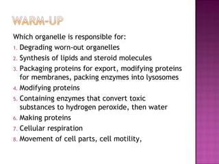
Anatomy & Physiology Lecture Notes - Ch. 3 cells - part 3
- 1. Which organelle is responsible for: 1. Degrading worn-out organelles 2. Synthesis of lipids and steroid molecules 3. Packaging proteins for export, modifying proteins for membranes, packing enzymes into lysosomes 4. Modifying proteins 5. Containing enzymes that convert toxic substances to hydrogen peroxide, then water 6. Making proteins 7. Cellular respiration 8. Movement of cell parts, cell motility,
- 2. Part 3: Cytoplasm & Nucleus
- 4. Between plasma membrane & nucleus Three elements: CCyyttoossooll: fluid Eg. water, proteins, salts, sugars OOrrggaanneelllleess: specific functions IInncclluussiioonnss: chemical substances that vary depending on cell type Eg. glycogen (liver), lipid droplets (fat cells), melanin (skin & hair)
- 5. “little organs” Specialized compartments specific functions Membranous = membrane-bound Mitochondria, peroxisomes, lysosomes, ER, Golgi apparatus Nonmembranous = no membrane cytoskeleton, centrioles, ribosomes
- 7. System of organelles that work to 1. Produce, store, export biological molecules 2. Degrade harmful substances Nuclear envelope, rough ER, smooth ER, Golgi apparatus, secretory vesicles, lysosomes
- 10. Control center contains DNA Most cells have only 1 nucleus Multinucleate: many nuclei (muscle, some liver cells) Anucleate: no nucleus (mature RBC) Three main structures: 1.Nuclear envelope 2.Nucleoli 3.Chromatin
- 11. Multinucleated Muscle Cells Multinucleated Liver Cells Anucleated Red Blood Cells
- 12. Double membrane barrier surrounds nucleus Outer part continuous with Rough ER Nuclear pores: control entry/exit of molecules
- 13. Dark-staining bodies in nucleus 1-2 per cell Site where ribosomes are made
- 14. CChhrroommaattiinn = DNA + Proteins Nucleosome = DNA wrapped around 8 histone proteins Histones allow for compact and orderly packing of long DNA molecules
- 16. During cell division, chromatin condenses to form chromosomes.
- 17. Make identical copies of DNA before a cell divides
- 18. Part of cell division Replicated DNA divided into 2 daughter cells Usually lasts about an hour Interphase prophase metaphase anaphase telophase & cytokinesis
- 19. Gene: segment of DNA that codes for 1 polypeptide Exon: part of DNA that codes for polypeptides Intron: part of DNA that is noncoding (not “junk”!)
- 21. Transcription: RNA formed from DNA Occurs in nucleus Types: mRNA, tRNA, rRNA Translation: protein synthesis polypeptide formed from mRNA Occurs in cytoplasm By ribosomes
- 23. Any substances outside cells 1. Body fluids (blood plasma, interstitial fluid) 2. Cellular secretions (saliva, mucus, gastric fluids) 3. Extracellular matrix (ECM): “glue” that holds cells together; jelly-like substance made of proteins (like collagen) and carbs