Downloaded 406 times
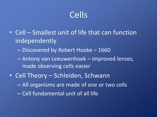
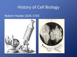
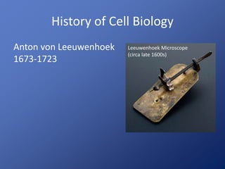
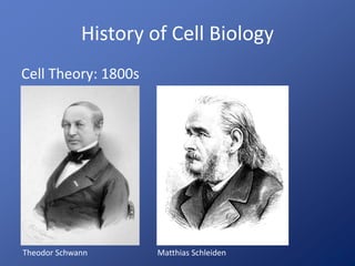
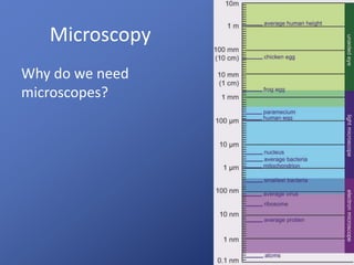
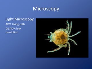
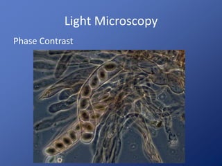
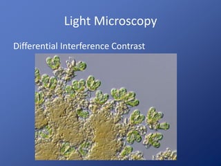
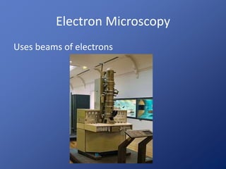
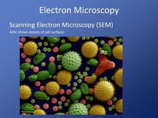
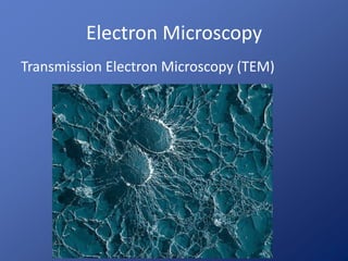
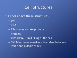
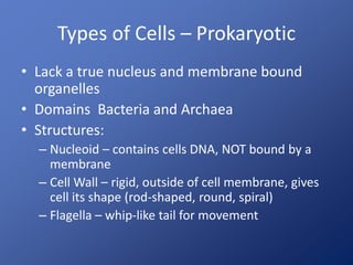
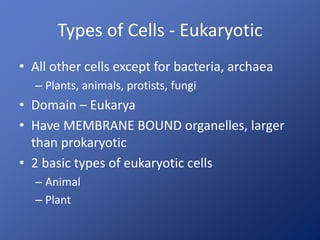
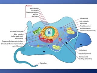
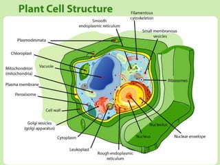
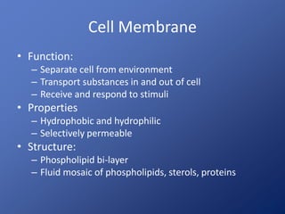
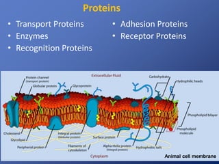
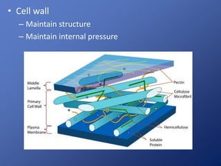
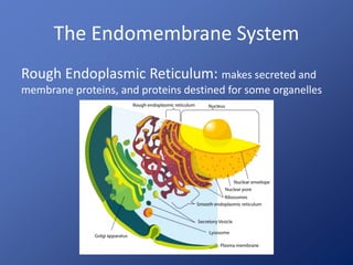
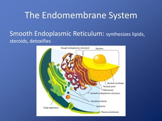
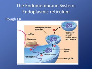
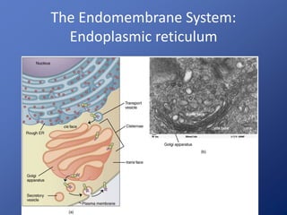
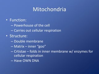
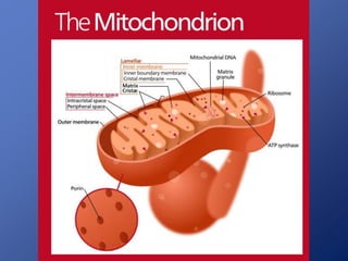
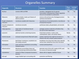

1) Cell biology is the study of cells, the fundamental unit of life. Key discoveries included Hooke observing cells in 1665 and van Leeuwenhoek improving microscopy. The cell theory established that cells are the basic unit of life. 2) Microscopy revolutionized cell biology by allowing observation of subcellular structures. Light microscopes use visible light while electron microscopes use beams of electrons for higher magnification. 3) Key cellular structures include the nucleus that houses DNA, organelles like mitochondria and chloroplasts that generate energy, and the endomembrane system involved in protein transport and modification.


![谷歌留痕技术 [ 𝙩𝙤𝙥 𝟮𝟯𝟯. 𝙘 𝙤𝙢 ]](https://cdn.slidesharecdn.com/ss_thumbnails/top233-260130174328-3833018c-thumbnail.jpg?width=640&height=640&fit=bounds)


