Blood is carried through the body via blood vessels. An artery is a blood vessel that carries blood away from the heart, where it branches into ever-smaller vessels.
The blood vessels are the components of the circulatory system that transport blood throughout the human body. These vessels transport blood cells, nutrients, and oxygen to the tissues of the body. They also take waste and carbon dioxide away from the tissues.
Blood vessels: Arteries, Veins and CapillariesAmir Rifaat
It is one of the circulatory systems. This explains the roles of arteries, veins and capillaries. It also differentiate between the arteries, veins and capillaries. This slide also explained the pulmonary circuit and systemic curcuit. This is an interesting notes and easy to be understand.
lymphatic system, a subsystem of the circulatory system in the vertebrate body that consists of a complex network of vessels, tissues, and organs. The lymphatic system helps maintain fluid balance in the body by collecting excess fluid and particulate matter from tissues and depositing them in the bloodstream
This presentation provides a clear understanding of the physiology of the circulatory system. It focus lies on the division and component of the circulatory system, the three major function of the circulatory system, blood composition, structure of the heart, blood circulation; pulmonary and systemic circuit, valves of the heart, the pathway of blood flow through the heart, the cardiac cycle, pressure changes during the cardiac cycle; systole and diastole, cardiac output, heart sounds among others.
This presentation was designed by Fasama H. Kollie and presented by Benetta N. Kekulah, Cordelia Capehart and Abraham Peters.
What is lymph ?Tissue fluid (interstitial fluid) that enters the lymphatic vessels
Reabsorbs excess interstitial fluid:returns it to the venous circulationmaintain blood volume levelsprevent interstitial fluid levels from rising out of control.Transport dietary lipids:transported through lactealsdrain into larger lymphatic vesselseventually into the bloodstream.lymphocyte development, and the immune response.
Cardiovascular System, Heart, Blood Vessel, ECG, Hypertension, Arrhythmia Audumbar Mali
Cardiovascular System,
Human Anatomy and Physiology-I,
The Blood Vessels,
The Heart,
The Electrocardiogram,
The Vascular Pathways,
As per PCI syllabus,
Atherosclerosis,
Coronary bypass operation,
Heart Transplants and Artificial Hearts
Circulatory system of human being its structure function types and comparison...Nabeel805998
It includes a comprehensive description of circulatory system, its structure function and types.
It also includes diagrammatic representation of various parts circuits and working of human circulation.
Hey, these are the slides me n my friends made... Use them if u want to... for viewing the videos used click on the links given ahead.
http://www.youtube.com/watch?v=jzOti_MtmBk
http://www.youtube.com/watch?v=N9MARqmqSf4
http://www.youtube.com/watch?v=yokcKhqq48c
http://www.youtube.com/watch?v=rJZVFRJmc9M
- Video recording of this lecture in English language: https://youtu.be/lK81BzxMqdo
- Video recording of this lecture in Arabic language: https://youtu.be/Ve4P0COk9OI
- Link to download the book free: https://nephrotube.blogspot.com/p/nephrotube-nephrology-books.html
- Link to NephroTube website: www.NephroTube.com
- Link to NephroTube social media accounts: https://nephrotube.blogspot.com/p/join-nephrotube-on-social-media.html
Ozempic: Preoperative Management of Patients on GLP-1 Receptor Agonists Saeid Safari
Preoperative Management of Patients on GLP-1 Receptor Agonists like Ozempic and Semiglutide
ASA GUIDELINE
NYSORA Guideline
2 Case Reports of Gastric Ultrasound
The prostate is an exocrine gland of the male mammalian reproductive system
It is a walnut-sized gland that forms part of the male reproductive system and is located in front of the rectum and just below the urinary bladder
Function is to store and secrete a clear, slightly alkaline fluid that constitutes 10-30% of the volume of the seminal fluid that along with the spermatozoa, constitutes semen
A healthy human prostate measures (4cm-vertical, by 3cm-horizontal, 2cm ant-post ).
It surrounds the urethra just below the urinary bladder. It has anterior, median, posterior and two lateral lobes
It’s work is regulated by androgens which are responsible for male sex characteristics
Generalised disease of the prostate due to hormonal derangement which leads to non malignant enlargement of the gland (increase in the number of epithelial cells and stromal tissue)to cause compression of the urethra leading to symptoms (LUTS
Title: Sense of Smell
Presenter: Dr. Faiza, Assistant Professor of Physiology
Qualifications:
MBBS (Best Graduate, AIMC Lahore)
FCPS Physiology
ICMT, CHPE, DHPE (STMU)
MPH (GC University, Faisalabad)
MBA (Virtual University of Pakistan)
Learning Objectives:
Describe the primary categories of smells and the concept of odor blindness.
Explain the structure and location of the olfactory membrane and mucosa, including the types and roles of cells involved in olfaction.
Describe the pathway and mechanisms of olfactory signal transmission from the olfactory receptors to the brain.
Illustrate the biochemical cascade triggered by odorant binding to olfactory receptors, including the role of G-proteins and second messengers in generating an action potential.
Identify different types of olfactory disorders such as anosmia, hyposmia, hyperosmia, and dysosmia, including their potential causes.
Key Topics:
Olfactory Genes:
3% of the human genome accounts for olfactory genes.
400 genes for odorant receptors.
Olfactory Membrane:
Located in the superior part of the nasal cavity.
Medially: Folds downward along the superior septum.
Laterally: Folds over the superior turbinate and upper surface of the middle turbinate.
Total surface area: 5-10 square centimeters.
Olfactory Mucosa:
Olfactory Cells: Bipolar nerve cells derived from the CNS (100 million), with 4-25 olfactory cilia per cell.
Sustentacular Cells: Produce mucus and maintain ionic and molecular environment.
Basal Cells: Replace worn-out olfactory cells with an average lifespan of 1-2 months.
Bowman’s Gland: Secretes mucus.
Stimulation of Olfactory Cells:
Odorant dissolves in mucus and attaches to receptors on olfactory cilia.
Involves a cascade effect through G-proteins and second messengers, leading to depolarization and action potential generation in the olfactory nerve.
Quality of a Good Odorant:
Small (3-20 Carbon atoms), volatile, water-soluble, and lipid-soluble.
Facilitated by odorant-binding proteins in mucus.
Membrane Potential and Action Potential:
Resting membrane potential: -55mV.
Action potential frequency in the olfactory nerve increases with odorant strength.
Adaptation Towards the Sense of Smell:
Rapid adaptation within the first second, with further slow adaptation.
Psychological adaptation greater than receptor adaptation, involving feedback inhibition from the central nervous system.
Primary Sensations of Smell:
Camphoraceous, Musky, Floral, Pepperminty, Ethereal, Pungent, Putrid.
Odor Detection Threshold:
Examples: Hydrogen sulfide (0.0005 ppm), Methyl-mercaptan (0.002 ppm).
Some toxic substances are odorless at lethal concentrations.
Characteristics of Smell:
Odor blindness for single substances due to lack of appropriate receptor protein.
Behavioral and emotional influences of smell.
Transmission of Olfactory Signals:
From olfactory cells to glomeruli in the olfactory bulb, involving lateral inhibition.
Primitive, less old, and new olfactory systems with different path
Title: Sense of Taste
Presenter: Dr. Faiza, Assistant Professor of Physiology
Qualifications:
MBBS (Best Graduate, AIMC Lahore)
FCPS Physiology
ICMT, CHPE, DHPE (STMU)
MPH (GC University, Faisalabad)
MBA (Virtual University of Pakistan)
Learning Objectives:
Describe the structure and function of taste buds.
Describe the relationship between the taste threshold and taste index of common substances.
Explain the chemical basis and signal transduction of taste perception for each type of primary taste sensation.
Recognize different abnormalities of taste perception and their causes.
Key Topics:
Significance of Taste Sensation:
Differentiation between pleasant and harmful food
Influence on behavior
Selection of food based on metabolic needs
Receptors of Taste:
Taste buds on the tongue
Influence of sense of smell, texture of food, and pain stimulation (e.g., by pepper)
Primary and Secondary Taste Sensations:
Primary taste sensations: Sweet, Sour, Salty, Bitter, Umami
Chemical basis and signal transduction mechanisms for each taste
Taste Threshold and Index:
Taste threshold values for Sweet (sucrose), Salty (NaCl), Sour (HCl), and Bitter (Quinine)
Taste index relationship: Inversely proportional to taste threshold
Taste Blindness:
Inability to taste certain substances, particularly thiourea compounds
Example: Phenylthiocarbamide
Structure and Function of Taste Buds:
Composition: Epithelial cells, Sustentacular/Supporting cells, Taste cells, Basal cells
Features: Taste pores, Taste hairs/microvilli, and Taste nerve fibers
Location of Taste Buds:
Found in papillae of the tongue (Fungiform, Circumvallate, Foliate)
Also present on the palate, tonsillar pillars, epiglottis, and proximal esophagus
Mechanism of Taste Stimulation:
Interaction of taste substances with receptors on microvilli
Signal transduction pathways for Umami, Sweet, Bitter, Sour, and Salty tastes
Taste Sensitivity and Adaptation:
Decrease in sensitivity with age
Rapid adaptation of taste sensation
Role of Saliva in Taste:
Dissolution of tastants to reach receptors
Washing away the stimulus
Taste Preferences and Aversions:
Mechanisms behind taste preference and aversion
Influence of receptors and neural pathways
Impact of Sensory Nerve Damage:
Degeneration of taste buds if the sensory nerve fiber is cut
Abnormalities of Taste Detection:
Conditions: Ageusia, Hypogeusia, Dysgeusia (parageusia)
Causes: Nerve damage, neurological disorders, infections, poor oral hygiene, adverse drug effects, deficiencies, aging, tobacco use, altered neurotransmitter levels
Neurotransmitters and Taste Threshold:
Effects of serotonin (5-HT) and norepinephrine (NE) on taste sensitivity
Supertasters:
25% of the population with heightened sensitivity to taste, especially bitterness
Increased number of fungiform papillae
HOT NEW PRODUCT! BIG SALES FAST SHIPPING NOW FROM CHINA!! EU KU DB BK substit...GL Anaacs
Contact us if you are interested:
Email / Skype : kefaya1771@gmail.com
Threema: PXHY5PDH
New BATCH Ku !!! MUCH IN DEMAND FAST SALE EVERY BATCH HAPPY GOOD EFFECT BIG BATCH !
Contact me on Threema or skype to start big business!!
Hot-sale products:
NEW HOT EUTYLONE WHITE CRYSTAL!!
5cl-adba precursor (semi finished )
5cl-adba raw materials
ADBB precursor (semi finished )
ADBB raw materials
APVP powder
5fadb/4f-adb
Jwh018 / Jwh210
Eutylone crystal
Protonitazene (hydrochloride) CAS: 119276-01-6
Flubrotizolam CAS: 57801-95-3
Metonitazene CAS: 14680-51-4
Payment terms: Western Union,MoneyGram,Bitcoin or USDT.
Deliver Time: Usually 7-15days
Shipping method: FedEx, TNT, DHL,UPS etc.Our deliveries are 100% safe, fast, reliable and discreet.
Samples will be sent for your evaluation!If you are interested in, please contact me, let's talk details.
We specializes in exporting high quality Research chemical, medical intermediate, Pharmaceutical chemicals and so on. Products are exported to USA, Canada, France, Korea, Japan,Russia, Southeast Asia and other countries.
MANAGEMENT OF ATRIOVENTRICULAR CONDUCTION BLOCK.pdfJim Jacob Roy
Cardiac conduction defects can occur due to various causes.
Atrioventricular conduction blocks ( AV blocks ) are classified into 3 types.
This document describes the acute management of AV block.
New Directions in Targeted Therapeutic Approaches for Older Adults With Mantl...i3 Health
i3 Health is pleased to make the speaker slides from this activity available for use as a non-accredited self-study or teaching resource.
This slide deck presented by Dr. Kami Maddocks, Professor-Clinical in the Division of Hematology and
Associate Division Director for Ambulatory Operations
The Ohio State University Comprehensive Cancer Center, will provide insight into new directions in targeted therapeutic approaches for older adults with mantle cell lymphoma.
STATEMENT OF NEED
Mantle cell lymphoma (MCL) is a rare, aggressive B-cell non-Hodgkin lymphoma (NHL) accounting for 5% to 7% of all lymphomas. Its prognosis ranges from indolent disease that does not require treatment for years to very aggressive disease, which is associated with poor survival (Silkenstedt et al, 2021). Typically, MCL is diagnosed at advanced stage and in older patients who cannot tolerate intensive therapy (NCCN, 2022). Although recent advances have slightly increased remission rates, recurrence and relapse remain very common, leading to a median overall survival between 3 and 6 years (LLS, 2021). Though there are several effective options, progress is still needed towards establishing an accepted frontline approach for MCL (Castellino et al, 2022). Treatment selection and management of MCL are complicated by the heterogeneity of prognosis, advanced age and comorbidities of patients, and lack of an established standard approach for treatment, making it vital that clinicians be familiar with the latest research and advances in this area. In this activity chaired by Michael Wang, MD, Professor in the Department of Lymphoma & Myeloma at MD Anderson Cancer Center, expert faculty will discuss prognostic factors informing treatment, the promising results of recent trials in new therapeutic approaches, and the implications of treatment resistance in therapeutic selection for MCL.
Target Audience
Hematology/oncology fellows, attending faculty, and other health care professionals involved in the treatment of patients with mantle cell lymphoma (MCL).
Learning Objectives
1.) Identify clinical and biological prognostic factors that can guide treatment decision making for older adults with MCL
2.) Evaluate emerging data on targeted therapeutic approaches for treatment-naive and relapsed/refractory MCL and their applicability to older adults
3.) Assess mechanisms of resistance to targeted therapies for MCL and their implications for treatment selection
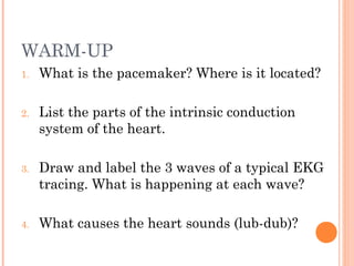
1. WARM-UP
1. What is the pacemaker? Where is it located?
2. List the parts of the intrinsic conduction
system of the heart.
3. Draw and label the 3 waves of a typical EKG
tracing. What is happening at each wave?
4. What causes the heart sounds (lub-dub)?
2. WARM-UP
1. Compare arteries, capillaries, & veins.
1. Imagine you are a red blood cell. List the
pathway you would travel through the body in a
complete circuit starting at a pinky toe.
2. Explain how blood pressure is measured.
3. WARM-UP
1. What is hypertension? What are possible
causes?
2. What is atherosclerosis?
3. What can you do to prevent atherosclerosis?
4. What treatment options are available for
patients with coronary atherosclerosis?
5. Vascular System: blood circulates inside
closed transport systems
Types of Blood Vessels:
Arteries (takes blood away from heart)
Arterioles
Capillary beds
Venules
Veins (return blood back to heart)
6. ANATOMY OF BLOOD VESSELS
Three coats (tunics):
1. Tunica intima: endothelium
lines the interior of vessels;
decreases friction as blood
flows
2. Tunica media: smooth muscle
& elastic tissue (dilates &
constricts vessels)
3. Tunica externa: fibrous
connective tissue on outside
supports and protects vessels
7.
8. ArteriesArteries CapillariesCapillaries VeinsVeins
• Blood away
from heart
• Thicker walls
• Withstand high
pressure
• Walls 1-cell
thick
• Exchange gases
between blood
and tissue cells
• Blood back to
heart
• Thinner walls
• Low pressure
• Large lumen
• ValvesValves: prevent
blood backflow
• Skeletal
muscles
enhance venous
return
9. VERICOSE VEINS
People stand for long periods of time inactivity
or pressure on veins
Blood pools in feet and legs
Valves weaken veins become twisted & dilated
Treatment: compression stockings, exercise, laser
treatment, surgery
10. VITAL SIGNS
Pulse: expansion &
recoil of an artery with
each beat of left
ventricle
Pressure points (eg.
carotid artery, radial
artery)
Normal resting: 70-76
beats/min
11. VITAL SIGNS
Blood pressure: pressure of blood on inner
walls of blood vessels
Systolic presure: peak of ventricular contraction
Diastolic pressure: ventricles relaxed
Written: Systolic/Diastolic
Normal: (120 mm Hg)/(70 mm Hg) or 120/70
14. USING A
SPHYGMOMANOMETER
Wrap cuff around upper arm
Place stethoscope on brachial artery
Inflate cuff to 180 mm Hg
Slowly release air listen for whooshing sounds
in brachial artery (Korotkoff sounds)
Systolic: when sound begin to appear
Diastolic: when sounds disappear