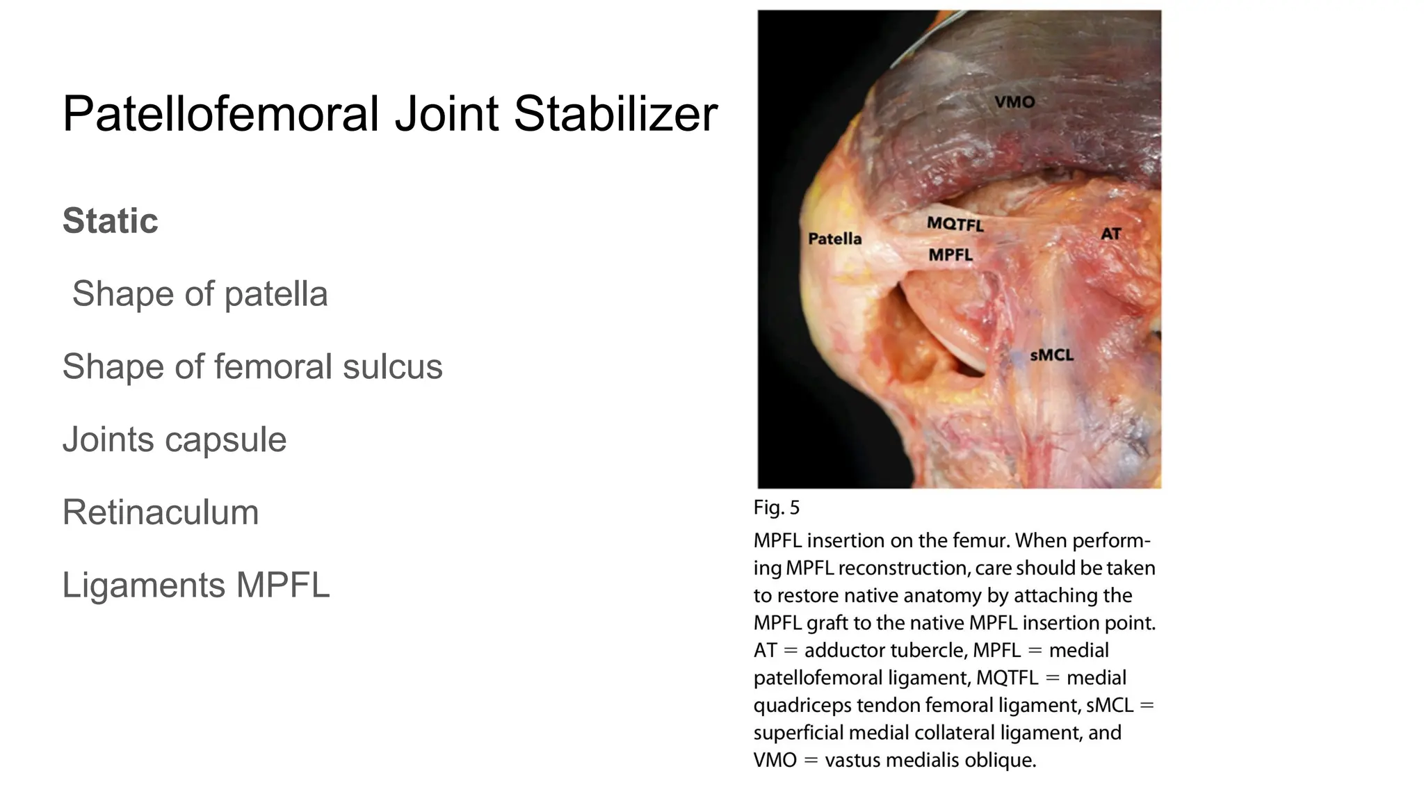
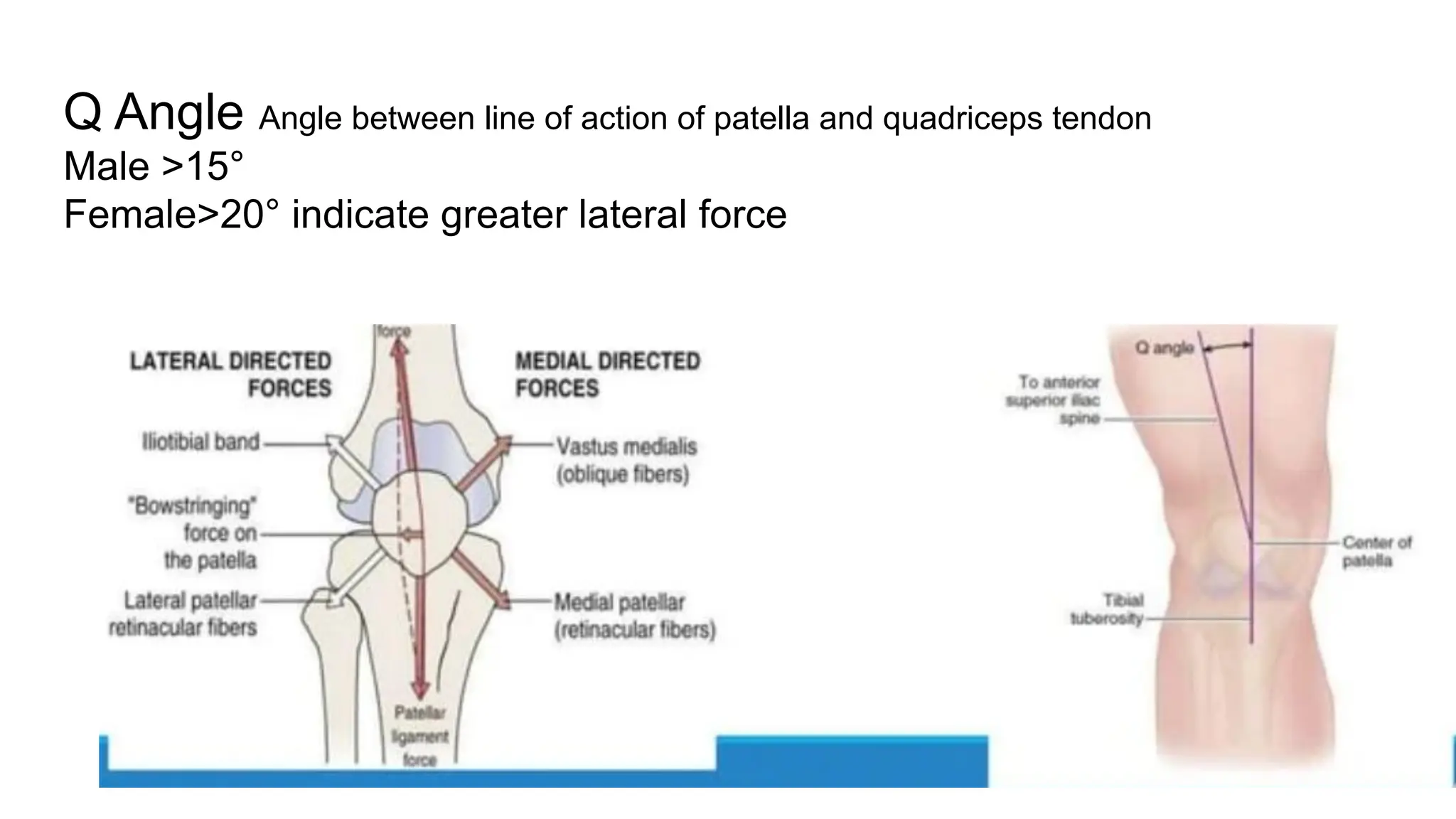
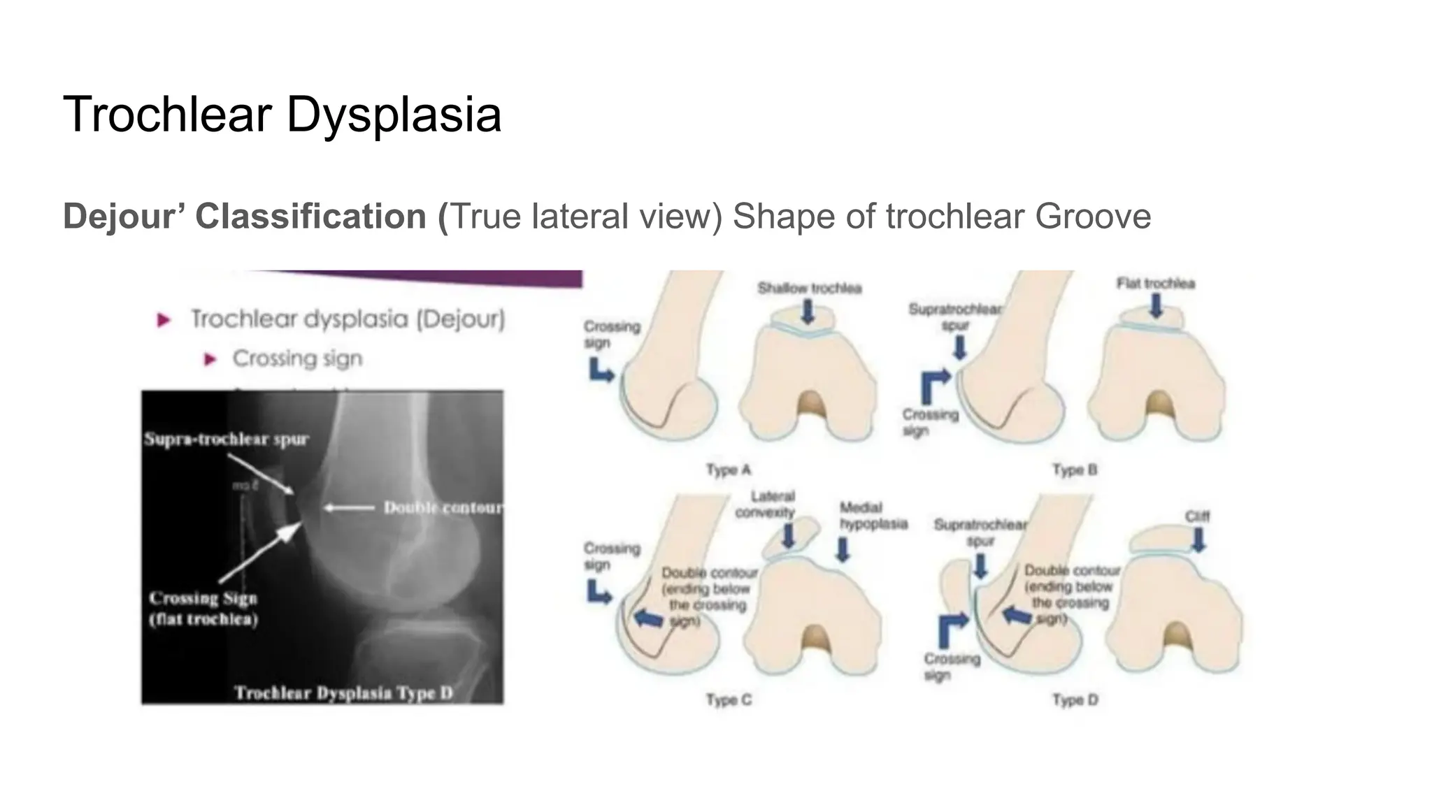
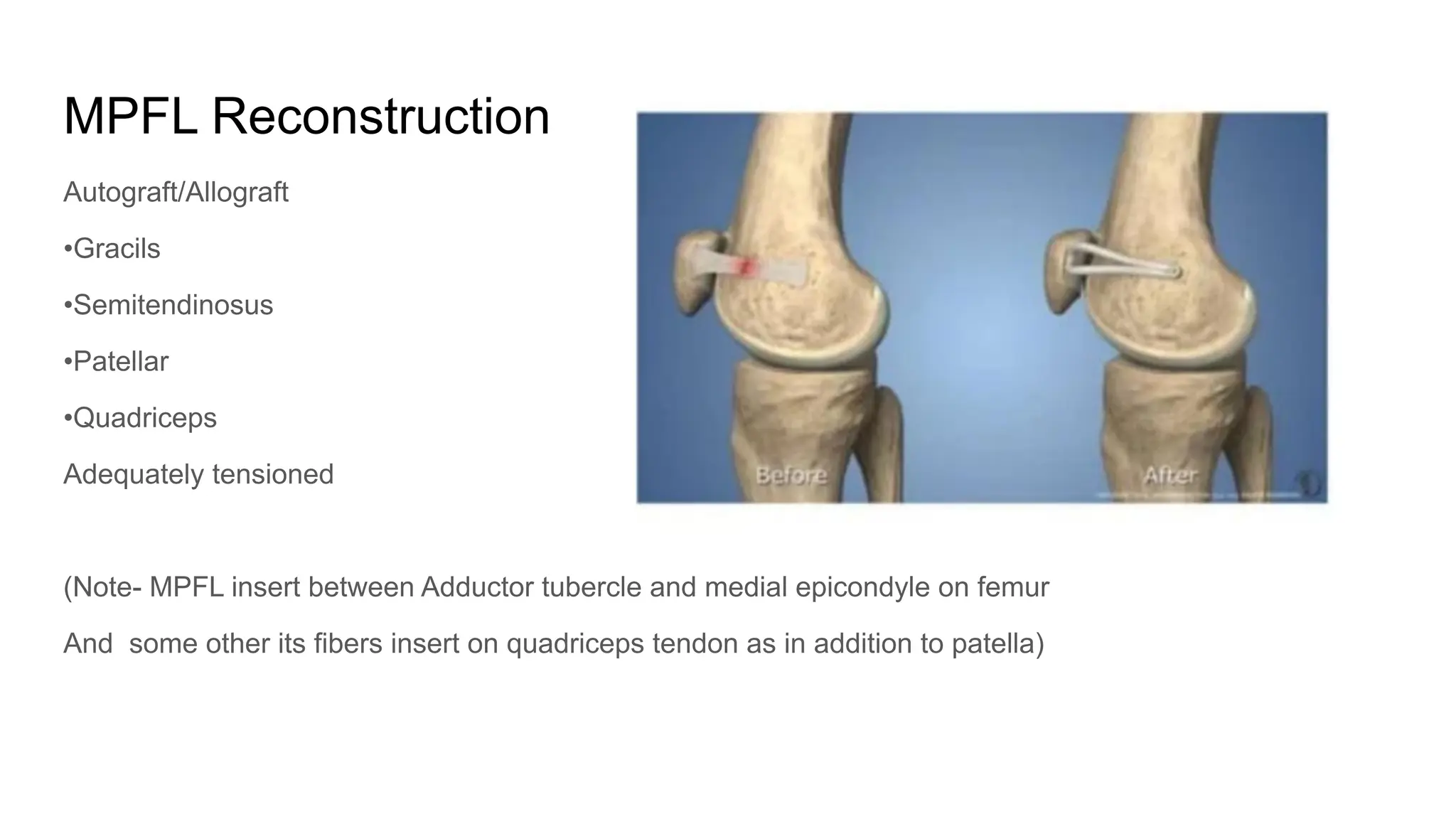
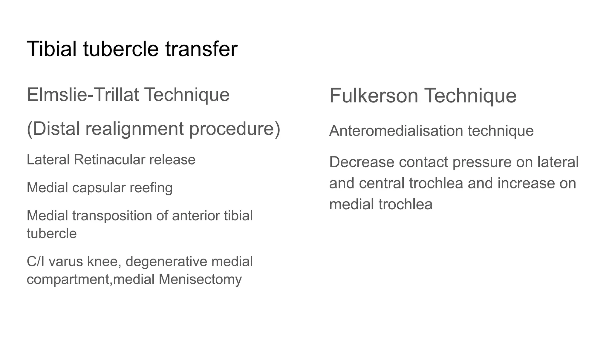
Patellar Instability
Allison K Perry
Bhargavi Maheshwer
Steven F DeFroda
Journal of Bone And Joint Surgery
(Nov 2022 Vol 10 issue 11)
Dr Nitin (Junior Resident)
Introduction
Patellar instability is a potentially devastating diagnosis with high recurrence rate
upto 71%
Return to preinjury level of sport 67% without surgery
After index instability episode, patients with a patellar dislocation have nearly 7
fold increased risk of recurrence
Risk Factors
General Factors
Ligamentous laxity
Previous traumatic event
Miserable Malalignment syndrome
Anatomic Factors
Patella Alta
Trochlear Dysplasia
Increased TT TG and TT PCL distance
Patellar Dysplasia
Excessive patellar tilt
Dysplastic VMO
Overpull of lateral structures:ITB, vastus
lateralis
Recurrence risk factors
Younger Age
Previous contralateral patellar dislocation
Skeletal immaturity
Lateral patellar tilt
Increased TT-TG distance
Patella alta
Trochlear Dysplasia
Classification
Acute - single episode after significant trauma
Almost always lateral
Recurrent- Repeated occasional dislocation
Occurs at interval pf weeks or months
Habitual - Patella dislocates every time the knee is flexed
(Critical Angle- Angle of flexion at which patella dislocates)
Patellofemoral Joint Stabilizer
Static
Shape of patella
Shape of femoral sulcus
Joints capsule
Retinaculum
Ligaments MPFL
Dynamic
Quadriceps Muscle
Vastus medialis obliqus
During full extension to 30° flexion main stabilizer MPFL
Beyond 30° main stabilizer trochlear bony anatomy
(Medial patellotibial and medial patellomeniscal ligament serve as secondary
stabilizer beyond 45°)
Examination
After an instability episode,lower limb
alignment,hypermobility and gait should be
assessed
Patellar height in seated position: high/low
riding patella
Dynamic patellar tracking :Positive J sign
:Patella move laterally on active knee
extension from flexed position.
hard/jumping snap - severe Dysplasia
Or soft glide - Malalignment
Q Angle Angle between line of action of patella and quadriceps tendon
Male >15°
Female>20° indicate greater lateral force
Q angle measured at 20° flexion
•Lateral Subluxation pf patella in full extension can give rise false low value
•External tibial torsion in full extension can increase the apparent Q angle
Factors Increasing Q Angle
Genu Valgum
Excessive femoral anteversiom
External tibial torsion
.lateral Tibial Tuberosity
Beighton Score
>4 out of 9 indicate ligamentous laxity
•passive little finger dorsiflexion >90°
•passive thumb opposition to flexor
aspect of the wrist
• Elbow Hyperextension >10°
•knee Hyperextension >10°
• Ability to flatten both palms on floor
with extended knee and flexed hip
Patellar Glide test
Patella is divided into 4 longitudinal
quadrants
Patella is displaced medially
Medial displacement of one quadrant or
less is suggestive of excessive
retinaculum tightness
If displacement ≥ 3 quadrants- Possible
injury to lateral or medial patellar stabilizer
Patellar Grind test
Pressure over patella
Displace it medially, laterally, superiorly
and inferiorly in the trochlear groove
Positive Test: Anterior knee pain with a
pathologic condition of Patellpfemoral
joint
Rotational profile of staheli
Craig Test (Femoral anteversion) normally 10-15°
Thigh foot angle (rotational deformity of tibia) >30°
Foot progression angle (ntoe gait)
Fairbanks apprehension test
Patellar tilt test: knee in 20 degree flexion
Inability to raise patella with thumb and finger
to the horizontal plane or slightly past indicates
excessive lateral retinacular tightness
Shape of patella
Wiberg classification of patellar shape.
Symptoms
Pain around knee aggregated on walking uphill/downhill or stairs
Feeling of insecurity and knee cap giving away
Patellar crepitation
Knee swelling
Investigations
Xray AP view
Xray knee lateral
Xray axial view
CT Scan
MRI
(MRI >CT)
Xray lateral view
Blumensaat Line
Insall Salvati index
Blackburn peel Ratio
Caton Deschampe index
AXIAL VIEW
Trochlear Dysplasia
Dejour’ Classification (True lateral view) Shape of trochlear Groove
Crossing Sign
The point at which the line of trochlear floor crosses the anterior contour of lateral
femoral condyle
As the crossing sign move inferiorly- More assymetrical condyles and more severe
trochlear Dysplasia
Trochlear Bump
>3mm abnormal
Sulcus Angle > 145° and trochlear depth< 4mm - Trochlear Dysplasia
Congruence angle > 16° - patellar Subluxation
CT Scan
TT TG Distance
(>20 mm is suggest Malalignment)
Patellar Tilt
Normal <20
MRI
Useful in assessment
of bony,cartilaginous
and ligamentous
lesions
eg MPFL
Associated chondral
injuries
MRI vs CT
MRI TT-TG values were 4.16 mm less than CT measurement
TT-PCL distance can be measured on MRI with excellent reliability
TT PCL >24 mm abnormal
(Represent true
tibial tubercle
lateralisation)
Treatment
Aim
Decrease swelling and relieve pain
Enhance restraints
Painfree ROM
Address the Pathoanatomy causing Dislocation
Acute Dislocation
Conservative
Immobilization for 6 Weeks : Stiffness
Immediate mobilization with brace : recurrence
Surgery indicated for: 1 osteochondral fractures
2 Loose bodies
3 Athlete with high level activity
MPFL reconstruction +VMO Repair
A systematic review of meta-analysis comparing nonoperative and operative management found that surgery have lower
rate of recurrent dislocation but does not improve functional outcome
A recent RCT demonstrated - MPFLR 6.7% instability rates
Non operative - 41.9% instability rates
Recurrent Dislocation
Surgery
No single universal surgical procedure
Choice of surgery depends on
Pathological anatomy
Patient demand
Skeletal maturity
Lateral release
Medial repair
MPFL reconstruction
Trochleoplasty
Proximal realignment procedure
Distal realignment procedure
(Patellar dislocation recurrence may occur after surgical stabilization 8.4% upto 5
years postop)
MPFL Reconstruction
Autograft/Allograft
•Gracils
•Semitendinosus
•Patellar
•Quadriceps
Adequately tensioned
(Note- MPFL insert between Adductor tubercle and medial epicondyle on femur
And some other its fibers insert on quadriceps tendon as in addition to patella)
Complications of MPFLR
Patellar fracture ( Transpatellar bone tunnel - provide greater stability than suture
repair but increase risk of iatrogenic fracture and other complication rate)
Postoperative instability
Flexion loss
Pain
Overall complication rate 26%
Tibial tubercle transfer
Elmslie-Trillat Technique
(Distal realignment procedure)
Lateral Retinacular release
Medial capsular reefing
Medial transposition of anterior tibial
tubercle
C/I varus knee, degenerative medial
compartment,medial Menisectomy
Fulkerson Technique
Anteromedialisation technique
Decrease contact pressure on lateral
and central trochlea and increase on
medial trochlea
Proximal realignment procedure
Done in skeletally immature individual
Lateral Retinaculum and capsule released
Quadriceps tendon split and suture to patella
Trochleoplasty
Indications
J sign with TT TG distance >20
mm
Dome shaped trochlea
Reduces risk of dislocation but
does not halt the progression of
arthritis
Summary
Presence of a preoperative J sign is predictive of recurrent instability after
operative management
Nonoperative treatment with patellar bracing and therapy best for primary patellar
dislocations
Aspiration of effusion acutely allows patient to regain quadriceps strength and
control
Loose body - arthroscopic removal / fixation +/- medial repair
Primary repair indicated when there is extensiveextensive medial side injury,
femoral avulsion of MPFL and avulsion of VMO
In case of recurrence treatment should be directed at correcting underlying
pathological condition
No role of isolated lateral release for patellar instability
MPFLR the choice of recurrent instability with or without trochlear Dysplasia with
normal TT TG distance and normal patellar height
Distal realignment- Choice for increased TT TG distance and patella alta
( standard medialisation of TT- Normal patellar height and trochlear anatomy and
increased TT TG
Distalisation- added if there is concomitant patella alta
Anteromedialisation of TT- Lateral and/or distal patellar facet Chondrosis)

Patellar Instability: Diagnosis Management

  • 1.
    Patellar Instability Allison KPerry Bhargavi Maheshwer Steven F DeFroda Journal of Bone And Joint Surgery (Nov 2022 Vol 10 issue 11) Dr Nitin (Junior Resident)
  • 2.
    Introduction Patellar instability isa potentially devastating diagnosis with high recurrence rate upto 71% Return to preinjury level of sport 67% without surgery After index instability episode, patients with a patellar dislocation have nearly 7 fold increased risk of recurrence
  • 3.
    Risk Factors General Factors Ligamentouslaxity Previous traumatic event Miserable Malalignment syndrome Anatomic Factors Patella Alta Trochlear Dysplasia Increased TT TG and TT PCL distance Patellar Dysplasia Excessive patellar tilt Dysplastic VMO Overpull of lateral structures:ITB, vastus lateralis
  • 4.
    Recurrence risk factors YoungerAge Previous contralateral patellar dislocation Skeletal immaturity Lateral patellar tilt Increased TT-TG distance Patella alta Trochlear Dysplasia
  • 5.
    Classification Acute - singleepisode after significant trauma Almost always lateral Recurrent- Repeated occasional dislocation Occurs at interval pf weeks or months Habitual - Patella dislocates every time the knee is flexed (Critical Angle- Angle of flexion at which patella dislocates)
  • 6.
    Patellofemoral Joint Stabilizer Static Shapeof patella Shape of femoral sulcus Joints capsule Retinaculum Ligaments MPFL
  • 7.
    Dynamic Quadriceps Muscle Vastus medialisobliqus During full extension to 30° flexion main stabilizer MPFL Beyond 30° main stabilizer trochlear bony anatomy (Medial patellotibial and medial patellomeniscal ligament serve as secondary stabilizer beyond 45°)
  • 8.
    Examination After an instabilityepisode,lower limb alignment,hypermobility and gait should be assessed Patellar height in seated position: high/low riding patella Dynamic patellar tracking :Positive J sign :Patella move laterally on active knee extension from flexed position. hard/jumping snap - severe Dysplasia Or soft glide - Malalignment
  • 9.
    Q Angle Anglebetween line of action of patella and quadriceps tendon Male >15° Female>20° indicate greater lateral force
  • 10.
    Q angle measuredat 20° flexion •Lateral Subluxation pf patella in full extension can give rise false low value •External tibial torsion in full extension can increase the apparent Q angle
  • 11.
    Factors Increasing QAngle Genu Valgum Excessive femoral anteversiom External tibial torsion .lateral Tibial Tuberosity
  • 12.
    Beighton Score >4 outof 9 indicate ligamentous laxity •passive little finger dorsiflexion >90° •passive thumb opposition to flexor aspect of the wrist • Elbow Hyperextension >10° •knee Hyperextension >10° • Ability to flatten both palms on floor with extended knee and flexed hip
  • 13.
    Patellar Glide test Patellais divided into 4 longitudinal quadrants Patella is displaced medially Medial displacement of one quadrant or less is suggestive of excessive retinaculum tightness If displacement ≥ 3 quadrants- Possible injury to lateral or medial patellar stabilizer
  • 14.
    Patellar Grind test Pressureover patella Displace it medially, laterally, superiorly and inferiorly in the trochlear groove Positive Test: Anterior knee pain with a pathologic condition of Patellpfemoral joint
  • 15.
    Rotational profile ofstaheli Craig Test (Femoral anteversion) normally 10-15° Thigh foot angle (rotational deformity of tibia) >30° Foot progression angle (ntoe gait)
  • 16.
    Fairbanks apprehension test Patellartilt test: knee in 20 degree flexion Inability to raise patella with thumb and finger to the horizontal plane or slightly past indicates excessive lateral retinacular tightness
  • 17.
    Shape of patella Wibergclassification of patellar shape.
  • 18.
    Symptoms Pain around kneeaggregated on walking uphill/downhill or stairs Feeling of insecurity and knee cap giving away Patellar crepitation Knee swelling
  • 19.
    Investigations Xray AP view Xrayknee lateral Xray axial view CT Scan MRI (MRI >CT)
  • 20.
  • 21.
    Insall Salvati index Blackburnpeel Ratio Caton Deschampe index
  • 22.
  • 23.
    Trochlear Dysplasia Dejour’ Classification(True lateral view) Shape of trochlear Groove
  • 24.
    Crossing Sign The pointat which the line of trochlear floor crosses the anterior contour of lateral femoral condyle As the crossing sign move inferiorly- More assymetrical condyles and more severe trochlear Dysplasia Trochlear Bump >3mm abnormal
  • 25.
    Sulcus Angle >145° and trochlear depth< 4mm - Trochlear Dysplasia Congruence angle > 16° - patellar Subluxation
  • 26.
    CT Scan TT TGDistance (>20 mm is suggest Malalignment)
  • 27.
  • 29.
    MRI Useful in assessment ofbony,cartilaginous and ligamentous lesions eg MPFL Associated chondral injuries
  • 30.
    MRI vs CT MRITT-TG values were 4.16 mm less than CT measurement TT-PCL distance can be measured on MRI with excellent reliability TT PCL >24 mm abnormal (Represent true tibial tubercle lateralisation)
  • 32.
    Treatment Aim Decrease swelling andrelieve pain Enhance restraints Painfree ROM Address the Pathoanatomy causing Dislocation
  • 33.
    Acute Dislocation Conservative Immobilization for6 Weeks : Stiffness Immediate mobilization with brace : recurrence Surgery indicated for: 1 osteochondral fractures 2 Loose bodies 3 Athlete with high level activity MPFL reconstruction +VMO Repair A systematic review of meta-analysis comparing nonoperative and operative management found that surgery have lower rate of recurrent dislocation but does not improve functional outcome A recent RCT demonstrated - MPFLR 6.7% instability rates Non operative - 41.9% instability rates
  • 34.
    Recurrent Dislocation Surgery No singleuniversal surgical procedure Choice of surgery depends on Pathological anatomy Patient demand Skeletal maturity
  • 35.
    Lateral release Medial repair MPFLreconstruction Trochleoplasty Proximal realignment procedure Distal realignment procedure (Patellar dislocation recurrence may occur after surgical stabilization 8.4% upto 5 years postop)
  • 36.
    MPFL Reconstruction Autograft/Allograft •Gracils •Semitendinosus •Patellar •Quadriceps Adequately tensioned (Note-MPFL insert between Adductor tubercle and medial epicondyle on femur And some other its fibers insert on quadriceps tendon as in addition to patella)
  • 37.
    Complications of MPFLR Patellarfracture ( Transpatellar bone tunnel - provide greater stability than suture repair but increase risk of iatrogenic fracture and other complication rate) Postoperative instability Flexion loss Pain Overall complication rate 26%
  • 38.
    Tibial tubercle transfer Elmslie-TrillatTechnique (Distal realignment procedure) Lateral Retinacular release Medial capsular reefing Medial transposition of anterior tibial tubercle C/I varus knee, degenerative medial compartment,medial Menisectomy Fulkerson Technique Anteromedialisation technique Decrease contact pressure on lateral and central trochlea and increase on medial trochlea
  • 39.
    Proximal realignment procedure Donein skeletally immature individual Lateral Retinaculum and capsule released Quadriceps tendon split and suture to patella
  • 40.
    Trochleoplasty Indications J sign withTT TG distance >20 mm Dome shaped trochlea Reduces risk of dislocation but does not halt the progression of arthritis
  • 41.
    Summary Presence of apreoperative J sign is predictive of recurrent instability after operative management Nonoperative treatment with patellar bracing and therapy best for primary patellar dislocations Aspiration of effusion acutely allows patient to regain quadriceps strength and control Loose body - arthroscopic removal / fixation +/- medial repair Primary repair indicated when there is extensiveextensive medial side injury, femoral avulsion of MPFL and avulsion of VMO
  • 42.
    In case ofrecurrence treatment should be directed at correcting underlying pathological condition No role of isolated lateral release for patellar instability MPFLR the choice of recurrent instability with or without trochlear Dysplasia with normal TT TG distance and normal patellar height Distal realignment- Choice for increased TT TG distance and patella alta ( standard medialisation of TT- Normal patellar height and trochlear anatomy and increased TT TG Distalisation- added if there is concomitant patella alta Anteromedialisation of TT- Lateral and/or distal patellar facet Chondrosis)