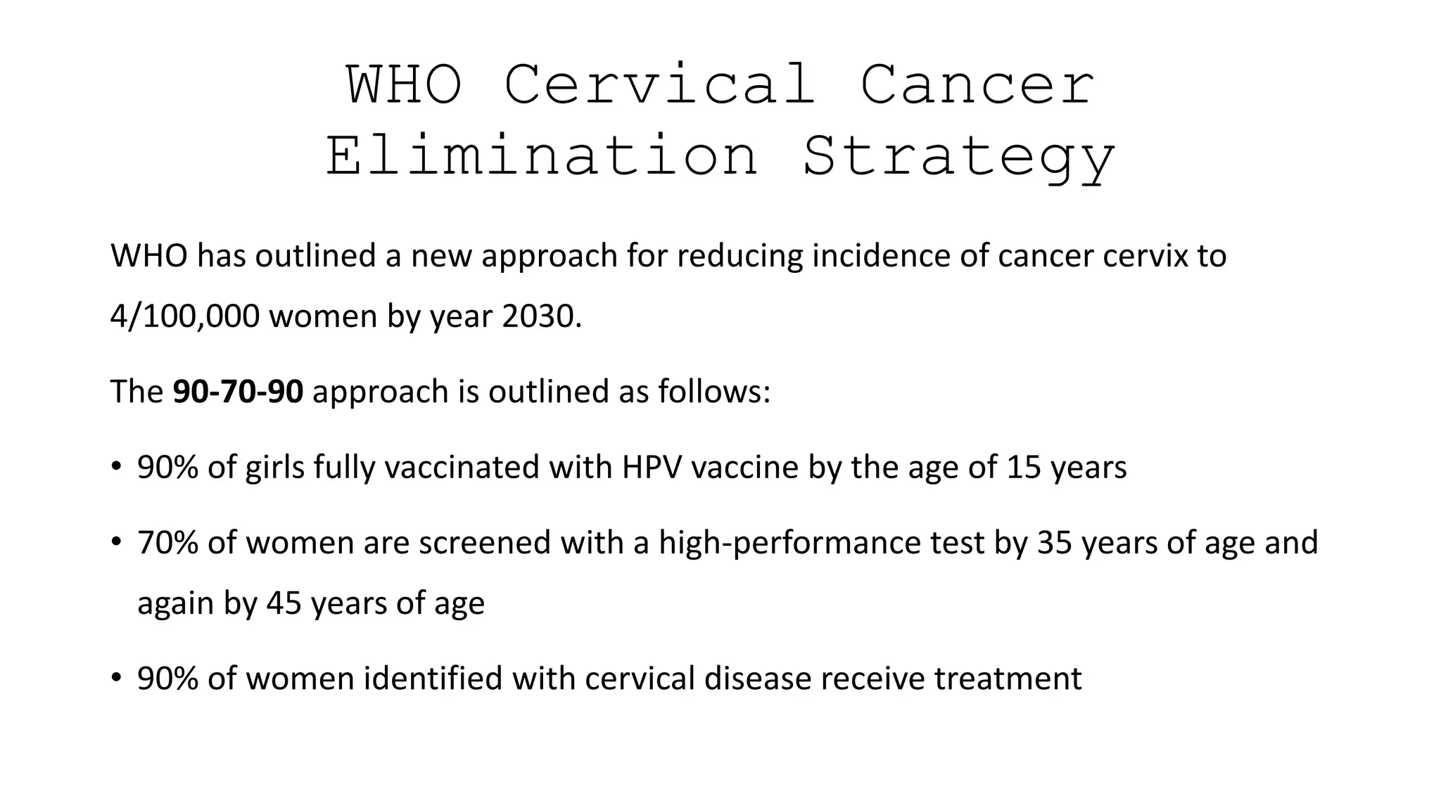
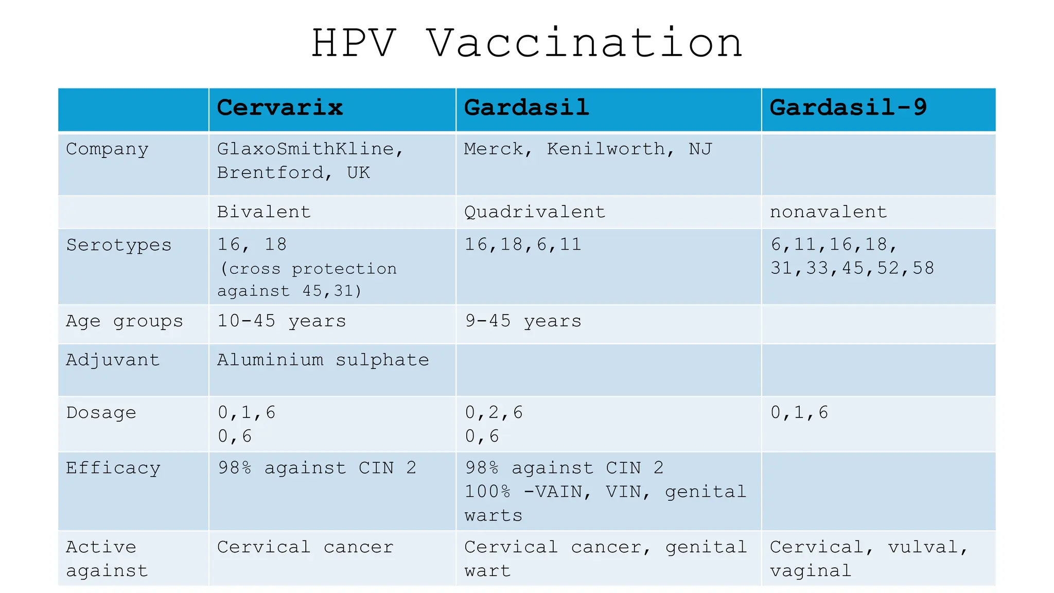
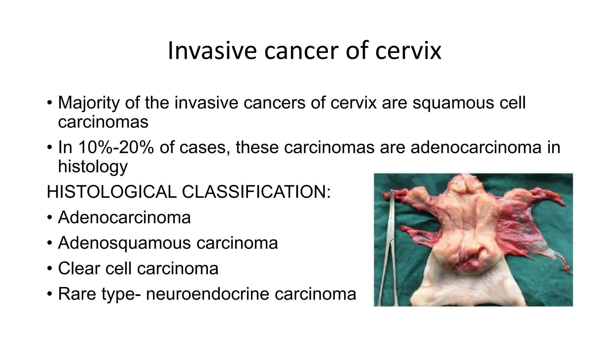
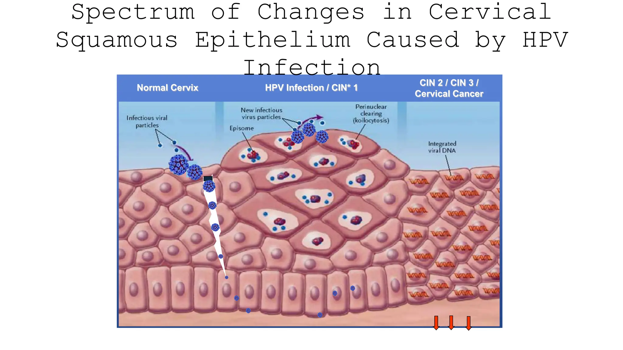
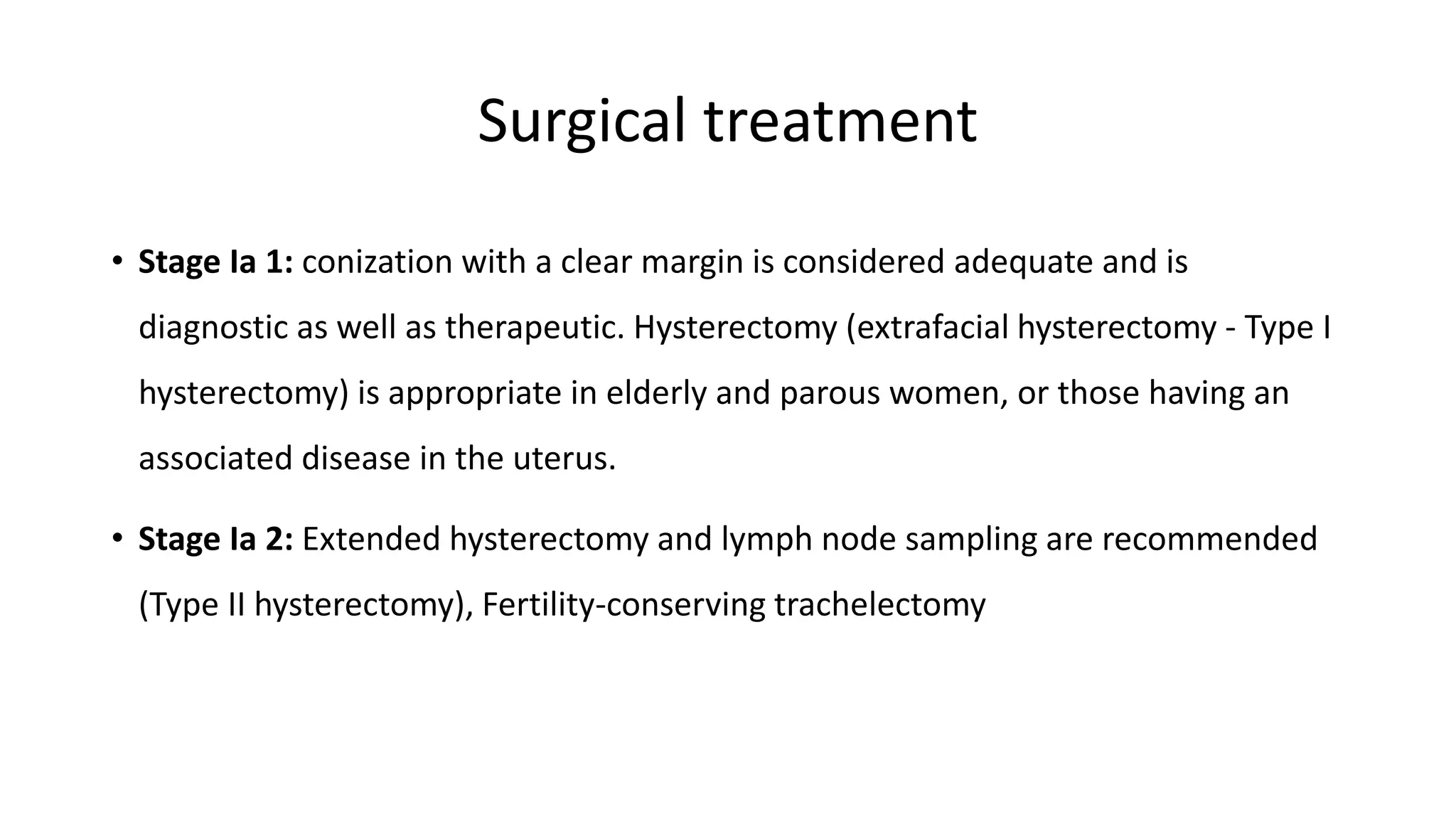
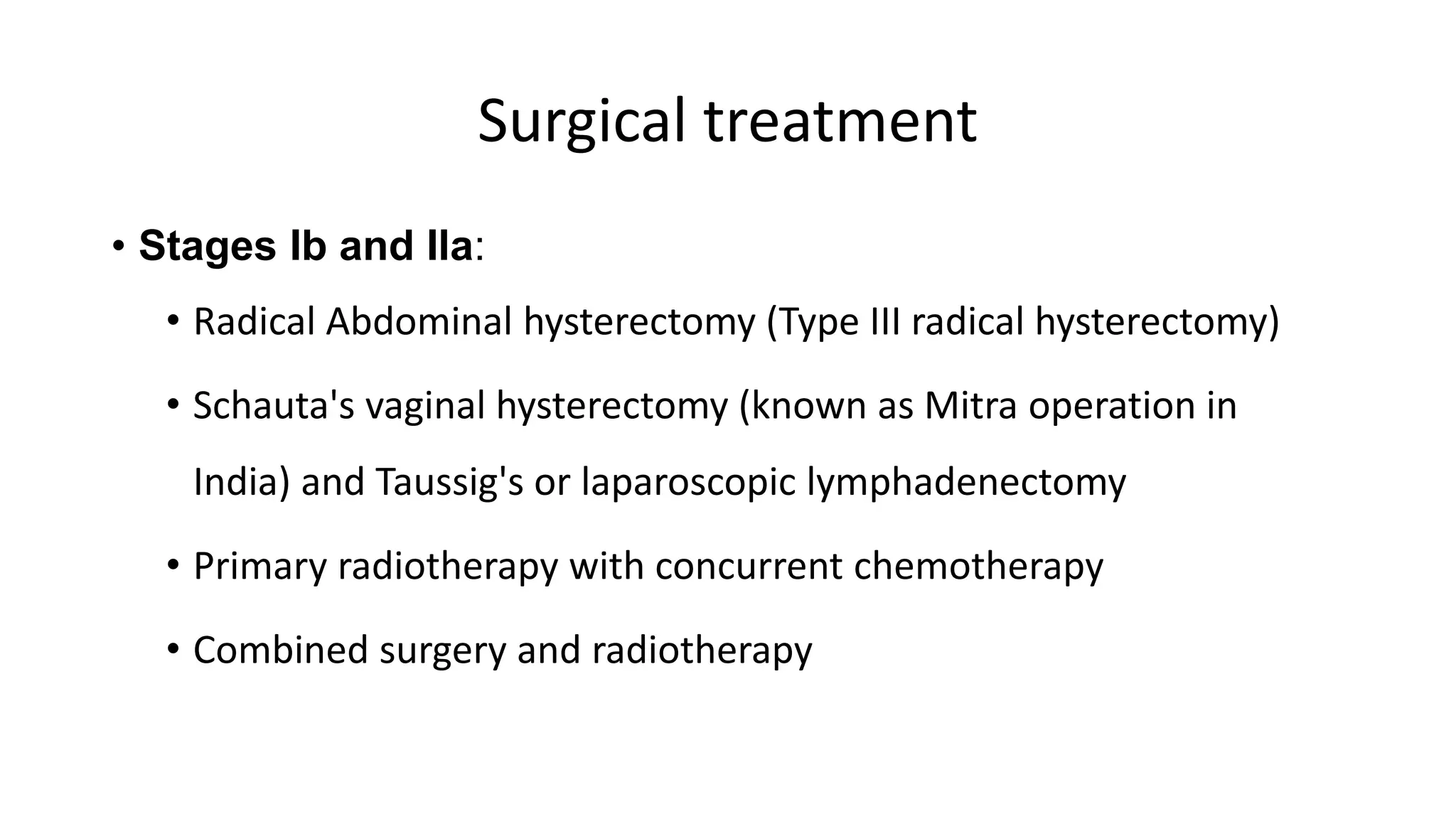
Cervical
Cancer
(Recent
advances)
Presenter: Dr Vinaya
Hugar
Introduction
• Carcinoma of the cervix is the fourth most common cancer among women worldwide and
is now attributable to infection with human papillomavirus (HPV)
• It is the most common genital cancer among women in India.
• It now ranks second to carcinoma of the breast amongst cancers in women
• The universal application of Pap smears in western communities has led to a drastic
decline in the number of invasive cancers of the cervix and a higher detection of
preinvasive lesions
Introduction
• Every year 530,000 new cases and 275,000 deaths are reported annually worldwide
• In India: 130,000 new cases occur with a death toll of 70,000 cases every year
• In India, the incidence is 20-35/100,000 women between 35 and 65 years
• In developed countries, where screening programmes are on, the incidence of
invasive cancer of cervix has fallen to 8/100,000 women.
Cancer Incidence : India
WHO Cervical Cancer
Elimination Strategy
WHO has outlined a new approach for reducing incidence of cancer cervix to
4/100,000 women by year 2030.
The 90-70-90 approach is outlined as follows:
• 90% of girls fully vaccinated with HPV vaccine by the age of 15 years
• 70% of women are screened with a high-performance test by 35 years of age and
again by 45 years of age
• 90% of women identified with cervical disease receive treatment
Predisposing factors for Ca Cervix
• HPV infection
• Coitus before the age of 18 years
• Multiple sexual partners
• Delivery of the first baby before the age of 20 years
• Multiparity with poor spacing between pregnancies
• Sexually transmitted diseases
• Poor personal hygiene
• Poor socioeconomic status
• Smoking and drug abuse, including alcohol
• Immunosuppressed individuals
• Women with preinvasive lesions of cervix
• Use of OCPs
CERVICAL INTRAEPITHELIAL NEOPLASIA (CIN)
• Most cancers of cervix begin in the region of squamocolumnar junction.
• Before actual development of cancer cervix, there are changes in the epithelium in the
region of transformation zone; these changes can be picked up on cytology
• These changes have been named cervical dysplasia, cervical intra epithelial neoplasia
(CIN) and lately as squamous intraepithelial lesions (SIL)
CERVICAL INTRAEPITHELIAL NEOPLASIA (CIN)
• Cervical dysplasia - cytological term
• CIN- histopathological description in which a part or the full thickness of the stratified
squamous epithelium is replaced by cells showing varying degrees of dysplasia
• The cells vary in size, shape and polarity.
• There is an alteration in the nuclear cytoplasmic ratio, and the cells reveal large, irregular,
hyperchromatic nuclei with marginal condensation of chromatin material and mitotic
figures
Dysplasia
• Mild dysplasia (CIN-1): undifferentiated cells
are confined to the lower one-third of the
epithelium.
• Moderate dysplasia (CIN-II): Undifferentiated
cells occupy the lower 50%-75% of the
epithelial thickness.
• Severe dysplasia and carcinoma in situ (CIN-
III): In this grade of dysplasia, the entire
thickness of the epithelium is replaced by
abnormal cells
Bethesda classification
• Low-grade squamous intraepithelial
lesions (LSIL): CIN-I
• High-grade squamous intraepithelial
lesions (HSIL): CIN-II and CIN-Ill
Pap smear screening
Comparison of Different Methods of
Treatment of Dysplasia and CIN
TREATMENT OF CERVICAL DYSPLASIA
Low- grade SIL
Management of CIN
PREVENTION OF CANCER OF THE CERVIX
• The success of screening programme world over shows that it is a preventable
cancer.
• Effective screening by Pap smear, VIA/VILI and HPV testing can detect most of
the lesions in preinvasive stage
• HPV vaccine is now available, although it is expensive as of today.
• Given to adolescents before exposure to the virus (before sexual activity begins),
a high protection rate is expected
Cytology (Pap Smear)
• Most commonly employed screening
method
• The Papanicolaou smear, a routine
screening test for cancer of the
uterine cervix, was reported in 1928,
and its efficacy was proved by 1941
• Reduced mortality by cervical cancer
by 70% in developed countries since
1950
Visual inspection methods
• Low cost and effective method of screening in low resource settings
• Offers immediate diagnosis & possibility of simultaneous treatment
• Comparable sensitivity to Pap smear
• Drawback is lower specificity
• Not reliable in post-menopausal women
Cervarix Gardasil Gardasil-9
Company GlaxoSmithKline,
Brentford, UK
Merck, Kenilworth, NJ
Bivalent Quadrivalent nonavalent
Serotypes 16, 18
(cross protection
against 45,31)
16,18,6,11 6,11,16,18,
31,33,45,52,58
Age groups 10-45 years 9-45 years
Adjuvant Aluminium sulphate
Dosage 0,1,6
0,6
0,2,6
0,6
0,1,6
Efficacy 98% against CIN 2 98% against CIN 2
100% -VAIN, VIN, genital
warts
Active
against
Cervical cancer Cervical cancer, genital
wart
Cervical, vulval,
vaginal
HPV Vaccination
Invasive cancer of cervix
• Majority of the invasive cancers of cervix are squamous cell
carcinomas
• In 10%-20% of cases, these carcinomas are adenocarcinoma in
histology
HISTOLOGICAL CLASSIFICATION:
• Adenocarcinoma
• Adenosquamous carcinoma
• Clear cell carcinoma
• Rare type- neuroendocrine carcinoma
HPV
>100 types identified
• ~ 15 oncogenic(high risk)
types, including 16, 18, 31,
33, 35, 39, 45, 51, 52, 58
• HPV-16 (54%) and -18 (13%)
account for majority of
cervical cancers worldwide
• Non oncogenic (low risk) types
include: 6, 11, 40, 42, 43,
44, 54
• HPV-6 and -11 associated
with 90% of genital warts
30–40 anogenital
Non enveloped double-
stranded DNA virus
HPV Oncogenesis
• HPV integration into host cells (not just infection) necessary
• Integration causes expression of E6 , E7 oncogenic proteins
• Uncontrolled degradation of p53 by E6 (thus inhibition of apoptosis) , E7 binds pRb
(leads to continuous activation of the cell cycle)
• Dr. Harald zur Hausen et al received the Nobel Prize in Medicine in 2008 for
isolating oncogenic HPV strains and elucidating the oncogenic process
Mechanism of HPV transmission
& Acquisition
• Sexual contact
• Through sexual intercourse
• Genital–genital, manual–genital, oral–genital
• Genital HPV infection in virgins rare, but may result
from non-penetrative sexual contact
• Proper condom use may reduce the risk, but not fully
protective against infection
• Nonsexual routes
• Mother to newborn (vertical transmission)
• Fomites (e.g., undergarments, surgical gloves, biopsy
forceps)- Hypothesized but not well documented; would
be rare
Spectrum of Changes in Cervical
Squamous Epithelium Caused by HPV
Infection
Normal Cervix HPV Infection / CIN* 1
CIN 2 / CIN 3 /
Cervical Cancer
Clinical features
• Irregular menses
• Menometrorrhagia
• Continuous bleeding
• Postcoital bleeding
• Leucorrhoea
• Blood-stained or offensive discharge.
DIFFERENTIAL DIAGNOSIS
• The cervical growth and ulcer may at times be mistaken for
• Tubercular and syphilitic ulcer
• Mucus and fibroid polyp
• Rarely sarcoma of the cervix
• Biopsy helps in ruling out other conditions
STAGING OF CANCER OF THE CERVIX
(REVISED FIGO STAGING 2018)
Treatment of invasive cancer
• Treatment depends on the stage of the disease.
• In case there is a need to preserve fertility, a conservative surgical
procedure is possible in early-stage disease
• Better understanding of early lesions has permitted a more
conservative surgical treatment without compromising the success
Surgical treatment
• Stage Ia 1: conization with a clear margin is considered adequate and is
diagnostic as well as therapeutic. Hysterectomy (extrafacial hysterectomy - Type I
hysterectomy) is appropriate in elderly and parous women, or those having an
associated disease in the uterus.
• Stage Ia 2: Extended hysterectomy and lymph node sampling are recommended
(Type II hysterectomy), Fertility-conserving trachelectomy
Surgical treatment
• Stages Ib and Ila:
• Radical Abdominal hysterectomy (Type III radical hysterectomy)
• Schauta's vaginal hysterectomy (known as Mitra operation in
India) and Taussig's or laparoscopic lymphadenectomy
• Primary radiotherapy with concurrent chemotherapy
• Combined surgery and radiotherapy
Radiotherapy
• Radiotherapy is applicable in stages such as Stages Ilb, Illa and Illb where surgery
is not feasible.
• Primary radiotherapy consists of intracavity brachytherapy and external radiation
to the pelvis. It yields the same 5-year cure rate as that of surgery, i.e. 80%-90%.
• Many surgical cases show positive lymph node metastasis which requires
additional postoperative radiotherapy anyway, and this combined therapy
increases the morbidity in the woman
Chemotherapy
• Addition of chemotherapy with cisplatin weekly to radiotherapy improves the
radiation effect, as cisplatin acts as a radiosensitizer agent.
• Current standard of radiation therapy is to combine it with weekly cisplatin when
the patient is undergoing external beam radiation.
Indications for Postoperative
Radiotherapy
• Positive lymph nodes for metastasis
• Positive resected margin of vagina or parametrium
• Evidence of lymphovascular invasion or deep stromal invasion
• Poorly differentiated tumour
Thank you

Recent advances on Cervical cancer .pptx

  • 1.
  • 2.
    Introduction • Carcinoma ofthe cervix is the fourth most common cancer among women worldwide and is now attributable to infection with human papillomavirus (HPV) • It is the most common genital cancer among women in India. • It now ranks second to carcinoma of the breast amongst cancers in women • The universal application of Pap smears in western communities has led to a drastic decline in the number of invasive cancers of the cervix and a higher detection of preinvasive lesions
  • 3.
    Introduction • Every year530,000 new cases and 275,000 deaths are reported annually worldwide • In India: 130,000 new cases occur with a death toll of 70,000 cases every year • In India, the incidence is 20-35/100,000 women between 35 and 65 years • In developed countries, where screening programmes are on, the incidence of invasive cancer of cervix has fallen to 8/100,000 women.
  • 4.
  • 5.
    WHO Cervical Cancer EliminationStrategy WHO has outlined a new approach for reducing incidence of cancer cervix to 4/100,000 women by year 2030. The 90-70-90 approach is outlined as follows: • 90% of girls fully vaccinated with HPV vaccine by the age of 15 years • 70% of women are screened with a high-performance test by 35 years of age and again by 45 years of age • 90% of women identified with cervical disease receive treatment
  • 6.
    Predisposing factors forCa Cervix • HPV infection • Coitus before the age of 18 years • Multiple sexual partners • Delivery of the first baby before the age of 20 years • Multiparity with poor spacing between pregnancies • Sexually transmitted diseases • Poor personal hygiene • Poor socioeconomic status • Smoking and drug abuse, including alcohol • Immunosuppressed individuals • Women with preinvasive lesions of cervix • Use of OCPs
  • 7.
    CERVICAL INTRAEPITHELIAL NEOPLASIA(CIN) • Most cancers of cervix begin in the region of squamocolumnar junction. • Before actual development of cancer cervix, there are changes in the epithelium in the region of transformation zone; these changes can be picked up on cytology • These changes have been named cervical dysplasia, cervical intra epithelial neoplasia (CIN) and lately as squamous intraepithelial lesions (SIL)
  • 8.
    CERVICAL INTRAEPITHELIAL NEOPLASIA(CIN) • Cervical dysplasia - cytological term • CIN- histopathological description in which a part or the full thickness of the stratified squamous epithelium is replaced by cells showing varying degrees of dysplasia • The cells vary in size, shape and polarity. • There is an alteration in the nuclear cytoplasmic ratio, and the cells reveal large, irregular, hyperchromatic nuclei with marginal condensation of chromatin material and mitotic figures
  • 9.
    Dysplasia • Mild dysplasia(CIN-1): undifferentiated cells are confined to the lower one-third of the epithelium. • Moderate dysplasia (CIN-II): Undifferentiated cells occupy the lower 50%-75% of the epithelial thickness. • Severe dysplasia and carcinoma in situ (CIN- III): In this grade of dysplasia, the entire thickness of the epithelium is replaced by abnormal cells Bethesda classification • Low-grade squamous intraepithelial lesions (LSIL): CIN-I • High-grade squamous intraepithelial lesions (HSIL): CIN-II and CIN-Ill
  • 10.
  • 11.
    Comparison of DifferentMethods of Treatment of Dysplasia and CIN
  • 12.
    TREATMENT OF CERVICALDYSPLASIA Low- grade SIL
  • 13.
  • 14.
    PREVENTION OF CANCEROF THE CERVIX • The success of screening programme world over shows that it is a preventable cancer. • Effective screening by Pap smear, VIA/VILI and HPV testing can detect most of the lesions in preinvasive stage • HPV vaccine is now available, although it is expensive as of today. • Given to adolescents before exposure to the virus (before sexual activity begins), a high protection rate is expected
  • 15.
    Cytology (Pap Smear) •Most commonly employed screening method • The Papanicolaou smear, a routine screening test for cancer of the uterine cervix, was reported in 1928, and its efficacy was proved by 1941 • Reduced mortality by cervical cancer by 70% in developed countries since 1950
  • 16.
    Visual inspection methods •Low cost and effective method of screening in low resource settings • Offers immediate diagnosis & possibility of simultaneous treatment • Comparable sensitivity to Pap smear • Drawback is lower specificity • Not reliable in post-menopausal women
  • 17.
    Cervarix Gardasil Gardasil-9 CompanyGlaxoSmithKline, Brentford, UK Merck, Kenilworth, NJ Bivalent Quadrivalent nonavalent Serotypes 16, 18 (cross protection against 45,31) 16,18,6,11 6,11,16,18, 31,33,45,52,58 Age groups 10-45 years 9-45 years Adjuvant Aluminium sulphate Dosage 0,1,6 0,6 0,2,6 0,6 0,1,6 Efficacy 98% against CIN 2 98% against CIN 2 100% -VAIN, VIN, genital warts Active against Cervical cancer Cervical cancer, genital wart Cervical, vulval, vaginal HPV Vaccination
  • 19.
    Invasive cancer ofcervix • Majority of the invasive cancers of cervix are squamous cell carcinomas • In 10%-20% of cases, these carcinomas are adenocarcinoma in histology HISTOLOGICAL CLASSIFICATION: • Adenocarcinoma • Adenosquamous carcinoma • Clear cell carcinoma • Rare type- neuroendocrine carcinoma
  • 20.
    HPV >100 types identified •~ 15 oncogenic(high risk) types, including 16, 18, 31, 33, 35, 39, 45, 51, 52, 58 • HPV-16 (54%) and -18 (13%) account for majority of cervical cancers worldwide • Non oncogenic (low risk) types include: 6, 11, 40, 42, 43, 44, 54 • HPV-6 and -11 associated with 90% of genital warts 30–40 anogenital Non enveloped double- stranded DNA virus
  • 21.
    HPV Oncogenesis • HPVintegration into host cells (not just infection) necessary • Integration causes expression of E6 , E7 oncogenic proteins • Uncontrolled degradation of p53 by E6 (thus inhibition of apoptosis) , E7 binds pRb (leads to continuous activation of the cell cycle) • Dr. Harald zur Hausen et al received the Nobel Prize in Medicine in 2008 for isolating oncogenic HPV strains and elucidating the oncogenic process
  • 22.
    Mechanism of HPVtransmission & Acquisition • Sexual contact • Through sexual intercourse • Genital–genital, manual–genital, oral–genital • Genital HPV infection in virgins rare, but may result from non-penetrative sexual contact • Proper condom use may reduce the risk, but not fully protective against infection • Nonsexual routes • Mother to newborn (vertical transmission) • Fomites (e.g., undergarments, surgical gloves, biopsy forceps)- Hypothesized but not well documented; would be rare
  • 23.
    Spectrum of Changesin Cervical Squamous Epithelium Caused by HPV Infection Normal Cervix HPV Infection / CIN* 1 CIN 2 / CIN 3 / Cervical Cancer
  • 24.
    Clinical features • Irregularmenses • Menometrorrhagia • Continuous bleeding • Postcoital bleeding • Leucorrhoea • Blood-stained or offensive discharge.
  • 25.
    DIFFERENTIAL DIAGNOSIS • Thecervical growth and ulcer may at times be mistaken for • Tubercular and syphilitic ulcer • Mucus and fibroid polyp • Rarely sarcoma of the cervix • Biopsy helps in ruling out other conditions
  • 26.
    STAGING OF CANCEROF THE CERVIX (REVISED FIGO STAGING 2018)
  • 27.
    Treatment of invasivecancer • Treatment depends on the stage of the disease. • In case there is a need to preserve fertility, a conservative surgical procedure is possible in early-stage disease • Better understanding of early lesions has permitted a more conservative surgical treatment without compromising the success
  • 28.
    Surgical treatment • StageIa 1: conization with a clear margin is considered adequate and is diagnostic as well as therapeutic. Hysterectomy (extrafacial hysterectomy - Type I hysterectomy) is appropriate in elderly and parous women, or those having an associated disease in the uterus. • Stage Ia 2: Extended hysterectomy and lymph node sampling are recommended (Type II hysterectomy), Fertility-conserving trachelectomy
  • 29.
    Surgical treatment • StagesIb and Ila: • Radical Abdominal hysterectomy (Type III radical hysterectomy) • Schauta's vaginal hysterectomy (known as Mitra operation in India) and Taussig's or laparoscopic lymphadenectomy • Primary radiotherapy with concurrent chemotherapy • Combined surgery and radiotherapy
  • 30.
    Radiotherapy • Radiotherapy isapplicable in stages such as Stages Ilb, Illa and Illb where surgery is not feasible. • Primary radiotherapy consists of intracavity brachytherapy and external radiation to the pelvis. It yields the same 5-year cure rate as that of surgery, i.e. 80%-90%. • Many surgical cases show positive lymph node metastasis which requires additional postoperative radiotherapy anyway, and this combined therapy increases the morbidity in the woman
  • 31.
    Chemotherapy • Addition ofchemotherapy with cisplatin weekly to radiotherapy improves the radiation effect, as cisplatin acts as a radiosensitizer agent. • Current standard of radiation therapy is to combine it with weekly cisplatin when the patient is undergoing external beam radiation.
  • 32.
    Indications for Postoperative Radiotherapy •Positive lymph nodes for metastasis • Positive resected margin of vagina or parametrium • Evidence of lymphovascular invasion or deep stromal invasion • Poorly differentiated tumour
  • 33.