Downloaded 134 times
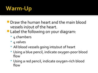

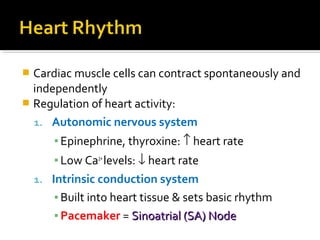
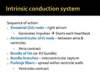
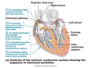


The document covers the anatomy and physiology of the human heart, detailing its chambers, valves, and blood vessels while explaining blood flow using color codes. It discusses the heart's electrical conduction system, regulation of heart rate, and various cardiac conditions, such as angina and myocardial infarction. The document also touches on concepts like cardiac output, heart murmurs, and the impact of various factors on heart health.


