Downloaded 193 times

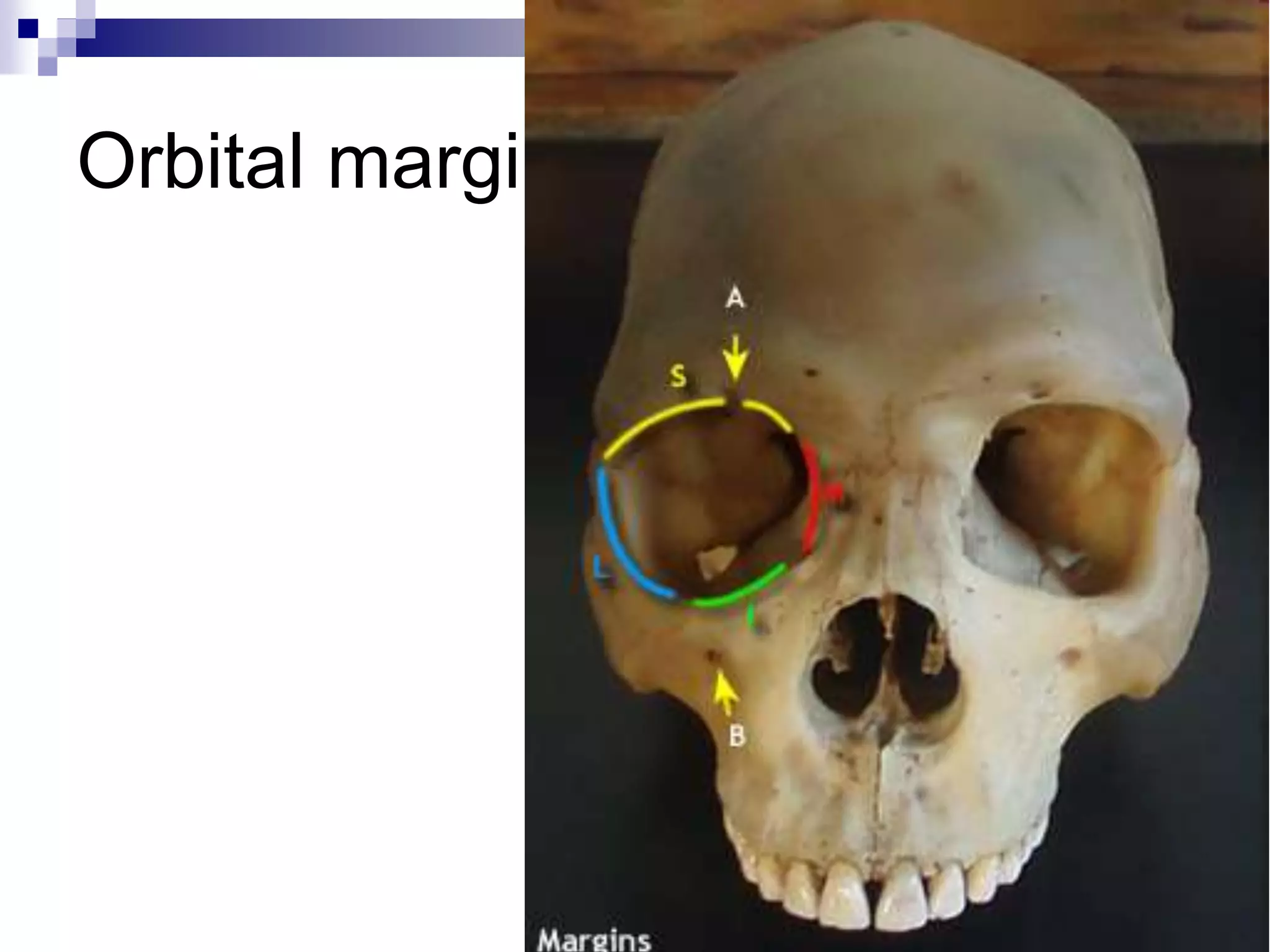
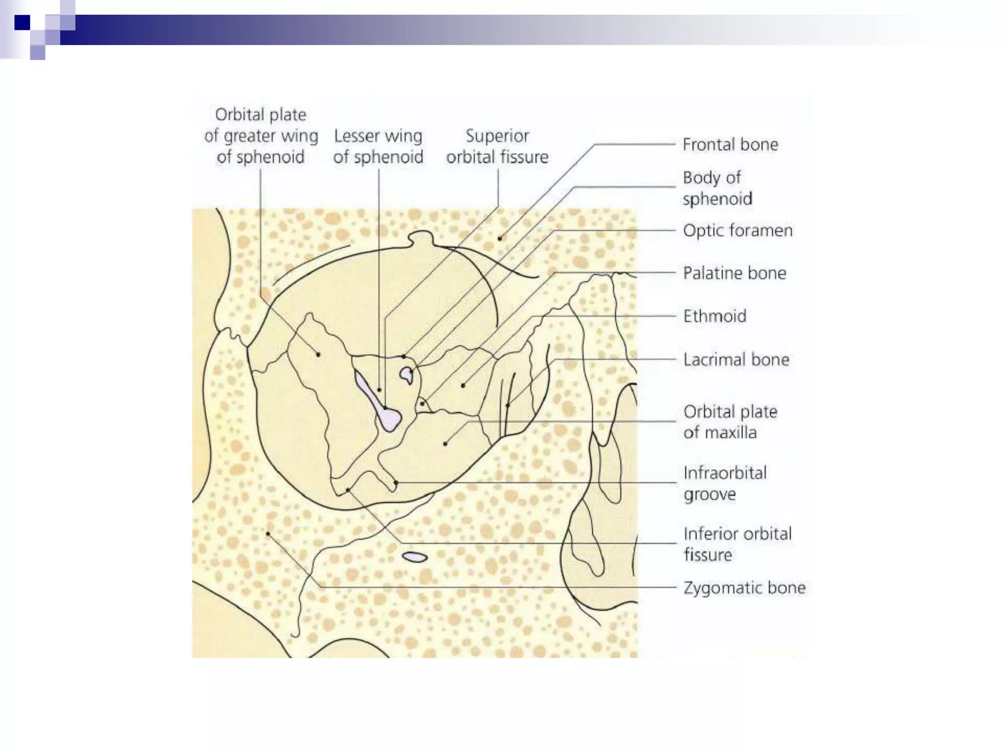


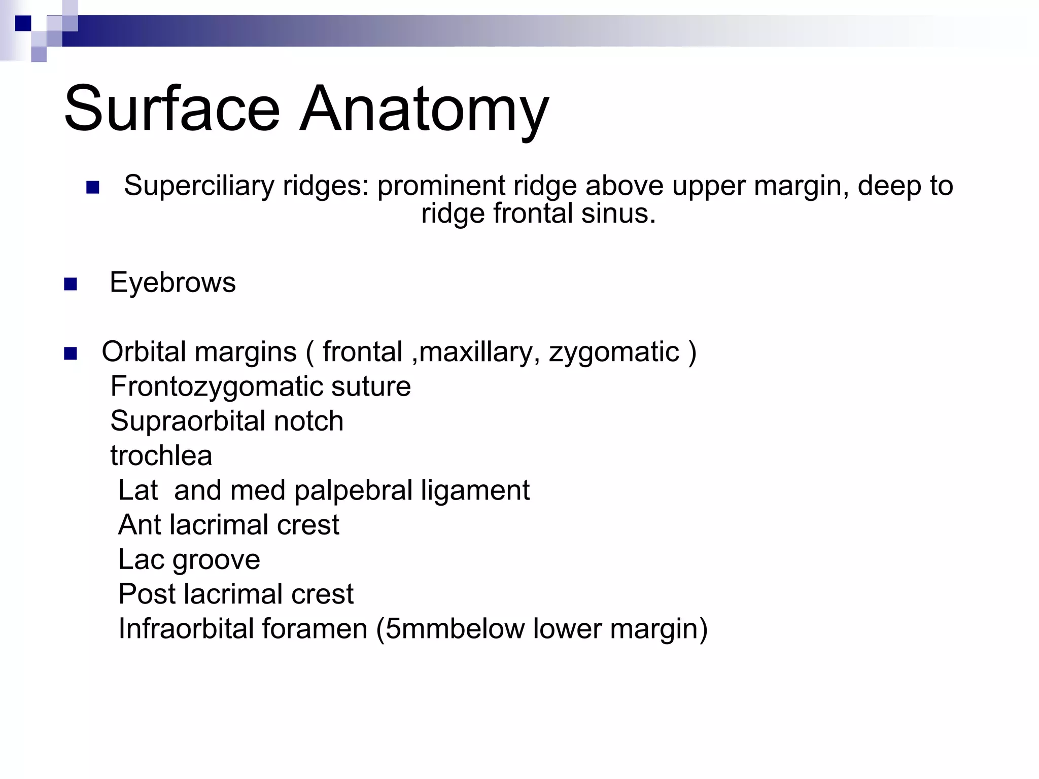


The orbital cavity contains the eyeball and surrounding structures. It is pear shaped with seven bones forming its walls. Key structures discussed include the openings like the superior and inferior orbital fissures which connect the orbit to the brain. The document outlines the anatomy, dimensions, walls, openings and relations of the orbital cavity. It also discusses variations with age and surface anatomy landmarks.


