Each eyelid contains a fibrous plate, called a tarsus, that gives it structure and shape; muscles, which move the eyelids; and meibomian (or tarsal) glands, which secrete lubricating fluids. The lids are covered with skin, lined with mucous membrane, and bordered with a fringe of hairs, the eyelashes.
1. Introduction Gross anatomy Layers Blood supply, drainage and nerve supply
2. INTRODUCTION • Sclera forms posterior 5/6th of external tunic , connective tissue coat of eyeball. • it continues with duramater and cornea • Its whole surface covered by tenon’s capsule • Anteriorly covered by- bulbar conjunctiva • Inner surface lies in contact with choroid • With a potential suprachoroidal space in between
3. Equa THICKNESS OF SCLERA
4. • Thickness varies with individual, with age • Thinner- children, elder, F> M • Thickest posteriorly • Gradually becomes thinner when traced anteriorly • Thin at insertion of extraocular muscle
Bony orbits are Quadrangular truncated pyramids with Anterior cranial fossa above and the maxillary sinuses below.
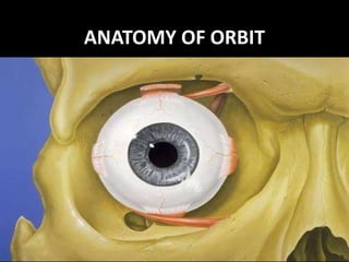
in this presentation we study the detailed anatomy of the arbit, the bones, relations of each wall, the contents, the apertures, orbital fissures and structures passing, fascia, septa and the surgical spaces of the orbit
Details about the anatomy with clinical importance. An easy guide for understanding the walls, surgical spaces, orbital contents, venous and arterial supply. Hope its helpful for your examinations too!!
Each eyelid contains a fibrous plate, called a tarsus, that gives it structure and shape; muscles, which move the eyelids; and meibomian (or tarsal) glands, which secrete lubricating fluids. The lids are covered with skin, lined with mucous membrane, and bordered with a fringe of hairs, the eyelashes.
1. Introduction Gross anatomy Layers Blood supply, drainage and nerve supply
2. INTRODUCTION • Sclera forms posterior 5/6th of external tunic , connective tissue coat of eyeball. • it continues with duramater and cornea • Its whole surface covered by tenon’s capsule • Anteriorly covered by- bulbar conjunctiva • Inner surface lies in contact with choroid • With a potential suprachoroidal space in between
3. Equa THICKNESS OF SCLERA
4. • Thickness varies with individual, with age • Thinner- children, elder, F> M • Thickest posteriorly • Gradually becomes thinner when traced anteriorly • Thin at insertion of extraocular muscle
Bony orbits are Quadrangular truncated pyramids with Anterior cranial fossa above and the maxillary sinuses below.
in this presentation we study the detailed anatomy of the arbit, the bones, relations of each wall, the contents, the apertures, orbital fissures and structures passing, fascia, septa and the surgical spaces of the orbit
Details about the anatomy with clinical importance. An easy guide for understanding the walls, surgical spaces, orbital contents, venous and arterial supply. Hope its helpful for your examinations too!!
Indian Dental Academy: will be one of the most relevant and exciting training center with best faculty and flexible training programs for dental professionals who wish to advance in their dental practice,Offers certified courses in Dental implants,Orthodontics,Endodontics,Cosmetic Dentistry, Prosthetic Dentistry, Periodontics and General Dentistry.
The orbits are conical or four-sided pyramidal cavities, which open into the midline of the face and point back into the head. Each consists of a base, an apex and four walls.[4]
Each orbit is formed by seven bones –
Frontal bone
Ethmoidal bone
Lacrimal bone
Palatine bone
Maxilla bone
Zygomatic bone
Sphenoid bone
Anatomy of Orbit and its clinical importanceAshish Gupta
It's a presentation of Anatomy of Bony Orbit and its applied aspects. It's been made by compiling images from many sources and includes almost all the information needed for a postgraduate .
Title: Sense of Taste
Presenter: Dr. Faiza, Assistant Professor of Physiology
Qualifications:
MBBS (Best Graduate, AIMC Lahore)
FCPS Physiology
ICMT, CHPE, DHPE (STMU)
MPH (GC University, Faisalabad)
MBA (Virtual University of Pakistan)
Learning Objectives:
Describe the structure and function of taste buds.
Describe the relationship between the taste threshold and taste index of common substances.
Explain the chemical basis and signal transduction of taste perception for each type of primary taste sensation.
Recognize different abnormalities of taste perception and their causes.
Key Topics:
Significance of Taste Sensation:
Differentiation between pleasant and harmful food
Influence on behavior
Selection of food based on metabolic needs
Receptors of Taste:
Taste buds on the tongue
Influence of sense of smell, texture of food, and pain stimulation (e.g., by pepper)
Primary and Secondary Taste Sensations:
Primary taste sensations: Sweet, Sour, Salty, Bitter, Umami
Chemical basis and signal transduction mechanisms for each taste
Taste Threshold and Index:
Taste threshold values for Sweet (sucrose), Salty (NaCl), Sour (HCl), and Bitter (Quinine)
Taste index relationship: Inversely proportional to taste threshold
Taste Blindness:
Inability to taste certain substances, particularly thiourea compounds
Example: Phenylthiocarbamide
Structure and Function of Taste Buds:
Composition: Epithelial cells, Sustentacular/Supporting cells, Taste cells, Basal cells
Features: Taste pores, Taste hairs/microvilli, and Taste nerve fibers
Location of Taste Buds:
Found in papillae of the tongue (Fungiform, Circumvallate, Foliate)
Also present on the palate, tonsillar pillars, epiglottis, and proximal esophagus
Mechanism of Taste Stimulation:
Interaction of taste substances with receptors on microvilli
Signal transduction pathways for Umami, Sweet, Bitter, Sour, and Salty tastes
Taste Sensitivity and Adaptation:
Decrease in sensitivity with age
Rapid adaptation of taste sensation
Role of Saliva in Taste:
Dissolution of tastants to reach receptors
Washing away the stimulus
Taste Preferences and Aversions:
Mechanisms behind taste preference and aversion
Influence of receptors and neural pathways
Impact of Sensory Nerve Damage:
Degeneration of taste buds if the sensory nerve fiber is cut
Abnormalities of Taste Detection:
Conditions: Ageusia, Hypogeusia, Dysgeusia (parageusia)
Causes: Nerve damage, neurological disorders, infections, poor oral hygiene, adverse drug effects, deficiencies, aging, tobacco use, altered neurotransmitter levels
Neurotransmitters and Taste Threshold:
Effects of serotonin (5-HT) and norepinephrine (NE) on taste sensitivity
Supertasters:
25% of the population with heightened sensitivity to taste, especially bitterness
Increased number of fungiform papillae
Ozempic: Preoperative Management of Patients on GLP-1 Receptor Agonists Saeid Safari
Preoperative Management of Patients on GLP-1 Receptor Agonists like Ozempic and Semiglutide
ASA GUIDELINE
NYSORA Guideline
2 Case Reports of Gastric Ultrasound
Tom Selleck Health: A Comprehensive Look at the Iconic Actor’s Wellness Journeygreendigital
Tom Selleck, an enduring figure in Hollywood. has captivated audiences for decades with his rugged charm, iconic moustache. and memorable roles in television and film. From his breakout role as Thomas Magnum in Magnum P.I. to his current portrayal of Frank Reagan in Blue Bloods. Selleck's career has spanned over 50 years. But beyond his professional achievements. fans have often been curious about Tom Selleck Health. especially as he has aged in the public eye.
Follow us on: Pinterest
Introduction
Many have been interested in Tom Selleck health. not only because of his enduring presence on screen but also because of the challenges. and lifestyle choices he has faced and made over the years. This article delves into the various aspects of Tom Selleck health. exploring his fitness regimen, diet, mental health. and the challenges he has encountered as he ages. We'll look at how he maintains his well-being. the health issues he has faced, and his approach to ageing .
Early Life and Career
Childhood and Athletic Beginnings
Tom Selleck was born on January 29, 1945, in Detroit, Michigan, and grew up in Sherman Oaks, California. From an early age, he was involved in sports, particularly basketball. which played a significant role in his physical development. His athletic pursuits continued into college. where he attended the University of Southern California (USC) on a basketball scholarship. This early involvement in sports laid a strong foundation for his physical health and disciplined lifestyle.
Transition to Acting
Selleck's transition from an athlete to an actor came with its physical demands. His first significant role in "Magnum P.I." required him to perform various stunts and maintain a fit appearance. This role, which he played from 1980 to 1988. necessitated a rigorous fitness routine to meet the show's demands. setting the stage for his long-term commitment to health and wellness.
Fitness Regimen
Workout Routine
Tom Selleck health and fitness regimen has evolved. adapting to his changing roles and age. During his "Magnum, P.I." days. Selleck's workouts were intense and focused on building and maintaining muscle mass. His routine included weightlifting, cardiovascular exercises. and specific training for the stunts he performed on the show.
Selleck adjusted his fitness routine as he aged to suit his body's needs. Today, his workouts focus on maintaining flexibility, strength, and cardiovascular health. He incorporates low-impact exercises such as swimming, walking, and light weightlifting. This balanced approach helps him stay fit without putting undue strain on his joints and muscles.
Importance of Flexibility and Mobility
In recent years, Selleck has emphasized the importance of flexibility and mobility in his fitness regimen. Understanding the natural decline in muscle mass and joint flexibility with age. he includes stretching and yoga in his routine. These practices help prevent injuries, improve posture, and maintain mobilit
micro teaching on communication m.sc nursing.pdfAnurag Sharma
Microteaching is a unique model of practice teaching. It is a viable instrument for the. desired change in the teaching behavior or the behavior potential which, in specified types of real. classroom situations, tends to facilitate the achievement of specified types of objectives.
Knee anatomy and clinical tests 2024.pdfvimalpl1234
This includes all relevant anatomy and clinical tests compiled from standard textbooks, Campbell,netter etc..It is comprehensive and best suited for orthopaedicians and orthopaedic residents.
Prix Galien International 2024 Forum ProgramLevi Shapiro
June 20, 2024, Prix Galien International and Jerusalem Ethics Forum in ROME. Detailed agenda including panels:
- ADVANCES IN CARDIOLOGY: A NEW PARADIGM IS COMING
- WOMEN’S HEALTH: FERTILITY PRESERVATION
- WHAT’S NEW IN THE TREATMENT OF INFECTIOUS,
ONCOLOGICAL AND INFLAMMATORY SKIN DISEASES?
- ARTIFICIAL INTELLIGENCE AND ETHICS
- GENE THERAPY
- BEYOND BORDERS: GLOBAL INITIATIVES FOR DEMOCRATIZING LIFE SCIENCE TECHNOLOGIES AND PROMOTING ACCESS TO HEALTHCARE
- ETHICAL CHALLENGES IN LIFE SCIENCES
- Prix Galien International Awards Ceremony
New Drug Discovery and Development .....NEHA GUPTA
The "New Drug Discovery and Development" process involves the identification, design, testing, and manufacturing of novel pharmaceutical compounds with the aim of introducing new and improved treatments for various medical conditions. This comprehensive endeavor encompasses various stages, including target identification, preclinical studies, clinical trials, regulatory approval, and post-market surveillance. It involves multidisciplinary collaboration among scientists, researchers, clinicians, regulatory experts, and pharmaceutical companies to bring innovative therapies to market and address unmet medical needs.
New Directions in Targeted Therapeutic Approaches for Older Adults With Mantl...i3 Health
i3 Health is pleased to make the speaker slides from this activity available for use as a non-accredited self-study or teaching resource.
This slide deck presented by Dr. Kami Maddocks, Professor-Clinical in the Division of Hematology and
Associate Division Director for Ambulatory Operations
The Ohio State University Comprehensive Cancer Center, will provide insight into new directions in targeted therapeutic approaches for older adults with mantle cell lymphoma.
STATEMENT OF NEED
Mantle cell lymphoma (MCL) is a rare, aggressive B-cell non-Hodgkin lymphoma (NHL) accounting for 5% to 7% of all lymphomas. Its prognosis ranges from indolent disease that does not require treatment for years to very aggressive disease, which is associated with poor survival (Silkenstedt et al, 2021). Typically, MCL is diagnosed at advanced stage and in older patients who cannot tolerate intensive therapy (NCCN, 2022). Although recent advances have slightly increased remission rates, recurrence and relapse remain very common, leading to a median overall survival between 3 and 6 years (LLS, 2021). Though there are several effective options, progress is still needed towards establishing an accepted frontline approach for MCL (Castellino et al, 2022). Treatment selection and management of MCL are complicated by the heterogeneity of prognosis, advanced age and comorbidities of patients, and lack of an established standard approach for treatment, making it vital that clinicians be familiar with the latest research and advances in this area. In this activity chaired by Michael Wang, MD, Professor in the Department of Lymphoma & Myeloma at MD Anderson Cancer Center, expert faculty will discuss prognostic factors informing treatment, the promising results of recent trials in new therapeutic approaches, and the implications of treatment resistance in therapeutic selection for MCL.
Target Audience
Hematology/oncology fellows, attending faculty, and other health care professionals involved in the treatment of patients with mantle cell lymphoma (MCL).
Learning Objectives
1.) Identify clinical and biological prognostic factors that can guide treatment decision making for older adults with MCL
2.) Evaluate emerging data on targeted therapeutic approaches for treatment-naive and relapsed/refractory MCL and their applicability to older adults
3.) Assess mechanisms of resistance to targeted therapies for MCL and their implications for treatment selection
Recomendações da OMS sobre cuidados maternos e neonatais para uma experiência pós-natal positiva.
Em consonância com os ODS – Objetivos do Desenvolvimento Sustentável e a Estratégia Global para a Saúde das Mulheres, Crianças e Adolescentes, e aplicando uma abordagem baseada nos direitos humanos, os esforços de cuidados pós-natais devem expandir-se para além da cobertura e da simples sobrevivência, de modo a incluir cuidados de qualidade.
Estas diretrizes visam melhorar a qualidade dos cuidados pós-natais essenciais e de rotina prestados às mulheres e aos recém-nascidos, com o objetivo final de melhorar a saúde e o bem-estar materno e neonatal.
Uma “experiência pós-natal positiva” é um resultado importante para todas as mulheres que dão à luz e para os seus recém-nascidos, estabelecendo as bases para a melhoria da saúde e do bem-estar a curto e longo prazo. Uma experiência pós-natal positiva é definida como aquela em que as mulheres, pessoas que gestam, os recém-nascidos, os casais, os pais, os cuidadores e as famílias recebem informação consistente, garantia e apoio de profissionais de saúde motivados; e onde um sistema de saúde flexível e com recursos reconheça as necessidades das mulheres e dos bebês e respeite o seu contexto cultural.
Estas diretrizes consolidadas apresentam algumas recomendações novas e já bem fundamentadas sobre cuidados pós-natais de rotina para mulheres e neonatos que recebem cuidados no pós-parto em unidades de saúde ou na comunidade, independentemente dos recursos disponíveis.
É fornecido um conjunto abrangente de recomendações para cuidados durante o período puerperal, com ênfase nos cuidados essenciais que todas as mulheres e recém-nascidos devem receber, e com a devida atenção à qualidade dos cuidados; isto é, a entrega e a experiência do cuidado recebido. Estas diretrizes atualizam e ampliam as recomendações da OMS de 2014 sobre cuidados pós-natais da mãe e do recém-nascido e complementam as atuais diretrizes da OMS sobre a gestão de complicações pós-natais.
O estabelecimento da amamentação e o manejo das principais intercorrências é contemplada.
Recomendamos muito.
Vamos discutir essas recomendações no nosso curso de pós-graduação em Aleitamento no Instituto Ciclos.
Esta publicação só está disponível em inglês até o momento.
Prof. Marcus Renato de Carvalho
www.agostodourado.com
2. REFERENCES
1. A K Khurana & Indu Khurana – Anatomy and
physiology of eye.
2. Parsons diseases of the EYE - by Ramanjit
sihota and Radhika tandon.
3. Comprehensive Ophthalmology by A K
Khurana.
3. > Bony orbit is formed from the
mesenchyme surrounding the
optic vesicle beginning at 06
weeks of embryonic age.
5. SHAPE
• The two bony orbits are quadrangular truncated
pyramids b/w anterior cranial fossa above and
maxillary sinuses below.
• The medial walls of two orbits are parallel to each
other.
• Lateral wall of each orbit lies at an angle
> 45 ’ with medial wall
> 90 ‘ with lateral wall of other orbit.
6. DIMENSIONS
• Depth – 42 mm along medial wall
50 mm along lateral wall.
. Base – 40 mm in width
35 mm in height.
. Inraorbital width – 25mm(medial margins)
. Extraorbital width – 100mm(lateral margins)
7. MEDIAL WALL
• Quadrilateral in shape.
• Formed by, from front to back-
1. frontal process of maxilla
2. the lacrimal bone
3. orbital plate of the ethamoid
4. body of sphenoid
8. Cont…
• The anterior part of medial wall bears the lacrimal
sac fossa. Medial to lacrimal fossa lie the anterior
ethamoidal sinus(upper part) and middle meatus of
nose(lower part).
• Attachments – posteriorly are
1. Horner’s muscle(lacrimal fibres of orbicularis)
2. Septum orbitale
3. Check ligament of the medial rectus muscle
9. RELATIONS
• Medial to the medial wall lie anterior ethamoidal
sinuses, middle meatus of nose, middle and
posterior ethamoidal sinuses and sphenoidal sinus.
• Orbital surface of medial wall is related to superior
oblique muscle in the upper part near the roof and
medial rectus muscle in the middle part. In
between these two muscles- anterior and
posterior ethamoidal nerve, infratrochlear nerve
and terminal branch of ophthalmic artery.
10.
11. CLINICAL APPLICATIONS
• Medial wall is the thinnest wall of orbit(0.2-0.4 mm)
and also called as lamina papyracea.
• Ethamoiditis being the commonest cause of orbital
cellulitis.
• Medila wall is frequently eroded by inflammatory
lesions, cysts and neoplasms that originate from
adjacent air sinuses.
• Easily fractured during injuries as well as orbitotomy
operations.
• During surgery along this wall, heamorrhage is most
troublesome due to injury to ethamoidal vessels.
• The medial wall can be easily visualised with routine
PA radiographs of orbit.
12. INFERIOR WALL (FLOOR)
• Triangular in shape, shortest of all walls and
extending about 35 to 40 mm from inf margin.
• Formed by 3 bones-
1. Maxillary bone medially
2. Zygomatic bone laterally and
3. Palatine bone posteriorly.
. It slopes downwards from posterior to anterior by
~ 20’.
. Posterior part of floor is separated from lateral
wall by inferior orbital fissure.
13. RELATIONS
• Below it is related to maxillary sinus and
palatine air cells.
• Above it is related to inferior rectus muscle,
inferior oblique muscle and nerve to inferior
oblique.
• Inferior oblique muscle originates just lateral
to opening of nasolacrimal duct.
14.
15. CLINICAL APPLICATIONS
• Orbital floor being thin is commonly involved
in ‘ blow-out fractures’ and also is easily
invaded by tumors of maxillary antrum.
• Floor is best visualised with standard
posteroanterior radiographs.
16. LATERAL WALL
• It is thickest , strongest, triangular in shape.
• It is formed by –
1. Zygomatic bone anteriorly
2. Greater wing of sphenoid bone posteriorly.
. On the posterior part of lateral wall – a bony projection –
spina recti lateralis.
. On anterior part – a bony projection – lateral orbital
tubercle of Whitnall.( att to check ligament of lateral
rectus muscle, and to suspensary ligament of eyeball,
lateral palpebral ligament and aponeurosis of levator
muscle.
. Posteriorly lateral wall is separated from the roof by
superior orbital fissure and from the floor by the inferior
orbital fissure.
17. RELATIONS
• Laterally , the lateral wall separates the orbit
from temporal fossa anteriorly and from the
middle cranial fossa posteriorly.
• Medially it is related to lateral rectus, lacrimal
nerve and vessels and zygomatic nerve.
18.
19. CLINICAL APPLICATIONS
• Lateral wall covers only posterior half of
eyeball. Anterior half is not covered by bone.
Hence ,palpation of retrobulbar tumors is easier
from lateral.
• Lateral orbital surgical approach is common.
• No foramina in lateral wall, so anterior part can
be breached without serious haemorrhgae.
20. ROOF (VAULT)
• Triangular in shape and formed by mainly
orbital plate of frontal bone and posteriorly by
the lesser wing of sphenoid bone at apex.
• Anterolateral part has a depression – fossa for
the lacrimal gland.
• Roof slopes backwards and downwards towards
apex.
• Trochlear fossa- small depression at the junction
of roof and medial wall- for the pulley of
superior oblique.
21. RELATIONS
• Above its related to frontal lobe of cerebrum,
meninges and frontal sinuses.
• Below are periorbita, frontal nerve, levator
palpebrae superioris, superior rectus, superior
oblique, trochlear nerve and lacrimal gland.
• At junction of roof and lateral wall is a gap
posteriorly- superior orbital fissure.
22.
23. CLINICAL APPLICATIONS
> Roof is thin throughout its extent and the
periorbita easily peels away from its
undersurface
> On the cranial side , the dura can be easily
lifted, because the roof is not perforated by
nerves or vessels
> Sharp objects injury to roof can penetrate
and damage the frontal lobe.
25. BASE OF ORBIT
• Anterior open end of orbit is base, bounded by
orbital margins.
• Margins are formed by a ring of compact bone,
gives attachment to the septum orbitale.
> Superior orbital margin – formed by orbital arch
of the frontal bone – lateral 2/3 is sharp and
medial 1/3 is rounded – at the junction is
supraorbital notch, which transmits supraorbital
nerve and artery.
10 mm medial to notch is supratrochlear groove
which transmits supratrochlear nerve and artery.
26. Cont….
> Lateral margin – its strongest , formed by
zygomatic process of frontal bone and zygomatic
bone. It is deficient anteriorly.
> Inferior orbital margin - formed by zygomatic
bone laterally and maxilla medially. 4-5mm
below margin is infraorbital foramen which
transmits infraorbital nerve and vessels.
> Medial orbital margin - formed by ant lacrimal
crest on frontal process of maxilla below and
frontal bone above. Its upper part becomes
continuous with the posterior lacrimal crest.
27. APEX OF ORBIT
• It is the posterior end of orbit. Where 4 walls
converge
• Apex has two orifices optic canal and sup
orbital fissure situated in sphenoid bone.
• At the apex jus below optic canal inf orbital
fissure joins the sup orbital fissure and is
contiguous with foramen rotundum .
28. OPTIC CANAL
• It connects orbit to middle cranial fossa.
• Transmits optic nerve and ophthalmic artery.
• Normal adult dimensions are attained by the age
of 4-5 years.
• Average length is 6-11mm.
• Orbital end is vertically oval, cranial end is
horizontally oval, and center is circular.
• Tumors like optic nerve glioma and meningiomas
may lead to enlargement of optic canal(detected
on X-ray films.
29. SUPERIOR ORBITAL FISSURE
• Comma shaped aperture in orbital cavity, bounded
by lesser and greater wing of sphenoid bone. It is
situated lateral to optic foramen.
• Fissure is divided into upper, middle and lower parts
by common tendinous ring(for origin of recti).
• Structures passing through - are
1. Lacrimal, frontal and trochlear nerve.
2. Sup ophthalmic vein and recurrent branch of
ophthalmic artery.
3. Sup and inf divisions of 3rd C N, nasociliary branch
of ophthalmic division of 5th and 6th C N.
4. Inf ophthalmic vein.
30. INFERIOR ORBITAL FISSURE
• Inf orbital fissure lies just below the sup orbital
fissure b/w lateral wall and floor of orbit, giving
access to the pterygopalatine and
inferotemporal fossae.
• Structures passing through – are
1. Infraorbital and zygomatic branches of the
maxillary div of 5th C N.
2. Orbital branch of pterygopalatine ganglion.
3. Branch of inf ophthalmic vein(communicates
with pterygoid plexus).
31. PERIORBITA
• It is the periosteum lining the orbital surface of
the bones of orbit.
• In the optic canal , the dural sheath of optic nerve
is closely adherent to periorbita.
• At the orbital margin , periorbia is thickened to
form the arcus marginale to which the septum
orbitale is attached.
• At the post lacrimal crest , periorbita splits into
two layers and encloses lacrimal sac and reunite
at ant lacrimal crest.
• At the apex the periorbita is thickened to form
the common tendinous ring of zinn.
32. FASCIA BULBI
• Fascia bulbi or tenon’s capsule is a dense,
ealastic, and vascular connective tissue that
envelops the globe from the limbus to the optic
disc.
• The lower part of fascia bulbi is thickened and
take part in the formation of a sling or hammock
on which the globe rests( suspensary ligament of
lockwood).
• Around the distal end of the optic nerve. The
fascia is fused with the dural sheath of the optic
nerve.
34. 1. SUBPERIOSTEAL SPACE
It is a potential space b/w orbital bones and
periorbita. Dermoid cyst, epidermoid cyst,
mucocele, subperiosteal abscess, myeloma,
osteomatous tumors, haematoma and fibrous
dysplasia are commonly seen in this space.
Plain X-ray are most useful in diagnosing the
tumors of this space.
35. 2. PERIPHERAL ORBITAL SPACE
• It is bounded peripherally by periorbita, internally
by four extraocular muscles with their
intermuscular septa and anteriorly bythe septum
orbitale.
• Tumors in this space produce eccentric proptosis
and can usually be palpated.
• Common tumors are – malignant lympoma,
capillary haemangioma of childhood, intrensic
neoplasms, of lacrimal gland and pseudotumors.
36. 3. CENTRAL SPACE
• Also called muscular cone or retrobulbar space.
• Bounded anteriorly by tenon’s capsule and peripherally
by recti muscles and their intermuscular septa.
• Contents – optic nerve and its meninges, divisions of 3rd
C N, 6th CN, nasociliary nerve, ciliary ganglion,
ophthalmic artery and sup ophthalmic vein.
• Tumors of this space includes - cavernous
haemangioma of adults, solitary neurofibroma,
neurilemomas, nodular orbital haemangiomas and optic
nerve gliomas . And usually produce axial proptosis.
37. 4. SUB-TENON’S SPACE
• It is a potential space around the eyeball b/w
the sclera and tenon’s capsule.
• Pus collected in this space is drained by
incision of tenon’s capsule through the
conjunctiva.
38. APERTURES AT THE BASE OF ORBIT
• The base of orbit is partially closed by the
globe and extraocular muscles with their facial
expansions . These expansions and two
oblique muscles bound about 5 orifices b/w
orbital margin and globe.
• Through these orifices fat may herniate from
orbit to come into contact with septum
orbitale.