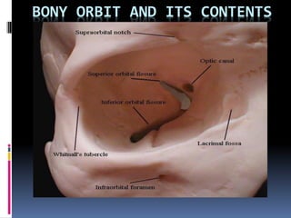
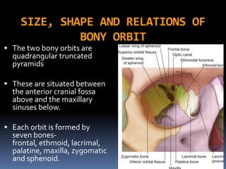
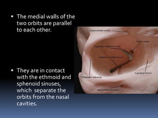
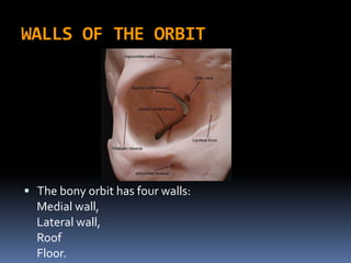
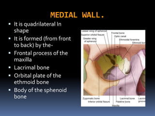
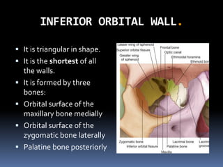
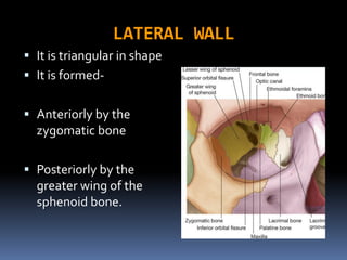
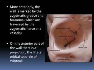
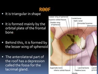
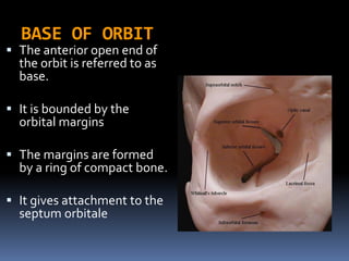
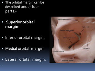
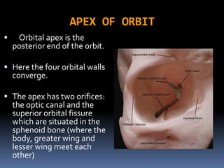
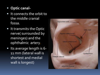
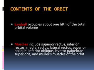
BONY ORBIT AND ITS CONTENTS
SIZE, SHAPE AND RELATIONS OF
BONY ORBIT
 The two bony orbits are
quadrangular truncated
pyramids
 These are situated between
the anterior cranial fossa
above and the maxillary
sinuses below.
 Each orbit is formed by
seven bones-
frontal, ethmoid, lacrimal,
palatine, maxilla, zygomatic
and sphenoid.
 The medial walls of the
two orbits are parallel
to each other.
 They are in contact
with the ethmoid and
sphenoid sinuses,
which separate the
orbits from the nasal
cavities.
WALLS OF THE ORBIT
 The bony orbit has four walls:
Medial wall,
Lateral wall,
Roof
Floor.
MEDIAL WALL.
 It is quadrilateral In
shape
 It is formed (from front
to back) by the-
 Frontal process of the
maxilla
 Lacrimal bone
 Orbital plate of the
ethmoid bone
 Body of the sphenoid
bone
INFERIOR ORBITAL WALL.
 It is triangular in shape.
 It is the shortest of all
the walls.
 It is formed by three
bones:
 Orbital surface of the
maxillary bone medially
 Orbital surface of the
zygomatic bone laterally
 Palatine bone posteriorly
 Clinical applications of the floor-
 The orbital floor being quite thin is commonly
involved in 'blow-out fractures' and is easily
invaded by tumours of the maxillary antrum.
 The floor of the orbit is best visualised with
standard postero anterior radiographs.
LATERAL WALL
 It is triangular in shape
 It is formed-
 Anteriorly by the
zygomatic bone
 Posteriorly by the
greater wing of the
sphenoid bone.
 More anteriorly, the
wall is marked by the
zygomatic groove and
foramina (which are
traversed by the
zygomatic nerve and
vessels)
 On the anterior part of
the wall there is a
projection, the lateral
orbital tubercle of
Whitnall.
 WHITNALL’S TUBERCLE:
 It gives attachment to the check ligament of
the lateral rectus muscle, Levator palpebral
superioris,Lateral palpebral ligament and to
the suspensory ligament of the eyeball
 The lateral wall posteriorly is separated from
the roof by the superior orbital fissure and
from the floor by the inferior orbital fissure
 Clinical applications of lateral wall-
 The lateral wall of the orbit protects only the
posterior half of the eyeball.
 The anterior half of globe is not covered by
bone on the lateral side.
 Hence palpation of retrobulbar tumours is
easier from the lateral rather than from the
nasal side of the eyeball.
R00F
 It is triangular in shape
 It is formed mainly by the
orbital plate of the frontal
bone.
 Behind this, it is formed by
the lesser wing of sphenoid.
 The anterolateral part of
the roof has a depression
called the fossa for the
lacrimal gland.
BASE OF ORBIT
 The anterior open end of
the orbit is referred to as
base.
 It is bounded by the
orbital margins
 The margins are formed
by a ring of compact bone.
 It gives attachment to the
septum orbitale
 The orbital margin can be
described under four
parts:-
 Superior orbital
margin-
 Inferior orbital margin.
 Medial orbital margin.
 Lateral orbital margin.
.
APEX OF ORBIT
 Orbital apex is the
posterior end of the orbit.
 Here the four orbital walls
converge.
 The apex has two orifices:
the optic canal and the
superior orbital fissure
which are situated in the
sphenoid bone (where the
body, greater wing and
lesser wing meet each
other)
 Optic canal-
 It connects the orbit to
the middle cranial
fossa.
 It transmits the Optic
nerve( surrounded by
meninges) and the
ophthalmic artery.
 Its average length is 6-
11 mm (lateral wall is
shortest and medial
wall is longest)
 Superior orbital fissure-
 It is a comma shaped aperture
in the orbital cavity.
 It is bounded by lesser and
greater wing of the sphenoid.
 It is situated lateral to the
optic foramen at the orbital
apex.
 The fissure is divided into
upper, middle and lower parts
by the common tendinous
ring (for origin of the recti).
 The structures passing through the upper and lateral part
are the lacrimal and frontal nerves (branches of
ophthalmic division ofVth nerve),trochlear nerve,
superior ophthalmic vein and recurrent branch of the
ophthalmic artery.
 The middle part of the fissure (within tendinous ring)
transmits the superior and inferior divisions of the
oculomotor nerve, the nasociliary branch of the ophthalmic
division of the trigeminal nerve and the abducent nerve.
 The lower and medial part of the fissure transmit the
inferior ophthalmic vein
PERIORBITA
 The periosteum lining the
surface of the orbital bones
is called the periorbita.
 Generally it is loosely
adherent to bone.
 However, it is firmly
adherent at the orbital
margin, superior and inferior
orbital fissures, the optic
canal, the lacrimal fossa and
at the sutures.
 At the orbital margin
periorbita is thickened to form
the arcus marginale to which
the septum orbitale is
attached.
 At the posterior lacrimal crest
the periorbita splits into two
layers which reunite at the
anterior lacrimal crest.
 These two layers enclose the
lacrimal sac (in the form of
lacrimal fascia).
 At the apex of orbit, the
periorbita is thickened to form
the common tendinous ring
of Zinn.
ORBITAL FASCIA
 It is a complex interwoven thin connective
tissue membrane joining the various
intraorbital content.
 Fascia bulbi,
 Membranous expansions of the extraocular
muscles,
 Ligament of Lockwood.
 Fascia bulbi-
 Fascia bulbi, or Tenon's
capsule is a loose
membranous sheath
envelops the eye ball from
the optic nerve to the
sclero-corneal junction.
 It is separated from the
sclera by the epi-scleral
space.
 The lower part of the
fascia bulbi is thickened and
takes part in the formation
of a sling or hammock on
which the globe rests
(suspensory ligament of
Lockwood).
 Around the distal end of
optic nerve the fascia is
fused with the dural sheath
of the optic nerve.
 Fascia bulbi is pierced
posteriorly
 The optic nerve,
 Ciliary nerves and vessels,
just behind the equator by
venae vorticosae,
 Anteriorly by six extraocular
muscles; where it becomes
continuous with the fascial
sheaths of these muscles.
 Suspensory ligament of
Lockwood-
It is a thickened sling or
hammock of fascial sheath
It extends from the
posterior lacrimal crest to
the lateral orbital tubercle,
on which rests the eyebal.
It is formed by fusion of
expansions from the
muscular sheaths of the
medial rectus, inferior
oblique, inferior rectus and
lateral rectus muscle
joined with the thickened
inferior part ofTenon's
capsule.
ORBITAL FAT
 Most of the orbital cavity is occupied by
orbital fat
 Which extends from the optic nerve to the
orbital wall and from the apex of the orbit to
the septum orbitale.
OPENINGS INTO THE ORBITAL
CAVITY
 Orbital Opening-
 Supra-orbital notch.
 Infra-orbital groove.
 Naso-lacrimal canal.
 Inferior orbital fissure.
 Superior orbital fissure.
 Optic canal.
 Zygomatico temporal and Zygomatico facial.
 Anterior and posterior Ethmoidal foramen.
1.EYEBALL and OPTIC NERVE
2. EXTRA-OCULAR MUSCLE
3. LACRIMAL GLAND and
LACRIMAL SAC
4. BLOODVESSELS and NERVES
5. FATS and FASCIAE
30
CONTENTS OF THE ORBIT
 Eyeball occupies about one fifth of the total
orbital volume
 Muscles include superior rectus, inferior
rectus, medial rectus, lateral rectus, superior
oblique, inferior oblique, levator palpebrae
superioris, and muller's muscles of the orbit
VASCULAR AND NERVE SUPPLY OF ORBIT
 ARTERY - ophthalmic artery
 VEINS – ophthalmic veins
 NERVES
MOTOR NERVES - IIIrd C.N., IVth C.N.
VIth C.N.
SENSORY NERVES- 1st and 2nd branches
of theVth .C.N.
 Gannglion-Cilliary ganglion.
 LYMPATIC – NONE
32
Ophthalmic artery
 Branch of internal carotid
 Arises when that vessel emerges through the roof of
cavernous sinus
 Enters orbit through optic canal infero-lateral to Optic
nerve.
34
Relations to optic nerve
 First infero lateral to optic nerve in a common sinovial
sheath of dura mater
 In the orbit it pierces dura, winds around and lies on the
lateral side of optic nerve
 Then Runs Forwards And medially above the optic nerve
between the superior ophthalmic vein in front and
nasociliary behind .I35
 In the medial wall of the orbit it lies between medial rectus and
superior oblique muscles
 At the medial end of upper eyelid the artery divides into its two
terminal branches:
1. supra trochlear
2. dorsal nasal
36
Branches:
Branche s are arranged in follwing groups
A. branches to eye ball
1. central artery of retina
2. posterior ciliary artery-long and short
B. Branches to orbital muscles
1. anterior ciliary arteries
37
 Is the first and most important branch
 First lie below the optic nerve
 Pierces the dural sheath and enters the substance
of the nerve
 Reach the optic disc divide into branches
38
CENTRAL ARTERY
OF RETINA
Branches- contd…….
C. Branches along the lateral wall:
1.Glandular branches.
2. lacrymal artery-two lateral palpabebral arteries,
3.Two zygomatic and
4. recurrent meningial artery
39
Branches- contd…….
D. Branches along the medial wall
1. posterior ethmoidal artery
2. anterior ethmoidal artery
3. medial palpebral arteries
4. Supra orbital and supra trochlear
arteries
5. dorsal nasal artery anastamoses with
terminal branch of facial artery
40
Ophthalmic veins
41
Superior ophthalmic vein:
 Lies in medial part of
upper eye lid
 Crosses above optic
nerve in company with
ophthalmic artery and
receives its tributaries
 Passes through
supraorbital fissure and
ends in cavernous sinus
 Devoid of valves
 At its commencement it
communicates with facial
vein through angular vein
Ophthalmic veins
42
Inferior ophthalmic
vein:
 Begins in the floor of
the orbit and drains in
the cavernous sinus
either directly or after
joining with superior
ophthalmic vein
 Communicates with
the pterygoid venous
plexus through inferior
orbital fissure
 Vorticose veins
Through superior orbital fissure
and drains to cav. sinus
43
Lymphatic drainage
No lymphatic vessels
 Because these may interfere with
movement with eyeballs
44
NERVE SUPPLY
 SENSORY.
 MOTOR.
OPTIC NERVE
 The optic nerve consists of more than 1 million axons
that originate in the ganglion cell layer of the retina. an
 It is not a peripheral nerve
 Prolongation of white matter of the brain ,because
it developed from the optic stalk
OPTIC NERVE
= (45 mm ) divided
into:
1. INTRAOCULAR
– 1 mm
2. ORBITAL
- 25 mm
3. I NTRACANAL
– 9 mm
4. INTRACRANIAL
– 10 mm
47
Motor Supply
• Lachrymal Nerve
• Frontal Nerve
• Trochlear Nerve
• Occulomotor Nerve
• Abducent Nerve
48
Cranial Nerve V (Trigeminal)
 The largest cranial nerve
 Possesses both sensory and motor divisionsThe
sensory portion subserves the greater part of the
scalp, forehead, face, eyelids, eye, lachrymal
gland, extra ocular muscles, ear, dura mater, and
tongue
 The motor portion innervates the muscles of
mastication through branches of the mandibular
division
Divisions of Cranial Nerve V
 Ophthalmic
 Frontal
 Lacrimal
 Nasociliary
 Maxillary
 Mandibular
51
Abducent Nerve
• Enters the orbit through lower part of the superior
orbital fissure
• Within the tendinous ring, it supplies the lateral rectus
muscle 52
 It is peripheral para
sympathetic ganglion
placed in the course of 3rd
nerve(NASO CILIARY)
 Topographically connected
to nasociliary(ophthalmic) .
 Functionally-
Oculomotor.(nerve to
inferior oblique).
 Lies in the apex(2mm)
of the orbit between optic
nerve and the tendon of
lateral rectus muscle.
53
CILIARY GANGLION.
 Sensory Root:
 Derived from Nasociliary .
 Consists sensory fibres
(PAIN,TOUCH,TEMPARATURE).-Pass through the
ganglion without relay.
 Sympathetic root:
 Derived from sympathetic plexus around internal
carotid artery.
Consists of Post ganglionic fibres from superior
sympathetic ganglion.
Pass through the ganglion without relay.
Then pass through Short ciliary nerves to supply the
Dilator pupillae and blood vessels of Eye ball.
Branches-8to 10 short ciliary nerves.
 SQ.
 1.Whitnall’s tubercle.
 2.Suspensory ligament of lockwood.
 3.Supra orbital fissure and structures passing
through it.
 4.Opthalmic artery and its branches.
 5.Central artery of Retina.
 6.Ciliary ganglion.
 Lateral orbital
margin-
 It is the strongest and
is formed by zygomatic
process of the frontal
bone and the
zygomatic bone.
 It does not reach as
far anterior as the
medial margin and
thus anterior half of
the globe is not
protected by the bone
laterally
 Inferior orbital margin-
 It is formed by the
zygomatic bone laterally
and maxilla medially,
almost in equal
proportion.
 It is slightly raised than
the floor.
 Medially it becomes
continuous with the
anterior lacrimal crest.
 The infraorbital foramen
transmitting infraorbital
nerves and vessels.
 Medial orbital margin-
 Below it is formed by
the anterior lacrimal
crest of the frontal
process of maxilla and
above by the frontal
bone.
 Its upper part becomes
continuous with the
posterior lacrimal
crest.
OPTIC NERVE
 The optic nerve may be divided
into the following topographic
areas.
 Intraocular portion of the optic
nerve: optic disc, or nerve
head; prelaminar; and laminar
portions
Intraorbital portion (located
within the muscle cone)
Intracanalicular portion
(located within the optic canal)
Intracranial portion (ending in
the optic chiasm)
Lachrymal Nerve
• Arises from the
ophthalmic division of
the trigeminal nerve
• Enters the orbit through
the upper part of
superior orbital fissure
• Enters the lacrimal gland
• Ends by supplying the
skin of lateral part of
upper eye lid
90

Bony orbit and its contents

  • 2.
    BONY ORBIT ANDITS CONTENTS
  • 3.
    SIZE, SHAPE ANDRELATIONS OF BONY ORBIT  The two bony orbits are quadrangular truncated pyramids  These are situated between the anterior cranial fossa above and the maxillary sinuses below.  Each orbit is formed by seven bones- frontal, ethmoid, lacrimal, palatine, maxilla, zygomatic and sphenoid.
  • 4.
     The medialwalls of the two orbits are parallel to each other.  They are in contact with the ethmoid and sphenoid sinuses, which separate the orbits from the nasal cavities.
  • 5.
    WALLS OF THEORBIT  The bony orbit has four walls: Medial wall, Lateral wall, Roof Floor.
  • 6.
    MEDIAL WALL.  Itis quadrilateral In shape  It is formed (from front to back) by the-  Frontal process of the maxilla  Lacrimal bone  Orbital plate of the ethmoid bone  Body of the sphenoid bone
  • 7.
    INFERIOR ORBITAL WALL. It is triangular in shape.  It is the shortest of all the walls.  It is formed by three bones:  Orbital surface of the maxillary bone medially  Orbital surface of the zygomatic bone laterally  Palatine bone posteriorly
  • 8.
     Clinical applicationsof the floor-  The orbital floor being quite thin is commonly involved in 'blow-out fractures' and is easily invaded by tumours of the maxillary antrum.  The floor of the orbit is best visualised with standard postero anterior radiographs.
  • 9.
    LATERAL WALL  Itis triangular in shape  It is formed-  Anteriorly by the zygomatic bone  Posteriorly by the greater wing of the sphenoid bone.
  • 10.
     More anteriorly,the wall is marked by the zygomatic groove and foramina (which are traversed by the zygomatic nerve and vessels)  On the anterior part of the wall there is a projection, the lateral orbital tubercle of Whitnall.
  • 11.
     WHITNALL’S TUBERCLE: It gives attachment to the check ligament of the lateral rectus muscle, Levator palpebral superioris,Lateral palpebral ligament and to the suspensory ligament of the eyeball  The lateral wall posteriorly is separated from the roof by the superior orbital fissure and from the floor by the inferior orbital fissure
  • 12.
     Clinical applicationsof lateral wall-  The lateral wall of the orbit protects only the posterior half of the eyeball.  The anterior half of globe is not covered by bone on the lateral side.  Hence palpation of retrobulbar tumours is easier from the lateral rather than from the nasal side of the eyeball.
  • 13.
    R00F  It istriangular in shape  It is formed mainly by the orbital plate of the frontal bone.  Behind this, it is formed by the lesser wing of sphenoid.  The anterolateral part of the roof has a depression called the fossa for the lacrimal gland.
  • 14.
    BASE OF ORBIT The anterior open end of the orbit is referred to as base.  It is bounded by the orbital margins  The margins are formed by a ring of compact bone.  It gives attachment to the septum orbitale
  • 15.
     The orbitalmargin can be described under four parts:-  Superior orbital margin-  Inferior orbital margin.  Medial orbital margin.  Lateral orbital margin. .
  • 16.
    APEX OF ORBIT Orbital apex is the posterior end of the orbit.  Here the four orbital walls converge.  The apex has two orifices: the optic canal and the superior orbital fissure which are situated in the sphenoid bone (where the body, greater wing and lesser wing meet each other)
  • 17.
     Optic canal- It connects the orbit to the middle cranial fossa.  It transmits the Optic nerve( surrounded by meninges) and the ophthalmic artery.  Its average length is 6- 11 mm (lateral wall is shortest and medial wall is longest)
  • 18.
     Superior orbitalfissure-  It is a comma shaped aperture in the orbital cavity.  It is bounded by lesser and greater wing of the sphenoid.  It is situated lateral to the optic foramen at the orbital apex.  The fissure is divided into upper, middle and lower parts by the common tendinous ring (for origin of the recti).
  • 19.
     The structurespassing through the upper and lateral part are the lacrimal and frontal nerves (branches of ophthalmic division ofVth nerve),trochlear nerve, superior ophthalmic vein and recurrent branch of the ophthalmic artery.
  • 20.
     The middlepart of the fissure (within tendinous ring) transmits the superior and inferior divisions of the oculomotor nerve, the nasociliary branch of the ophthalmic division of the trigeminal nerve and the abducent nerve.  The lower and medial part of the fissure transmit the inferior ophthalmic vein
  • 22.
    PERIORBITA  The periosteumlining the surface of the orbital bones is called the periorbita.  Generally it is loosely adherent to bone.  However, it is firmly adherent at the orbital margin, superior and inferior orbital fissures, the optic canal, the lacrimal fossa and at the sutures.
  • 23.
     At theorbital margin periorbita is thickened to form the arcus marginale to which the septum orbitale is attached.  At the posterior lacrimal crest the periorbita splits into two layers which reunite at the anterior lacrimal crest.  These two layers enclose the lacrimal sac (in the form of lacrimal fascia).  At the apex of orbit, the periorbita is thickened to form the common tendinous ring of Zinn.
  • 24.
    ORBITAL FASCIA  Itis a complex interwoven thin connective tissue membrane joining the various intraorbital content.  Fascia bulbi,  Membranous expansions of the extraocular muscles,  Ligament of Lockwood.
  • 25.
     Fascia bulbi- Fascia bulbi, or Tenon's capsule is a loose membranous sheath envelops the eye ball from the optic nerve to the sclero-corneal junction.  It is separated from the sclera by the epi-scleral space.  The lower part of the fascia bulbi is thickened and takes part in the formation of a sling or hammock on which the globe rests (suspensory ligament of Lockwood).
  • 26.
     Around thedistal end of optic nerve the fascia is fused with the dural sheath of the optic nerve.  Fascia bulbi is pierced posteriorly  The optic nerve,  Ciliary nerves and vessels, just behind the equator by venae vorticosae,  Anteriorly by six extraocular muscles; where it becomes continuous with the fascial sheaths of these muscles.
  • 27.
     Suspensory ligamentof Lockwood- It is a thickened sling or hammock of fascial sheath It extends from the posterior lacrimal crest to the lateral orbital tubercle, on which rests the eyebal. It is formed by fusion of expansions from the muscular sheaths of the medial rectus, inferior oblique, inferior rectus and lateral rectus muscle joined with the thickened inferior part ofTenon's capsule.
  • 28.
    ORBITAL FAT  Mostof the orbital cavity is occupied by orbital fat  Which extends from the optic nerve to the orbital wall and from the apex of the orbit to the septum orbitale.
  • 29.
    OPENINGS INTO THEORBITAL CAVITY  Orbital Opening-  Supra-orbital notch.  Infra-orbital groove.  Naso-lacrimal canal.  Inferior orbital fissure.  Superior orbital fissure.  Optic canal.  Zygomatico temporal and Zygomatico facial.  Anterior and posterior Ethmoidal foramen.
  • 30.
    1.EYEBALL and OPTICNERVE 2. EXTRA-OCULAR MUSCLE 3. LACRIMAL GLAND and LACRIMAL SAC 4. BLOODVESSELS and NERVES 5. FATS and FASCIAE 30 CONTENTS OF THE ORBIT
  • 31.
    CONTENTS OF THEORBIT  Eyeball occupies about one fifth of the total orbital volume  Muscles include superior rectus, inferior rectus, medial rectus, lateral rectus, superior oblique, inferior oblique, levator palpebrae superioris, and muller's muscles of the orbit
  • 32.
    VASCULAR AND NERVESUPPLY OF ORBIT  ARTERY - ophthalmic artery  VEINS – ophthalmic veins  NERVES MOTOR NERVES - IIIrd C.N., IVth C.N. VIth C.N. SENSORY NERVES- 1st and 2nd branches of theVth .C.N.  Gannglion-Cilliary ganglion.  LYMPATIC – NONE 32
  • 34.
    Ophthalmic artery  Branchof internal carotid  Arises when that vessel emerges through the roof of cavernous sinus  Enters orbit through optic canal infero-lateral to Optic nerve. 34
  • 35.
    Relations to opticnerve  First infero lateral to optic nerve in a common sinovial sheath of dura mater  In the orbit it pierces dura, winds around and lies on the lateral side of optic nerve  Then Runs Forwards And medially above the optic nerve between the superior ophthalmic vein in front and nasociliary behind .I35
  • 36.
     In themedial wall of the orbit it lies between medial rectus and superior oblique muscles  At the medial end of upper eyelid the artery divides into its two terminal branches: 1. supra trochlear 2. dorsal nasal 36
  • 37.
    Branches: Branche s arearranged in follwing groups A. branches to eye ball 1. central artery of retina 2. posterior ciliary artery-long and short B. Branches to orbital muscles 1. anterior ciliary arteries 37
  • 38.
     Is thefirst and most important branch  First lie below the optic nerve  Pierces the dural sheath and enters the substance of the nerve  Reach the optic disc divide into branches 38 CENTRAL ARTERY OF RETINA
  • 39.
    Branches- contd……. C. Branchesalong the lateral wall: 1.Glandular branches. 2. lacrymal artery-two lateral palpabebral arteries, 3.Two zygomatic and 4. recurrent meningial artery 39
  • 40.
    Branches- contd……. D. Branchesalong the medial wall 1. posterior ethmoidal artery 2. anterior ethmoidal artery 3. medial palpebral arteries 4. Supra orbital and supra trochlear arteries 5. dorsal nasal artery anastamoses with terminal branch of facial artery 40
  • 41.
    Ophthalmic veins 41 Superior ophthalmicvein:  Lies in medial part of upper eye lid  Crosses above optic nerve in company with ophthalmic artery and receives its tributaries  Passes through supraorbital fissure and ends in cavernous sinus  Devoid of valves  At its commencement it communicates with facial vein through angular vein
  • 42.
    Ophthalmic veins 42 Inferior ophthalmic vein: Begins in the floor of the orbit and drains in the cavernous sinus either directly or after joining with superior ophthalmic vein  Communicates with the pterygoid venous plexus through inferior orbital fissure
  • 43.
     Vorticose veins Throughsuperior orbital fissure and drains to cav. sinus 43
  • 44.
    Lymphatic drainage No lymphaticvessels  Because these may interfere with movement with eyeballs 44
  • 45.
  • 46.
    OPTIC NERVE  Theoptic nerve consists of more than 1 million axons that originate in the ganglion cell layer of the retina. an  It is not a peripheral nerve  Prolongation of white matter of the brain ,because it developed from the optic stalk
  • 47.
    OPTIC NERVE = (45mm ) divided into: 1. INTRAOCULAR – 1 mm 2. ORBITAL - 25 mm 3. I NTRACANAL – 9 mm 4. INTRACRANIAL – 10 mm 47
  • 48.
    Motor Supply • LachrymalNerve • Frontal Nerve • Trochlear Nerve • Occulomotor Nerve • Abducent Nerve 48
  • 49.
    Cranial Nerve V(Trigeminal)  The largest cranial nerve  Possesses both sensory and motor divisionsThe sensory portion subserves the greater part of the scalp, forehead, face, eyelids, eye, lachrymal gland, extra ocular muscles, ear, dura mater, and tongue  The motor portion innervates the muscles of mastication through branches of the mandibular division
  • 50.
    Divisions of CranialNerve V  Ophthalmic  Frontal  Lacrimal  Nasociliary  Maxillary  Mandibular
  • 51.
  • 52.
    Abducent Nerve • Entersthe orbit through lower part of the superior orbital fissure • Within the tendinous ring, it supplies the lateral rectus muscle 52
  • 53.
     It isperipheral para sympathetic ganglion placed in the course of 3rd nerve(NASO CILIARY)  Topographically connected to nasociliary(ophthalmic) .  Functionally- Oculomotor.(nerve to inferior oblique).  Lies in the apex(2mm) of the orbit between optic nerve and the tendon of lateral rectus muscle. 53 CILIARY GANGLION.
  • 55.
     Sensory Root: Derived from Nasociliary .  Consists sensory fibres (PAIN,TOUCH,TEMPARATURE).-Pass through the ganglion without relay.  Sympathetic root:  Derived from sympathetic plexus around internal carotid artery. Consists of Post ganglionic fibres from superior sympathetic ganglion. Pass through the ganglion without relay. Then pass through Short ciliary nerves to supply the Dilator pupillae and blood vessels of Eye ball. Branches-8to 10 short ciliary nerves.
  • 58.
     SQ.  1.Whitnall’stubercle.  2.Suspensory ligament of lockwood.  3.Supra orbital fissure and structures passing through it.  4.Opthalmic artery and its branches.  5.Central artery of Retina.  6.Ciliary ganglion.
  • 86.
     Lateral orbital margin- It is the strongest and is formed by zygomatic process of the frontal bone and the zygomatic bone.  It does not reach as far anterior as the medial margin and thus anterior half of the globe is not protected by the bone laterally
  • 87.
     Inferior orbitalmargin-  It is formed by the zygomatic bone laterally and maxilla medially, almost in equal proportion.  It is slightly raised than the floor.  Medially it becomes continuous with the anterior lacrimal crest.  The infraorbital foramen transmitting infraorbital nerves and vessels.
  • 88.
     Medial orbitalmargin-  Below it is formed by the anterior lacrimal crest of the frontal process of maxilla and above by the frontal bone.  Its upper part becomes continuous with the posterior lacrimal crest.
  • 89.
    OPTIC NERVE  Theoptic nerve may be divided into the following topographic areas.  Intraocular portion of the optic nerve: optic disc, or nerve head; prelaminar; and laminar portions Intraorbital portion (located within the muscle cone) Intracanalicular portion (located within the optic canal) Intracranial portion (ending in the optic chiasm)
  • 90.
    Lachrymal Nerve • Arisesfrom the ophthalmic division of the trigeminal nerve • Enters the orbit through the upper part of superior orbital fissure • Enters the lacrimal gland • Ends by supplying the skin of lateral part of upper eye lid 90