Downloaded 2,954 times

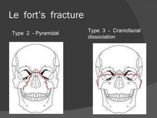
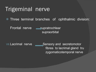
![Roof of orbit Frontal bone [Orbital plate] & lesser wing of sphenoid Separated from frontal sinus and anterior cranial fossa above Lacrimal gland fossa and trochlear fossa behind orbital rim](https://image.slidesharecdn.com/orbit-anatomy-120131061741-phpapp02/85/Orbit-anatomy-9-320.jpg)
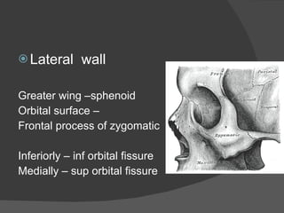
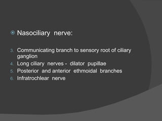
![Medial wall Body of sphenoid Ethmoid Lacrimal Maxilla[frontal process]](https://image.slidesharecdn.com/orbit-anatomy-120131061741-phpapp02/85/Orbit-anatomy-11-320.jpg)
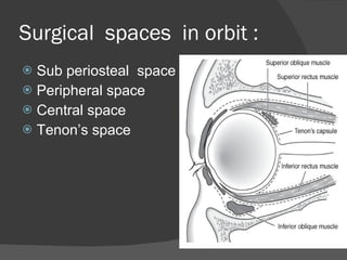
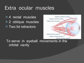


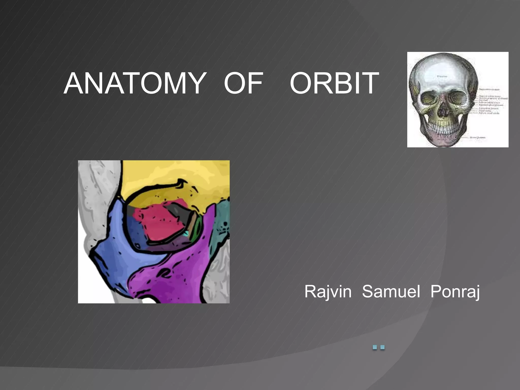
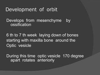
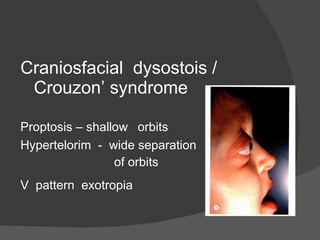
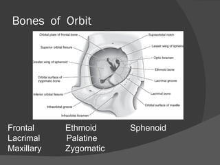
The document discusses the anatomy of the orbit, including: 1. The orbit develops from mesenchyme between the 6th and 7th week, with bones forming around the optic vesicle. 2. The bones that make up the orbit include the frontal, ethmoid, sphenoid, lacrimal, palatine, maxillary, and zygomatic bones. 3. The orbit contains the eyeball, orbital fat, connective tissue system, blood vessels, and nerves including the extraocular muscles.


![CASE_PRESENTATION_ON_subdural_hematoma(SDH)[1 FINAL PPT]-1.pptx](https://cdn.slidesharecdn.com/ss_thumbnails/casepresentationonsubduralhematomasdh1finalppt-1-260129172522-d405d375-thumbnail.jpg?width=640&height=640&fit=bounds)


