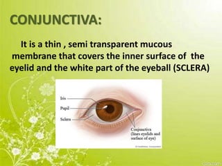
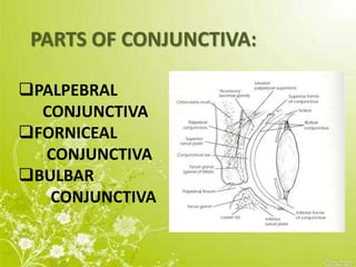
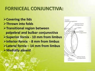
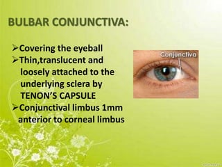
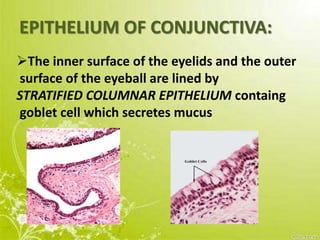
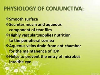
CONJUNCTIVA –
ANATOMY AND PHYSIOLOGY
-AARTHY.M.R.S.
CONJUNCTIVA:
It is a thin , semi transparent mucous
membrane that covers the inner surface of the
eyelid and the white part of the eyeball (SCLERA)
PARTS OF CONJUNCTIVA:
PALPEBRAL
CONJUNCTIVA
FORNICEAL
CONJUNCTIVA
BULBAR
CONJUNCTIVA
PALPEBRAL CONJUNCTIVA:
Covering the lids
Firmly adherent
Highly vascular
Extremely thin
Strongly bound to the
tarsal plate
FORNICEAL CONJUNCTIVA:
Covering the lids
Thrown into folds
Transitional region between
palpebral and bulbar conjunvctiva
Superior fornix - 10 mm from limbus
Inferior fornix - 8 mm from limbus
Lateral fornix – 14 mm from limbus
Medially absent
BULBAR CONJUNCTIVA:
Covering the eyeball
Thin,translucent and
loosely attached to the
underlying sclera by
TENON’S CAPSULE
Conjunctival limbus 1mm
anterior to corneal limbus
EPITHELIUM OF CONJUNCTIVA:
The inner surface of the eyelids and the outer
surface of the eyeball are lined by
STRATIFIED COLUMNAR EPITHELIUM containg
goblet cell which secretes mucus
At limbus,change into stratified
squamous non-keratinized epithelium
At lid margin,change into stratified
squamous keratinized epithelium
Presence of accessory lacrimal glands
The epithelial layer contains
blood vessels,Fibrous tissue and
lymphatic channel
BLOOD SUPPLY:
Anterior conjunctival arteries derived from the
Ant.ciliary arteries
Posterior conjunctival arteries derived from the
Arterial arcade of lids which is formed by palpebral
Branches of nasal and lacrimal arteries of lids
ARTERIAL SUPPLY:
VENOUS DRAINAGE:
Palpebral and ophthalmic veins
NERVE SUPPLY:
BULBAR CONJUNCTIVA:
long ciliary nerve
Naso ciliary nerve
Ophthalmic division of trigeminal nerve
SUP.PALPEBRAL AND FORNICEAL CONJUNCTIVA:
Frontal and lacrimal branches of
ophthalmic division of trigeminal nerve
INFERIOR PALPEBRAL AND FORNICEAL
CONJUNCTIVA:
Laterally-lacrimal branches of ophthalmic
division of trigeminal nerve
Medially-infraorbital nerve
maxillary division of trigeminal nerve
SYMPATHETIC:
Superior cervical sympatics to blood vessels
LYMPHATIC DRAINAGE:
Lymph vessels are arranged as a deep and
a superficial plexes in submucosa
PHYSIOLOGY OF CONJUNCTIVA:
Smooth surface
Secretes mucin and aqueous
component of tear flim
Highly vascular:supplies nutrition
to the peripheral cornea
Aqueous veins drain from ant.chamber
for the maintanence of IOP
Helps to prevent the entry of microbes
into the eye
Conjunctiva

Conjunctiva

  • 1.
    CONJUNCTIVA – ANATOMY ANDPHYSIOLOGY -AARTHY.M.R.S.
  • 2.
    CONJUNCTIVA: It is athin , semi transparent mucous membrane that covers the inner surface of the eyelid and the white part of the eyeball (SCLERA)
  • 3.
  • 6.
    PALPEBRAL CONJUNCTIVA: Covering thelids Firmly adherent Highly vascular Extremely thin Strongly bound to the tarsal plate
  • 7.
    FORNICEAL CONJUNCTIVA: Covering thelids Thrown into folds Transitional region between palpebral and bulbar conjunvctiva Superior fornix - 10 mm from limbus Inferior fornix - 8 mm from limbus Lateral fornix – 14 mm from limbus Medially absent
  • 8.
    BULBAR CONJUNCTIVA: Covering theeyeball Thin,translucent and loosely attached to the underlying sclera by TENON’S CAPSULE Conjunctival limbus 1mm anterior to corneal limbus
  • 9.
    EPITHELIUM OF CONJUNCTIVA: Theinner surface of the eyelids and the outer surface of the eyeball are lined by STRATIFIED COLUMNAR EPITHELIUM containg goblet cell which secretes mucus
  • 10.
    At limbus,change intostratified squamous non-keratinized epithelium At lid margin,change into stratified squamous keratinized epithelium Presence of accessory lacrimal glands The epithelial layer contains blood vessels,Fibrous tissue and lymphatic channel
  • 11.
    BLOOD SUPPLY: Anterior conjunctivalarteries derived from the Ant.ciliary arteries Posterior conjunctival arteries derived from the Arterial arcade of lids which is formed by palpebral Branches of nasal and lacrimal arteries of lids ARTERIAL SUPPLY: VENOUS DRAINAGE: Palpebral and ophthalmic veins
  • 13.
    NERVE SUPPLY: BULBAR CONJUNCTIVA: longciliary nerve Naso ciliary nerve Ophthalmic division of trigeminal nerve SUP.PALPEBRAL AND FORNICEAL CONJUNCTIVA: Frontal and lacrimal branches of ophthalmic division of trigeminal nerve
  • 14.
    INFERIOR PALPEBRAL ANDFORNICEAL CONJUNCTIVA: Laterally-lacrimal branches of ophthalmic division of trigeminal nerve Medially-infraorbital nerve maxillary division of trigeminal nerve SYMPATHETIC: Superior cervical sympatics to blood vessels
  • 15.
    LYMPHATIC DRAINAGE: Lymph vesselsare arranged as a deep and a superficial plexes in submucosa
  • 16.
    PHYSIOLOGY OF CONJUNCTIVA: Smoothsurface Secretes mucin and aqueous component of tear flim Highly vascular:supplies nutrition to the peripheral cornea Aqueous veins drain from ant.chamber for the maintanence of IOP Helps to prevent the entry of microbes into the eye