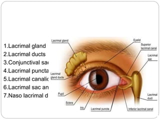
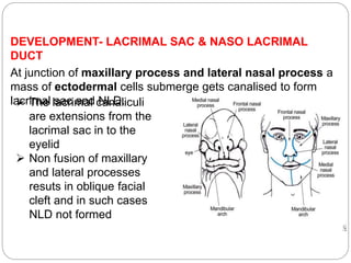
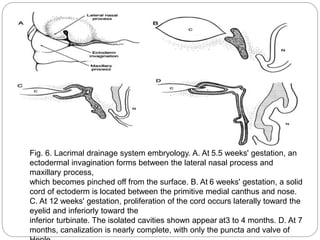
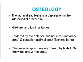
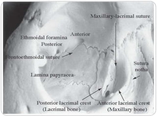
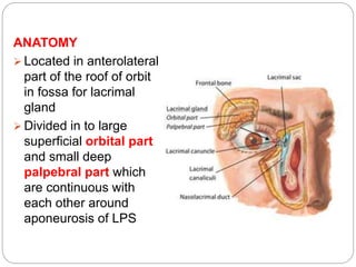
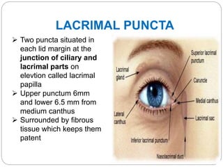
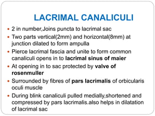
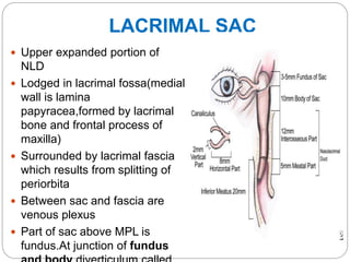
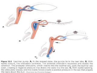
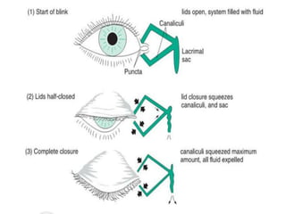
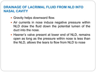
The lacrimal apparatus consists of the lacrimal gland, lacrimal drainage system, and associated structures that produce and drain tears from the eye.
The lacrimal gland develops from surface ectoderm and is located above and lateral to the eye. Tears produced by the gland drain through puncta and canaliculi into the lacrimal sac, then through the nasolacrimal duct into the nose.
The lacrimal sac is lodged in the lacrimal fossa of the medial orbital wall. It connects to the nasolacrimal duct, which courses posteriorly and laterally through bone to drain into the nasal cavity. Coordinated blinking and pressure differences aid
The lacrimal apparatus is the physiological system containing the orbital structures for tear production and drainage. It consists of: The lacrimal gland, which secretes the tears, and its excretory ducts, which convey the fluid to the surface of the human eye;it is a serous gland located in lacrimal fossa.
1-IT IS A MIDDLE VASCULAR COAT OF EYEBALL.
2-IT MAINLY CONSIST OF THREE PARTS IRIS, CHOROID, CILIARY BODY.
3- CILIARY BODY CAN HOLD THE LENS AND PLAY IMPORTANT ROLE IN ACCOMODATION.
Each eyelid contains a fibrous plate, called a tarsus, that gives it structure and shape; muscles, which move the eyelids; and meibomian (or tarsal) glands, which secrete lubricating fluids. The lids are covered with skin, lined with mucous membrane, and bordered with a fringe of hairs, the eyelashes.
Diagnosis, Management, and Surgery by Adam J. Cohen, Michael Mercandetti & Brian G. Brazzo. The dry eye , a practical approach by Sudi Patel & Kenny J Blades. Jack J Kanski’s clinical ophthalmology Clinical Anatomy of the Eye by Richard S. Snell & Michael A. Lemp.
3. It is concerned with the tear formation & transport. Lacrimal passage includes : Lacrimal gland Conjunctival sac Lacrimal puncta Lacrimal canaliculi Lacrimal sac Nasolacrimal duct
4. The following components of the lacrimal apparatus are discussed : Embryology Osteology Secretory system Excretory system Physiology
5. Ectodermal origin Solid epithelial buds(first 2 months) Supero
These lecture slides, by Dr Sidra Arshad, offer a quick overview of physiological basis of a normal electrocardiogram.
Learning objectives:
1. Define an electrocardiogram (ECG) and electrocardiography
2. Describe how dipoles generated by the heart produce the waveforms of the ECG
3. Describe the components of a normal electrocardiogram of a typical bipolar leads (limb II)
4. Differentiate between intervals and segments
5. Enlist some common indications for obtaining an ECG
Study Resources:
1. Chapter 11, Guyton and Hall Textbook of Medical Physiology, 14th edition
2. Chapter 9, Human Physiology - From Cells to Systems, Lauralee Sherwood, 9th edition
3. Chapter 29, Ganong’s Review of Medical Physiology, 26th edition
4. Electrocardiogram, StatPearls - https://www.ncbi.nlm.nih.gov/books/NBK549803/
5. ECG in Medical Practice by ABM Abdullah, 4th edition
6. ECG Basics, http://www.nataliescasebook.com/tag/e-c-g-basics
Ethanol (CH3CH2OH), or beverage alcohol, is a two-carbon alcohol
that is rapidly distributed in the body and brain. Ethanol alters many
neurochemical systems and has rewarding and addictive properties. It
is the oldest recreational drug and likely contributes to more morbidity,
mortality, and public health costs than all illicit drugs combined. The
5th edition of the Diagnostic and Statistical Manual of Mental Disorders
(DSM-5) integrates alcohol abuse and alcohol dependence into a single
disorder called alcohol use disorder (AUD), with mild, moderate,
and severe subclassifications (American Psychiatric Association, 2013).
In the DSM-5, all types of substance abuse and dependence have been
combined into a single substance use disorder (SUD) on a continuum
from mild to severe. A diagnosis of AUD requires that at least two of
the 11 DSM-5 behaviors be present within a 12-month period (mild
AUD: 2–3 criteria; moderate AUD: 4–5 criteria; severe AUD: 6–11 criteria).
The four main behavioral effects of AUD are impaired control over
drinking, negative social consequences, risky use, and altered physiological
effects (tolerance, withdrawal). This chapter presents an overview
of the prevalence and harmful consequences of AUD in the U.S.,
the systemic nature of the disease, neurocircuitry and stages of AUD,
comorbidities, fetal alcohol spectrum disorders, genetic risk factors, and
pharmacotherapies for AUD.
Pulmonary Thromboembolism - etilogy, types, medical- Surgical and nursing man...VarunMahajani
Disruption of blood supply to lung alveoli due to blockage of one or more pulmonary blood vessels is called as Pulmonary thromboembolism. In this presentation we will discuss its causes, types and its management in depth.
MANAGEMENT OF ATRIOVENTRICULAR CONDUCTION BLOCK.pdfJim Jacob Roy
Cardiac conduction defects can occur due to various causes.
Atrioventricular conduction blocks ( AV blocks ) are classified into 3 types.
This document describes the acute management of AV block.
- Video recording of this lecture in English language: https://youtu.be/lK81BzxMqdo
- Video recording of this lecture in Arabic language: https://youtu.be/Ve4P0COk9OI
- Link to download the book free: https://nephrotube.blogspot.com/p/nephrotube-nephrology-books.html
- Link to NephroTube website: www.NephroTube.com
- Link to NephroTube social media accounts: https://nephrotube.blogspot.com/p/join-nephrotube-on-social-media.html
Title: Sense of Taste
Presenter: Dr. Faiza, Assistant Professor of Physiology
Qualifications:
MBBS (Best Graduate, AIMC Lahore)
FCPS Physiology
ICMT, CHPE, DHPE (STMU)
MPH (GC University, Faisalabad)
MBA (Virtual University of Pakistan)
Learning Objectives:
Describe the structure and function of taste buds.
Describe the relationship between the taste threshold and taste index of common substances.
Explain the chemical basis and signal transduction of taste perception for each type of primary taste sensation.
Recognize different abnormalities of taste perception and their causes.
Key Topics:
Significance of Taste Sensation:
Differentiation between pleasant and harmful food
Influence on behavior
Selection of food based on metabolic needs
Receptors of Taste:
Taste buds on the tongue
Influence of sense of smell, texture of food, and pain stimulation (e.g., by pepper)
Primary and Secondary Taste Sensations:
Primary taste sensations: Sweet, Sour, Salty, Bitter, Umami
Chemical basis and signal transduction mechanisms for each taste
Taste Threshold and Index:
Taste threshold values for Sweet (sucrose), Salty (NaCl), Sour (HCl), and Bitter (Quinine)
Taste index relationship: Inversely proportional to taste threshold
Taste Blindness:
Inability to taste certain substances, particularly thiourea compounds
Example: Phenylthiocarbamide
Structure and Function of Taste Buds:
Composition: Epithelial cells, Sustentacular/Supporting cells, Taste cells, Basal cells
Features: Taste pores, Taste hairs/microvilli, and Taste nerve fibers
Location of Taste Buds:
Found in papillae of the tongue (Fungiform, Circumvallate, Foliate)
Also present on the palate, tonsillar pillars, epiglottis, and proximal esophagus
Mechanism of Taste Stimulation:
Interaction of taste substances with receptors on microvilli
Signal transduction pathways for Umami, Sweet, Bitter, Sour, and Salty tastes
Taste Sensitivity and Adaptation:
Decrease in sensitivity with age
Rapid adaptation of taste sensation
Role of Saliva in Taste:
Dissolution of tastants to reach receptors
Washing away the stimulus
Taste Preferences and Aversions:
Mechanisms behind taste preference and aversion
Influence of receptors and neural pathways
Impact of Sensory Nerve Damage:
Degeneration of taste buds if the sensory nerve fiber is cut
Abnormalities of Taste Detection:
Conditions: Ageusia, Hypogeusia, Dysgeusia (parageusia)
Causes: Nerve damage, neurological disorders, infections, poor oral hygiene, adverse drug effects, deficiencies, aging, tobacco use, altered neurotransmitter levels
Neurotransmitters and Taste Threshold:
Effects of serotonin (5-HT) and norepinephrine (NE) on taste sensitivity
Supertasters:
25% of the population with heightened sensitivity to taste, especially bitterness
Increased number of fungiform papillae
Tom Selleck Health: A Comprehensive Look at the Iconic Actor’s Wellness Journeygreendigital
Tom Selleck, an enduring figure in Hollywood. has captivated audiences for decades with his rugged charm, iconic moustache. and memorable roles in television and film. From his breakout role as Thomas Magnum in Magnum P.I. to his current portrayal of Frank Reagan in Blue Bloods. Selleck's career has spanned over 50 years. But beyond his professional achievements. fans have often been curious about Tom Selleck Health. especially as he has aged in the public eye.
Follow us on: Pinterest
Introduction
Many have been interested in Tom Selleck health. not only because of his enduring presence on screen but also because of the challenges. and lifestyle choices he has faced and made over the years. This article delves into the various aspects of Tom Selleck health. exploring his fitness regimen, diet, mental health. and the challenges he has encountered as he ages. We'll look at how he maintains his well-being. the health issues he has faced, and his approach to ageing .
Early Life and Career
Childhood and Athletic Beginnings
Tom Selleck was born on January 29, 1945, in Detroit, Michigan, and grew up in Sherman Oaks, California. From an early age, he was involved in sports, particularly basketball. which played a significant role in his physical development. His athletic pursuits continued into college. where he attended the University of Southern California (USC) on a basketball scholarship. This early involvement in sports laid a strong foundation for his physical health and disciplined lifestyle.
Transition to Acting
Selleck's transition from an athlete to an actor came with its physical demands. His first significant role in "Magnum P.I." required him to perform various stunts and maintain a fit appearance. This role, which he played from 1980 to 1988. necessitated a rigorous fitness routine to meet the show's demands. setting the stage for his long-term commitment to health and wellness.
Fitness Regimen
Workout Routine
Tom Selleck health and fitness regimen has evolved. adapting to his changing roles and age. During his "Magnum, P.I." days. Selleck's workouts were intense and focused on building and maintaining muscle mass. His routine included weightlifting, cardiovascular exercises. and specific training for the stunts he performed on the show.
Selleck adjusted his fitness routine as he aged to suit his body's needs. Today, his workouts focus on maintaining flexibility, strength, and cardiovascular health. He incorporates low-impact exercises such as swimming, walking, and light weightlifting. This balanced approach helps him stay fit without putting undue strain on his joints and muscles.
Importance of Flexibility and Mobility
In recent years, Selleck has emphasized the importance of flexibility and mobility in his fitness regimen. Understanding the natural decline in muscle mass and joint flexibility with age. he includes stretching and yoga in his routine. These practices help prevent injuries, improve posture, and maintain mobilit
The prostate is an exocrine gland of the male mammalian reproductive system
It is a walnut-sized gland that forms part of the male reproductive system and is located in front of the rectum and just below the urinary bladder
Function is to store and secrete a clear, slightly alkaline fluid that constitutes 10-30% of the volume of the seminal fluid that along with the spermatozoa, constitutes semen
A healthy human prostate measures (4cm-vertical, by 3cm-horizontal, 2cm ant-post ).
It surrounds the urethra just below the urinary bladder. It has anterior, median, posterior and two lateral lobes
It’s work is regulated by androgens which are responsible for male sex characteristics
Generalised disease of the prostate due to hormonal derangement which leads to non malignant enlargement of the gland (increase in the number of epithelial cells and stromal tissue)to cause compression of the urethra leading to symptoms (LUTS
Lung Cancer: Artificial Intelligence, Synergetics, Complex System Analysis, S...Oleg Kshivets
RESULTS: Overall life span (LS) was 2252.1±1742.5 days and cumulative 5-year survival (5YS) reached 73.2%, 10 years – 64.8%, 20 years – 42.5%. 513 LCP lived more than 5 years (LS=3124.6±1525.6 days), 148 LCP – more than 10 years (LS=5054.4±1504.1 days).199 LCP died because of LC (LS=562.7±374.5 days). 5YS of LCP after bi/lobectomies was significantly superior in comparison with LCP after pneumonectomies (78.1% vs.63.7%, P=0.00001 by log-rank test). AT significantly improved 5YS (66.3% vs. 34.8%) (P=0.00000 by log-rank test) only for LCP with N1-2. Cox modeling displayed that 5YS of LCP significantly depended on: phase transition (PT) early-invasive LC in terms of synergetics, PT N0—N12, cell ratio factors (ratio between cancer cells- CC and blood cells subpopulations), G1-3, histology, glucose, AT, blood cell circuit, prothrombin index, heparin tolerance, recalcification time (P=0.000-0.038). Neural networks, genetic algorithm selection and bootstrap simulation revealed relationships between 5YS and PT early-invasive LC (rank=1), PT N0—N12 (rank=2), thrombocytes/CC (3), erythrocytes/CC (4), eosinophils/CC (5), healthy cells/CC (6), lymphocytes/CC (7), segmented neutrophils/CC (8), stick neutrophils/CC (9), monocytes/CC (10); leucocytes/CC (11). Correct prediction of 5YS was 100% by neural networks computing (area under ROC curve=1.0; error=0.0).
CONCLUSIONS: 5YS of LCP after radical procedures significantly depended on: 1) PT early-invasive cancer; 2) PT N0--N12; 3) cell ratio factors; 4) blood cell circuit; 5) biochemical factors; 6) hemostasis system; 7) AT; 8) LC characteristics; 9) LC cell dynamics; 10) surgery type: lobectomy/pneumonectomy; 11) anthropometric data. Optimal diagnosis and treatment strategies for LC are: 1) screening and early detection of LC; 2) availability of experienced thoracic surgeons because of complexity of radical procedures; 3) aggressive en block surgery and adequate lymph node dissection for completeness; 4) precise prediction; 5) adjuvant chemoimmunoradiotherapy for LCP with unfavorable prognosis.
HOT NEW PRODUCT! BIG SALES FAST SHIPPING NOW FROM CHINA!! EU KU DB BK substit...GL Anaacs
Contact us if you are interested:
Email / Skype : kefaya1771@gmail.com
Threema: PXHY5PDH
New BATCH Ku !!! MUCH IN DEMAND FAST SALE EVERY BATCH HAPPY GOOD EFFECT BIG BATCH !
Contact me on Threema or skype to start big business!!
Hot-sale products:
NEW HOT EUTYLONE WHITE CRYSTAL!!
5cl-adba precursor (semi finished )
5cl-adba raw materials
ADBB precursor (semi finished )
ADBB raw materials
APVP powder
5fadb/4f-adb
Jwh018 / Jwh210
Eutylone crystal
Protonitazene (hydrochloride) CAS: 119276-01-6
Flubrotizolam CAS: 57801-95-3
Metonitazene CAS: 14680-51-4
Payment terms: Western Union,MoneyGram,Bitcoin or USDT.
Deliver Time: Usually 7-15days
Shipping method: FedEx, TNT, DHL,UPS etc.Our deliveries are 100% safe, fast, reliable and discreet.
Samples will be sent for your evaluation!If you are interested in, please contact me, let's talk details.
We specializes in exporting high quality Research chemical, medical intermediate, Pharmaceutical chemicals and so on. Products are exported to USA, Canada, France, Korea, Japan,Russia, Southeast Asia and other countries.
4. EMBRYOLOGY
DEVELOPMENT-LACRIMAL GLAND
surface ectoderm
Initially solid cords formed from supero lateral
conjunctiva,but by 3months central cells vacuolte and
lumina appear
Full differentiation by 3-4 yrs postnatally
Composed of ectodermal glandular units and
mesodermal myoepidermal cells and fibrous
tissue.
Functions 6wks after birth.so no tears in new born
when crying.
5. DEVELOPMENT- LACRIMAL SAC & NASO LACRIMAL
DUCT
At junction of maxillary process and lateral nasal process a
mass of ectodermal cells submerge gets canalised to form
lacrimal sac and NLD. The lacrimal canaliculi
are extensions from the
lacrimal sac in to the
eyelid
Non fusion of maxillary
and lateral processes
resuts in oblique facial
cleft and in such cases
NLD not formed
6. Fig. 6. Lacrimal drainage system embryology. A. At 5.5 weeks' gestation, an
ectodermal invagination forms between the lateral nasal process and
maxillary process,
which becomes pinched off from the surface. B. At 6 weeks' gestation, a solid
cord of ectoderm is located between the primitive medial canthus and nose.
C. At 12 weeks' gestation, proliferation of the cord occurs laterally toward the
eyelid and inferiorly toward the
inferior turbinate. The isolated cavities shown appear at3 to 4 months. D. At 7
months, canalization is nearly complete, with only the puncta and valve of
7. OSTEOLOGY
The lacrimal sac fossa is a depression in the
inferomedial orbital rim,
Maxillary and lacrimal bones.
Bordered by the anterior lacrimal crest (maxillary
bone) & posterior lacrimal crest (lacrimal bone).
The fossa is approximately 16-mm high, 4- to 9-
mm wide, and 2-mm deep.
8.
9. The medial orbital wall : Frontal process of
maxilla, lacrimal ,ethmoid , lesser wing of
sphenoid bone.
The frontoethmoidal suture is important in
lacrimal surgery
It marks the roof of the ethmoid sinus. Bony
dissection superior to this suture may expose the
dura of the cranial cavity.
10. The nasolacrimal canal originates at base of
lacrimal fossa.
Formed by the maxillary bone laterally and the
lacrimal and inferior turbinate bones medially.
The width of superior opening is 4–6 mm.
The duct courses posteriorly and laterally in the
bone for 12 mm to drain into the inferior meatus
of the nasal cavity.
11. SECRETORY SYSTEM
It includes lacrimal gland, accessory glands
Lacrimal gland is above & anterolateral to globe.
Secretes tears into superior fornix.
Tears moisten & lubricates the : cornea ,
conjunctiva.
12. LACRIMAL GLAND
DEVELOPMENT
Develops from surface ectoderm
Develops as epithelial bud evaginating from basal
cells of conjunctiva in supratemporal portion of
embryonic fornix
Initially solid cords formed,but by 3months central cells
vacuolte and lumina appear
Full differentiation by 3-4 yrs postnatally
Composed of ectodermal glandular units and
mesodermal myoepidermal cells and fibrous
tissue.
Functions 6wks after birth.so no tears in new born
when crying.
13. ANATOMY
Located in anterolateral
part of the roof of orbit
in fossa for lacrimal
gland
Divided in to large
superficial orbital part
and small deep
palpebral part which
are continuous with
each other around
aponeurosis of LPS
14. ORBITAL PART
Almond shaped
Two surfaces (superior and inferior) two borders
(anterior and posterior and two extremities (medial
and lateral)
superior surface convex and related to orbit roof
Inferior surface concave and related to LPS
Anterior border limited by orbital septum
Posterior border related to orbital pad of fat
Medial extremity related to LPS
Lateral extremity rests on lateral rectus
15. PALPEBRAL PART
1/3RD size of orbital part
Superiorly related to
LPS and inferiorly to
superior fornix
When lid is everted the
gland can be seen in
superior fornix of
conjunctiva
16. ANCHORS
1.Above by suspensory ligament
2.Below by fibrous attachment to the zygomatic bone
3.Behind by fascial condensation around lacrimal
nerves and vessels
4.Internally by fascial expansion of ocular muscles
17. LACRIMAL DUCTS
10-12 ducts
Ducts arising from the orbital part passes through
palpebral part and opens in to superior fornix of
conjunctiva
Additional ducts from palpebral part open directly in
to conjunctiva
Removal or damage to palpebral part of the gland
will stop secrections reaching the fornix
So biopsy of gland always done in orbital part of
lobe
18. STRUCTURE OF LACRIMAL GLAND
Lobulated tubulo acinar gland
Microscopically has Glandular tissue,Stroma and
Septa
-Glandular tissue consists of
acini and ducts arranged in
lobes and lobules seperated by
Septa
-acini has pyramidal cells which
secrete the tears expelled by the
contraction of myofibrils
-Stroma formed by mesodermal
tissue which has connective
tissue,lymphoid cells,plasma
cells,rich nerve terminals and
19. BLOOD SUPPLY-
Internal carotid artery Angular vein
Ophthalmic artery Superior
ophthalmic vein
Lacrimal artery Lacrimal vein
Some times by infraorbital artery(Br of maxillary artery)
LYMPHATIC DRAINAGE-
Pre auricular group
20. NERVE SUPPLY
Parasympathetic secretomotor fibres(efferent) from
superior salivatory nucleus
Sympathetic nerve supply from carotid plexus
Sensory supply(afferent) from lacrimal nerve Br of
ophthalmic division of fifth nerve
22. CONJUNCTIVAL SAC
Conjunctiva stretches from lid margin to limbus
and encloses a potential space conjunctival sac
which opens at palpebral fissure
Sac is closed only when lids are approximated
23. LACRIMAL PUNCTA
Two puncta situated in
each lid margin at the
junction of ciliary and
lacrimal parts on
elevtion called lacrimal
papilla
Upper punctum 6mm
and lower 6.5 mm from
medium canthus
Surrounded by fibrous
tissue which keeps them
patent
24. LACRIMAL CANALICULI
2 in number,Joins puncta to lacrimal sac
Two parts vertical(2mm) and horizontal(8mm) at
junction dilated to form ampulla
Pierce lacrimal fascia and unite to form common
canaliculi opens in to lacrimal sinus of maier
At opening in to sac protected by valve of
rosenmuller
Surrounded by fibres of pars lacrimalis of orbicularis
oculi muscle
During blink canaliculi pulled medially,shortened and
compressed by pars lacrimalis.also helps in dilatation
of lacrimal sac
25. LACRIMAL SAC
Upper expanded portion of
NLD
Lodged in lacrimal fossa(medial
wall is lamina
papyracea,formed by lacrimal
bone and frontal process of
maxilla)
Surrounded by lacrimal fascia
which results from splitting of
periorbita
Between sac and fascia are
venous plexus
Part of sac above MPL is
fundus.At junction of fundus
26. RELATIONS
Anteriorly to medial palpebral ligament
Posteriorly to posterior lacrimal crest and orbicularis
oculi
Medially to middle meatus and ant ethmoidal sinus
Laterally to skin,fascia and orbicularis oculi(lacrimal
part)
ANGULAR VEIN and ANGULAR ARETRY crosses
MPL about 8mm from the medial canthus.many times
a tributary runs 3mm from medial canthus.so to avoid
profuse bleeding during sac surgery incison should
be made within 3mm medial to medial canthus
27.
28. Extends from lacrimal
sac to inferior meatus of
nose
18 mm in length and
3mm diameter
Upper end is the
narrowest
Runs
downward,backward
and laterally
Lined by two layers of
coloumnar epithelium
Has intraosseus and
intra mural part
NASO LACRIMAL DUCT
29. Intraosseus part lodged in naso lacrimal Canal
formed by maxilla anterolaterally,lacrimal bone
and inferior nasal concha postero medially
Intramural part variable in length and lies in inferior
meatus.
NLD opens below in to anterior part of inferior
meatus.
opening guarded by a fold of mucosa-valve of
hasner.prevents air from entering the sac when air
blown out of closed nose
In infants some times canalisation is delayed or do
not occur causing epiphora and cong dacrocystitis
Duct is surrounded by rich plexus of veins,forming a
erectile tissue .engorgement leads to obstruction of
NLD and epiphora
30. BLOOD SUPPLY
ARTERIAL SUPPLY
Superior and inferior palpebral
A.
Angular A.
Infraorbital A.
Nasal br. Of sphenopalatine A.
VENOUS DRIANAGE
Angular vein
Infraorbital vein
Nasal vein
LYMPHATICS
Sub mandibular group
Deep cervical group
NERVE SUPPLY
Infra trochlear nerve
Anterior superior alveolar N.
31. ELIMINATION OF TEARS
Lacrimal fluid over the preocular surfacemarginal
tear stripLacus lacrimalisinner canthus lacrimal
passages nasal cavity
Lacrimal pump mechanism:- fibres of the pretarsal &
preseptal portion of the Orbicularis which arise from
the lacrimal fascia & posterior lacrimal crest.
This LPM operates with the blinking movements of
the eyelids as follows:-
32.
33.
34.
35. DRAINAGE OF LACRIMAL FLUID FROM NLD INTO
NASAL CAVITY
Gravity helps downward flow.
Air currents in nose induce negative pressure within
NLD draw the fluid down the potential lumen of the
duct into the nose.
Hasner’s valve present at lower end of NLD, remains
open as long as the pressure within nose is less than
the NLD, allows the tears to flow from NLD to nose