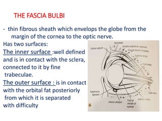
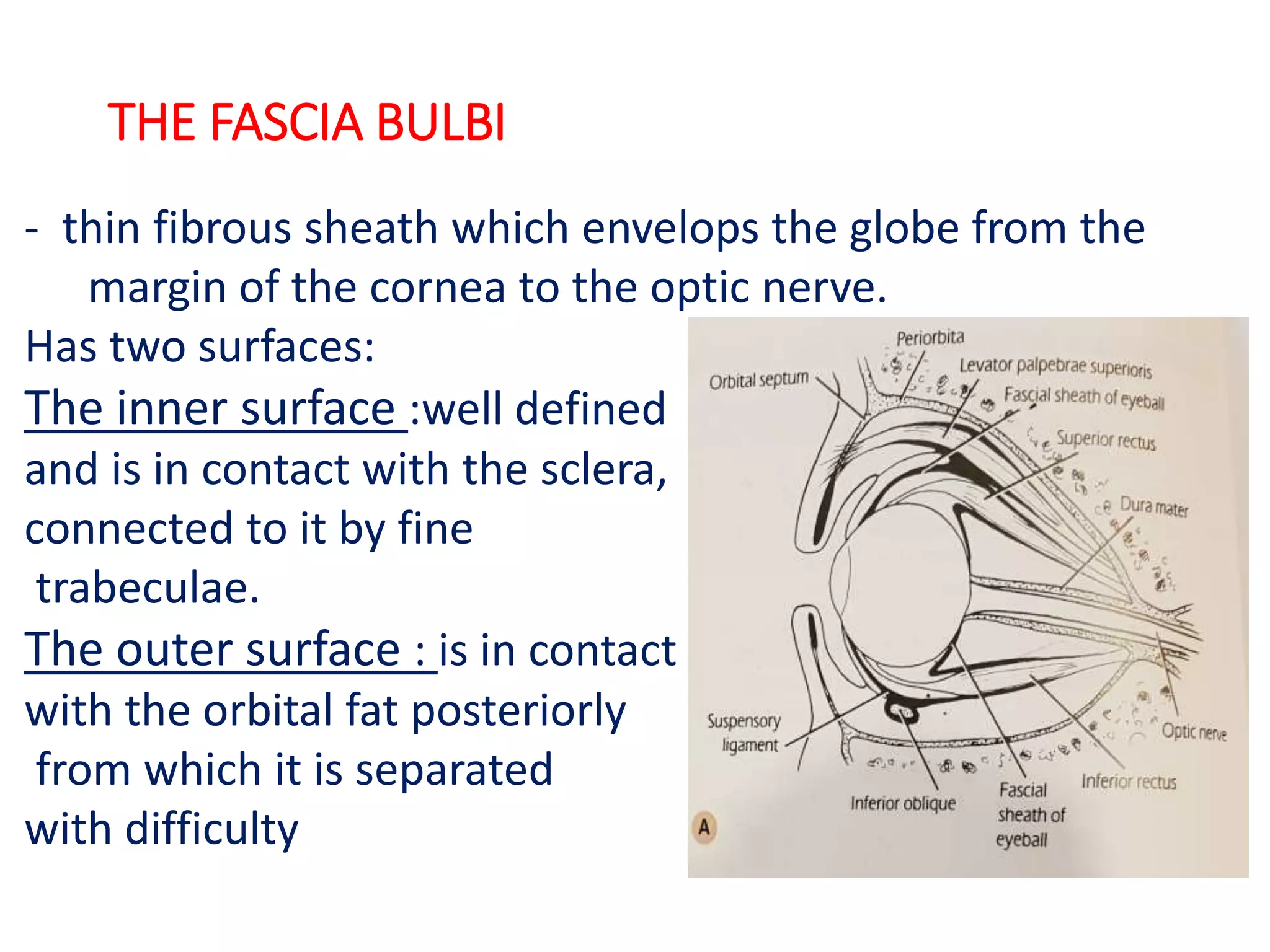
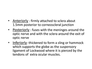
The fascia bulbi is a thin fibrous sheath that envelops the globe from the cornea to the optic nerve. It has two surfaces - an inner surface firmly attached to the sclera, and an outer surface in contact with the orbital fat. Its main functions are to position and support the globe within the orbital cavity and allow movements of the extrinsic eye muscles. It is important during eye surgery and enucleation to preserve the fascia bulbi to serve as a socket for a prosthesis.
Each eyelid contains a fibrous plate, called a tarsus, that gives it structure and shape; muscles, which move the eyelids; and meibomian (or tarsal) glands, which secrete lubricating fluids. The lids are covered with skin, lined with mucous membrane, and bordered with a fringe of hairs, the eyelashes.
This presentation gives a brief idea about angle of anterior chamber along with its structures and diagnostic methods to grade and visualize the structures.
1. Introduction Gross anatomy Layers Blood supply, drainage and nerve supply
2. INTRODUCTION • Sclera forms posterior 5/6th of external tunic , connective tissue coat of eyeball. • it continues with duramater and cornea • Its whole surface covered by tenon’s capsule • Anteriorly covered by- bulbar conjunctiva • Inner surface lies in contact with choroid • With a potential suprachoroidal space in between
3. Equa THICKNESS OF SCLERA
4. • Thickness varies with individual, with age • Thinner- children, elder, F> M • Thickest posteriorly • Gradually becomes thinner when traced anteriorly • Thin at insertion of extraocular muscle
The tear film is a complex mixture of substances secreted from multiple sources on the ocular surface, including the lacrimal gland, the accessory lacrimal glands, the meibomian glands, and the goblet cells.
LIMBUS… • The limbus forms the border between the transparent cornea and opaque sclera, contains the pathways of aqueous humour outflow, and is the site of surgical incisions for cataract and glaucoma
2. Anatomical Limbus: Circumcorneal transitional zone of the conjunctivocorneal & corneoscleral junction Conjunctivo-corneal junction: • Bulbar conjunctiva is firmly adherent to underlying structures • Substantia propria of the conjunctiva stops here but its epithelium continues with that of the cornea. Sclero-corneal junction: • Transparent corneal lamellae become continuous • With the oblique, circular and opaque fibres of sclera
3. CONTINUE…. • In the area near limbus, the conjunctiva, tenon’s capsule & the episcleral tissue are fused into a dense tissue which is strongly adherent to corneo scleral junction.It is preferred site for obtaining a firm hold of the eyeball during ocular surgery. • The limbus is a common site for the occurrence of corneal epithelial neoplasm. • The Limbus contains radially oriented fibrovascular ridge known as the palisades of Vogt that may harbour a stem cell population. The palisades of Vogt are more common in the superior and inferior quadrants around the eye
Diseases of sclera
2. anatomy • Sclera posterior 5/6th opaque part of the external fibrous tunic of the eyeball.
3. • outer surface }covered by Tenon's capsule. • anterior part } covered by bulbar conjunctiva.
4. Its inner surface lies in contact with choroid with a potential suprachoroidal space in between
5. Thickness of sclera. • thinner }children and in females Sclera • thickest} posteriorly (1mm) • gradually becomes thin when traced anteriorly. • thinnest } insertion of extraocular muscles (0.3 mm). • Lamina cribrosa is a sieve-like sclera from which fibres of optic nerve pass.
6. Apertures of sclera • Anterior • Anterior ciliary vessels • Middle • four vortex veins (vena verticosae) • Posterior • Optic nerve • Long & short ciliary nerves
7. Layers of sclera sclera episclera Sclera proper Lamina fusca thin, dense vascularised layer of connective tissue fibroblasts, macrophages and lymphocytes avascular structure dense bundles of collagen fibres. innermost blends with suprachoroidal and supraciliary laminae of the uveal tract. brownish in colour presence of pigmented cells.
The choroid, also known as the choroidea or choroid coat, is a part of the uvea, the vascular layer of the eye. It contains connective tissues, and lies between the retina and the sclera. The human choroid is thickest at the far extreme rear of the eye (at 0.2 mm), while in the outlying areas it narrows to 0.1 mm. The choroid provides oxygen and nourishment to the outer layers of the retin
Dr, Kathirvel Gopalakrishnan
M.D.S (OMFS)
Presentation on Anatomy of orbit which helps for a quick refresh.
Applied aspects described well and slides contains images for easy understanding of the subject.
QA Paediatric dentistry department, Hospital Melaka 2020Azreen Aj
QA study - To improve the 6th monthly recall rate post-comprehensive dental treatment under general anaesthesia in paediatric dentistry department, Hospital Melaka
Navigating the Health Insurance Market_ Understanding Trends and Options.pdfEnterprise Wired
From navigating policy options to staying informed about industry trends, this comprehensive guide explores everything you need to know about the health insurance market.
India Clinical Trials Market: Industry Size and Growth Trends [2030] Analyzed...Kumar Satyam
According to TechSci Research report, "India Clinical Trials Market- By Region, Competition, Forecast & Opportunities, 2030F," the India Clinical Trials Market was valued at USD 2.05 billion in 2024 and is projected to grow at a compound annual growth rate (CAGR) of 8.64% through 2030. The market is driven by a variety of factors, making India an attractive destination for pharmaceutical companies and researchers. India's vast and diverse patient population, cost-effective operational environment, and a large pool of skilled medical professionals contribute significantly to the market's growth. Additionally, increasing government support in streamlining regulations and the growing prevalence of lifestyle diseases further propel the clinical trials market.
Growing Prevalence of Lifestyle Diseases
The rising incidence of lifestyle diseases such as diabetes, cardiovascular diseases, and cancer is a major trend driving the clinical trials market in India. These conditions necessitate the development and testing of new treatment methods, creating a robust demand for clinical trials. The increasing burden of these diseases highlights the need for innovative therapies and underscores the importance of India as a key player in global clinical research.
Global launch of the Healthy Ageing and Prevention Index 2nd wave – alongside...ILC- UK
The Healthy Ageing and Prevention Index is an online tool created by ILC that ranks countries on six metrics including, life span, health span, work span, income, environmental performance, and happiness. The Index helps us understand how well countries have adapted to longevity and inform decision makers on what must be done to maximise the economic benefits that comes with living well for longer.
Alongside the 77th World Health Assembly in Geneva on 28 May 2024, we launched the second version of our Index, allowing us to track progress and give new insights into what needs to be done to keep populations healthier for longer.
The speakers included:
Professor Orazio Schillaci, Minister of Health, Italy
Dr Hans Groth, Chairman of the Board, World Demographic & Ageing Forum
Professor Ilona Kickbusch, Founder and Chair, Global Health Centre, Geneva Graduate Institute and co-chair, World Health Summit Council
Dr Natasha Azzopardi Muscat, Director, Country Health Policies and Systems Division, World Health Organisation EURO
Dr Marta Lomazzi, Executive Manager, World Federation of Public Health Associations
Dr Shyam Bishen, Head, Centre for Health and Healthcare and Member of the Executive Committee, World Economic Forum
Dr Karin Tegmark Wisell, Director General, Public Health Agency of Sweden
Medical Technology Tackles New Health Care Demand - Research Report - March 2...pchutichetpong
M Capital Group (“MCG”) predicts that with, against, despite, and even without the global pandemic, the medical technology (MedTech) industry shows signs of continuous healthy growth, driven by smaller, faster, and cheaper devices, growing demand for home-based applications, technological innovation, strategic acquisitions, investments, and SPAC listings. MCG predicts that this should reflects itself in annual growth of over 6%, well beyond 2028.
According to Chris Mouchabhani, Managing Partner at M Capital Group, “Despite all economic scenarios that one may consider, beyond overall economic shocks, medical technology should remain one of the most promising and robust sectors over the short to medium term and well beyond 2028.”
There is a movement towards home-based care for the elderly, next generation scanning and MRI devices, wearable technology, artificial intelligence incorporation, and online connectivity. Experts also see a focus on predictive, preventive, personalized, participatory, and precision medicine, with rising levels of integration of home care and technological innovation.
The average cost of treatment has been rising across the board, creating additional financial burdens to governments, healthcare providers and insurance companies. According to MCG, cost-per-inpatient-stay in the United States alone rose on average annually by over 13% between 2014 to 2021, leading MedTech to focus research efforts on optimized medical equipment at lower price points, whilst emphasizing portability and ease of use. Namely, 46% of the 1,008 medical technology companies in the 2021 MedTech Innovator (“MTI”) database are focusing on prevention, wellness, detection, or diagnosis, signaling a clear push for preventive care to also tackle costs.
In addition, there has also been a lasting impact on consumer and medical demand for home care, supported by the pandemic. Lockdowns, closure of care facilities, and healthcare systems subjected to capacity pressure, accelerated demand away from traditional inpatient care. Now, outpatient care solutions are driving industry production, with nearly 70% of recent diagnostics start-up companies producing products in areas such as ambulatory clinics, at-home care, and self-administered diagnostics.
The dimensions of healthcare quality refer to various attributes or aspects that define the standard of healthcare services. These dimensions are used to evaluate, measure, and improve the quality of care provided to patients. A comprehensive understanding of these dimensions ensures that healthcare systems can address various aspects of patient care effectively and holistically. Dimensions of Healthcare Quality and Performance of care include the following; Appropriateness, Availability, Competence, Continuity, Effectiveness, Efficiency, Efficacy, Prevention, Respect and Care, Safety as well as Timeliness.
How many patients does case series should have In comparison to case reports.pdfpubrica101
Pubrica’s team of researchers and writers create scientific and medical research articles, which may be important resources for authors and practitioners. Pubrica medical writers assist you in creating and revising the introduction by alerting the reader to gaps in the chosen study subject. Our professionals understand the order in which the hypothesis topic is followed by the broad subject, the issue, and the backdrop.
https://pubrica.com/academy/case-study-or-series/how-many-patients-does-case-series-should-have-in-comparison-to-case-reports/
CRISPR-Cas9, a revolutionary gene-editing tool, holds immense potential to reshape medicine, agriculture, and our understanding of life. But like any powerful tool, it comes with ethical considerations.
Unveiling CRISPR: This naturally occurring bacterial defense system (crRNA & Cas9 protein) fights viruses. Scientists repurposed it for precise gene editing (correction, deletion, insertion) by targeting specific DNA sequences.
The Promise: CRISPR offers exciting possibilities:
Gene Therapy: Correcting genetic diseases like cystic fibrosis.
Agriculture: Engineering crops resistant to pests and harsh environments.
Research: Studying gene function to unlock new knowledge.
The Peril: Ethical concerns demand attention:
Off-target Effects: Unintended DNA edits can have unforeseen consequences.
Eugenics: Misusing CRISPR for designer babies raises social and ethical questions.
Equity: High costs could limit access to this potentially life-saving technology.
The Path Forward: Responsible development is crucial:
International Collaboration: Clear guidelines are needed for research and human trials.
Public Education: Open discussions ensure informed decisions about CRISPR.
Prioritize Safety and Ethics: Safety and ethical principles must be paramount.
CRISPR offers a powerful tool for a better future, but responsible development and addressing ethical concerns are essential. By prioritizing safety, fostering open dialogue, and ensuring equitable access, we can harness CRISPR's power for the benefit of all. (2998 characters)
1. THE FASCIA BULBI
- thin fibrous sheath which envelops the globe from the
margin of the cornea to the optic nerve.
Has two surfaces:
The inner surface :well defined
and is in contact with the sclera,
connected to it by fine
trabeculae.
The outer surface : is in contact
with the orbital fat posteriorly
from which it is separated
with difficulty
2. • Anteriorly : firmly attached to sclera about
1.5mm posterior to corneoscleral junction
• Posteriorly : fuses with the meninges around the
optic nerve and with the sclera around the exit of
optic nerve
• Inferiorly: thickened to form a sling or hammock
which supports the globe as the suspensory
ligament of Lockwood where it is pierced by the
tendons of extra ocular muscles.
3. MAIN FUNCTION
•To position and support the globe within the orbital
cavity
•To permit the actions of extrinsic muscles to
produce movement of eyeball.
4. APPLIED ANATOMY OF FASCIA BULBI
•During enucleation of the eyeball the fascial sheath
should be preserved to serve as a socket for the
prosthesis
•Close relationship exists between the suspensory
ligament of lockwood and the inferior rectus and
the inferior oblique muscle making operations on
these muscles very difficult.
5. CONTD…
•Even after extensive removal of maxilla, eyeball
does not sag down because the suspensory
ligament is strong enough to provide the eyeball
with adequate support from below.
•Extension of the fascial sheath through the orbital
fat to the bony walls of orbital cavity assists the
orbital septum in preventing herniation of fat into
the lids.
6. EMBRYOLOGY OF SCLERA
•The human sclera differentiates from neural crest and
mesoderm7 week
•The majority of the
sclera differentiates
from neural crest
that surrounds the
optic cup of
Neuroectoderm
• a small temporal portion of the sclera differentiates
from mesoderm
8. •consists almost entirely of the collagen( chiefly with type 1
and moderately with type 3) within a lesser amount of the
ground substance and scanty fibrocytes.
•Viscoelastic
•relatively avascular
•thicker in males than in females
9. •Scleral collagen fibrils are highly variable in their diameter,
•lamellae vary in thickness, irregular with respect to
neighbouring lamella
•water content of the sclera 68%
12. SPECIAL REGION OF SCLERA
•Both the internal and external aspects of sclera at the
sclerocorneal junction project more anteriorly than the main
body of sclera- concave cirumferential groove - Internal
scleral sulcus(occupied by trabecular meshwork)
•Just posterior to the limbus and lying within the sclera is
circular running canal called the canal of schlemn.
13. SCLERAL SPUR
•Circular flang of the anterior
most part of sclera lies
deep to Schlemm’s canal
•Meridional fibres of ciliary
muscle attached to SS.
14. LAMINA CRIBOSA
• thin, sieve-like portion of
sclera at the base of the
optic disc through which
optic nerve passes.
•Concave at intraocular
aspect
•Holes in the network
remain relatively aligned
with each other providing
unobstructed passage for
bundle of nerve fibers
16. INSERTION
• The medial rectus:
5.5mm
• The inferior rectus:
6.5mm
• The lateral rectus:
6.9mm
• The superior rectus:
7.7mm
• The insertion of the
superior and the inferior
oblique are posterior to
the scleral equator.
17. APERTURES
•Sclera is pierced by two potential openings
•Anterior scleral foramen: where sclera meets
and anatomically converges with cornea
•Posterior scleral foramen: Provides an exit for
the optic nerve
18. EMISSERIA
Channels through which vessels and nerves pass
through the sclera.
•Anterior emissaria
•Middle emissaria:
•Posterior emissaria:
19. BLOOD SUPPLY
•Anteriorly by the
anterior cilliary artery.
•Posteriorly by short
ciliary artery
•Episcleral plexus
•Underlying choroid
21. APPLIED ANATOMY
•Profuse sensory innervation of sclera results in dull
aching pain associated with inflammations of sclera.
The pain is worse during ocular movement
•Emissaria provides pathway for extraocular spread
of intraocular tumors. Most common site for
extension is along optic nerve
22. CONT…
•Scleral rupture following blunt trauma can occur at
a number of sites:
-in a circumferential arc parallel to the corneal limbus
opposite the site of impact,
-at the insertion of rectus muscles or at the equator
of the globe.
-The most common site is the superonasal quadrant
near the limbus.
23. CONT…
•As the scleral is thin the strabismus and retinal
detachment surgery require careful placement of
the suture.
•In infantile glaucoma, the viscid slow stretch in
response to changes in IOP results in buphthalmic
globe.
24. CONT..
•Progressive Myopia is characterized by scleral
thinning and ocular elongation. Defects in scleral
ECM remodeling lead to myopia
•In glaucoma the raised IOP causes lamina cribrosa
to bulge outwards – resultant cupping of disc in
chronic glaucoma
25. CONT…
change in colour of sclera with age and with disease
•In elderly - yellowish colour
•In jaundice - yellow discolouration
•In osteogenesis imperfecta, Ehlers- Danlos syndrome,
Pseudoxanthoma elasticum and other collagen diseases
thin and blue
26. EPISCLERITIS
•Immununologically mediated recurrent inflammation of the
tissue that lies between the deep conjunctival stroma and
superficial scleral lamellae
•Presence of deep hyperemia is benign, short-lived not
associated with tenderness, ciliary pain or flare and cell in
the anterior chamber
•Caused by allergy to food,
airborne allergen.
27. SCLERITIS
• Immunologically mediated inflammation of the sclera
• always associated with the secondary inflammation of the
episclera
• deep hyperemia, tenderness,
ciliary pain, photophobia and
flare and cells in the anterior
chamber
• Causes:
auto-immune collagen vascular
disease like SLE, Scleroderma,
granulomatous diseases like syphillis,
tuberculosis, gout.
• 50% is idiopathic
28. PIGMENTATIONS
•Nerve loop of Axenfeld: branch of long ciliary nerve
accompanying the anterior ciliary artery form a loop in the
sclera; often carry some pigments producing blue black spot
in superficial sclera.
29. OCULAR MELANOCYTOSIS
•slate gray patches of scleral and episcleral
pigmentations, usually associated with nevus of
ota/oculodremal melanocytosis (ipsilateral
hyperpigmentation of the iris, fundus and
periocular skin).
30. STAPHYLOMA
An ectasia of the outer coats(cornea, or sclera or
both) of the eye with an incarceration of the uveal
tissue.
31. THE LIMBAL TRANSITION ZONE
• Junctional zone between the cornea and sclera.
• 1.5mm wide in horizontal plane and 2mm wide in vertical
plane
• Internal edge; corneal limbus
• External edge; scleral limbus
32. • Scleral limbus
Defined by a line perpendicular to the surface passing
through the scleral spur.
• Corneal Limbus
demonstrated by the line joining the termination of
Bowman’s layer to the termination of Descemet’s
membrane
33. AT THE LIMBUS
• The corneal epithelium becomes continuous with the
epithelium of bulbar conjunctiva
• Bowman's membrane becomes continuous with the
lamina propria of the conjunctiva and tenon's capsule.
• Stroma becomes sclera
• Descemet's membrane becomes schwalbe's line.
• Endothelium lines the trabecular meshwork and
becomes continuous with the anterior surface of the
epithelium
• Pallisades of Vogt :folds of epithelial cells that run
radially into the cornea
34. THE ANATOMICAL LIMBUS
•The anatomical limbus takes up an arc as it
traverses the tissues in an anterior to posterior
manner
•Schwalbe’s line marks the posterior limit to the
anatomical limbus.
37. THE CATARACT INCISION & THE SURGICAL
LIMBUS
•Anterior limbal incision
-
At blue limbal zone
-traverses Descemet’s membrane,may cause
stripping
•Clear corneal incision
-
infront of the anterior limbal line
-chances of induced astigmatism and Descemet’s
membrane stripping
38. •Scleral incision
- posterior to the posterior Limbal border
-excessive bleeding and hyphaema
•Posterior limbal incision
-at white limbal zone
-injures trabecular meshwork
•Mid-limbal incision
-at mid limbal line
-corresponds to schwalbe’s line
-safest
39. REFERENCES
• Anthony J Bron, Ramesh C Tripathi, Brenda J Tripathi, Wolff’s Anatomy of the
eye and orbit, 8th edition
• External Disease and Cornea,Basic and Clinical Science Course, American
Academy Of Ophthalmology
• Practical Ophtahlmology, A Manual For Beginning Residents, American
Academy of Ophthalmology
• Snell, Richard s. and Michael A. lemp, Clinical Anatomy of the eye,2nd Edition,
India:Blackwell science,1998.
• Jack J kanski, Brad Bowling, Clinical Ophthalmology, 7th edition
• A.K. Khurana Anatomy and Physiology of eye ( third edition)
• Internet Resources: www.oculist.com
: www.eophtha .com
Editor's Notes
EOM have fascial sleeves that are continuous with the sheath of the eyeball, so the socket moves when the muscles contracts
Human beings are the only primates with white sclera
The term sclera is derived from Greek word scleros meaning "hard".
opaque, fibrous and protective outer layer of eyeball
Protects the intraocular contents from injury and displacement, contains the intraocular pressure and prevents deformation of the eyeball.
This white appearance is because of the scattering of all wavelengths of light by dense irregular bundles of collagen in sclera
Often described as viscoelastic as it exhibits biphasic response when suddenly deformed, elasticrapid but bried lengthening,, viscid slow streching
Lamellar organization of the human sclera. Scleral fibroblasts (F) can be seen between irregularly arranged collagenous lamella (L). Within each lamella, collagen fibrils are oriented in the same general direction, with some running longitudinally in the plane of section (arrow), and some running perpendicular to the plane of section and seen in cross section (asterisk). The black bar indicates the width of a lamella
Thickest : 1mm near the optic nerve
Thinnest : 0.3mm at the insertion of the recti
At equator : 0.6mm
From the recti muscles insertions towards limbus there is gradual increase in thickness up to 0.8mm
Schwalbe's line is the anatomical line found on the interior surface of the eye's cornea, and delineates the outer limit of the corneal endothelium layer. Specifically, it represents the termination of Descemet's membrane.[1] In many cases it can be seen via gonioscopy.
maintain the pressure gradient between the inside of the eye and the surrounding tissue. Increase in posterior curvature producing glaucomatous cupping disc
Epi thin,dense vascularized layer of connective tissue. Ant: continuous with tenon’s capsule. Capillary network in ant part of episcera Ciliary flush. Umyelinated nerve fibre, keratocyte, melanocytes.ant: ant ciliary artery post: post ciliary artery
Sp:avascular, type I collagen crossing each other in all directionopaque.increasing age increased lipids deposit and sclera becomes yellow
LF: Lamina fusca is the innermost layer of sclera. It is characterised by abundance of pigmented cells or melanocytes, mostly migrated from choroid. The connective tissue of this layer is loosely arranged than rest of the sclera. Lamina fusca is separated from choroid by a thin potential space known as suprachoroidal or perichoroidal space.
All 4 rectus muscle are inserted in sclera at different distance from limbus
>Ant ciliary arteries-2 in number except in lateral rectus (1 in number)
>the largest branch of this vessel also enters the ciliary body to form major arterial aracade
>for 1 ant ciliary artery - 2 ciliary veins in the ciliary body and is accompanied by 1 post. Ciliary nerve. (nerve loop of axenfeld)
The vortex veins pierce sclera 4 mm posterior to the equator
Choroidal capillaries are fenestrated and the sclera doesn’t present a major barrier to the diffusion of even a larger molecules like albumin to choroid. Hence, subtenon or subconjunctivally injected drugs can reach the internal tunics of eyeball.
The termination of Bowman’s layer is indicated on the biomicroscopy by the internal limit of the marginal arcade of the corneal vessels.
The termination of Descemet’s membrane is visible on gonioscopy as the most anterior landmark of the drainage angle, Schwalbe’s line ( hypertrophied in the anterior embryotoxon when it is visible as a fine internal ridge.
Three landmarks
Anterior limbal border: overlies the termination of Bowman’s layer.
Mid limbal line: overlies the termination of Descemet’s membrane.
Posterior limbal border: overlies the scleral spur