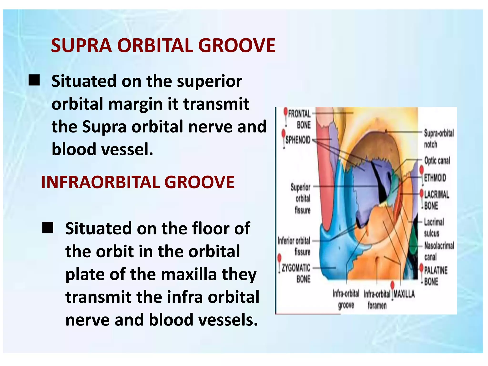
Anatomy Presentation
Group members
AQSA SHAHZAD TAYYABA SAEED
Topic : ORBIT
THE ORBIT
 Two bony cavities in facial skeleton situated
on each side of root of nose.
 Inferior to anterior part of cranial cavity.
 Apex
 Base
 Four walls
Relations
 Superiorly :Anterior cranial fossal
 Medially :Nasal cavity &
Ethmoidal air sinuses
 Inferiorly - Maxillary sinus
 Laterally - Middle cranial fossa &
Temporal fossa
CONTENT OF ORBIT
 Optic Nerve
 Ocular muscle
 Nerves
 Fat
 Vessels
 Lacrimal gland
 Conjunctival sac
 Eyeball
Volume : 30cm3
Rim: Horizontally-4cm
Vertically-3.5cm
Intra orbital width: 2.5cm
Extra orbital width: 10cm
DIMENSIONS OF ORBIT
CHANGES IN ORBIT WITH AGE
Shape Height Width Index
Fetus Oval 14mm 18mm 77.7
Newborns Round 27mm 27mm 100
7years Pyramidal 28mm 33mm 84 4
Adults Pyramidal 35mm 40mm 89.2
BONY ORBIT
 7 bones make up the bony orbit :
①Frontal bone
②Zygomatic bone
③Maxilla bone
④Ethmoid bone
⑤Sphenoid bone
⑥Lacrimal bone
⑦Palatine bone
APEX OF ORBIT
 Apex of orbit positioned at the craniofacial
junction located where the four orbital wall
converge.
 It is also the point where the
extraocular muscles derive their
origins.
BASE OF ORBIT
Formed by :
1. frontal 2. maxilla 3. zygomatic bones.
Upper margin: Frontal bone.
Lateral Margin : Process of frontal and zygomatic
bones.
Inferior Margin : Zygomatic bone and maxilla.
Medial Margin : Process of maxilla and frontal bone.
ORBITAL WALLS
Bony orbit has four walls:
Superior wall (roof) Inferior wall (floor)
Lateral wall Medial wall
Superior wall (roof)
Formed by :
 Orbital plate of frontal Bone.
 Lesser wing of Sphenoid Bone.
 Separates the orbit from the
anterior cranial fossa.
Anteriolateral
ly:
Slight depression (
lacrimal fossa ) for
orbital part of
lacrimal gland.
 It consists of:-

 Maxillary Bone : Medially
 Zygomatic bone : Laterally
 Palatine bone : posteriorly
 Separate orbit And maxillary sinus

 The roof of the maxillary sinus.

INFERIOR WALL
Medial wall
 Frontal process of maxilla,
 Lacrimal bone
 orbital plate of ethmoid (which separates
orbital cavity from ethmoid sinuses) &
 Body of sphenoid.
Formed by
 The weakest part of
the medial wall of
bony orbit is the
lamina papyracea.
Lateral wall
Formed by :
 zygomatic bone
 greater wing of sphenoid.
 It is separated posteriorly from roof by
superior orbital fissure.
 Strongest orbital wall.
 It give attachment to
check ligaments of
rectus muscle and to
suspensory
ligaments of eye ball.
 It is a projection on
orbital surface of
zygomatic bone.
WHITNEL'S TUBERCLE
OPENINGS INTO ORBITAL CAVITY
Orbital opening : orbital opening is the large
opening lie anteriorly.
 Approximately one sixth of the eye is exposed
the remainder is protected by the wall of orbit.
 Supra orbital groove
 Superior orbital
fissure
 Inferior orbital fissure
 Nasolacrimal canal  Optical canal
 infra orbital groove and
canal
 Situated on the superior
orbital margin it transmit
the Supra orbital nerve and
blood vessel.
SUPRA ORBITAL GROOVE
INFRAORBITAL GROOVE
 Situated on the floor of
the orbit in the orbital
plate of the maxilla they
transmit the infra orbital
nerve and blood vessels.
NASOLACRIMAL CANAL
 Located anteriorly on the medial wall
 it communicate with the inferior meatus of
the nose and transmit the nasolacrimal duct
 Located posteriorly between the maxilla and the
greater wing of sphenoid.
 it communicate with the pterygopalatine fossa and
transmit the maxillary nerve and its zygomatic branch
the inferior ophthalmic nerve and sympathetic nerve.
INFERIOR ORBITAL FISSURE
SUPERIOR ORBITAL FISSURE
 Located posteriorly between the greater or
lesser wings of the sphenoid .
 IT communicate with the middle cranial fossa
and transmit the lacrimal nerve frontal nerve
trochlearnerve the oculomotor nerve the
abducent nerve the nasociliary nerve and the
superior opthalmic vein.
OPTICAL CANAL
 Located posteriorly in the lesserwing of the
sphenoid.
 It communicate with the middle cranial fossa
and transmit the optic nerve and the
ophthalamic artery.
There are two types of fractures of bony orbit
.
1.Orbital rim fracture.
 This is the fracture of the bones forming the
outer rim of the bony orbit.
 it usually occur at the suture joining the three
bones of the orbital rim the maxilla,
zygomatic and frontal.

Fractures of the bony Orbit
2.Blow out fracture
 This refer to partial herniation of the
orbital contact through one of its wall.
 This usually occur via force trauma to the
eye the medial and the inferior walls are
the weakest with the contacts herniating
in to ethmoid and maxillary sinuses
respectively
 Any fracture of the orbit will result in
intra orbital pressure raising the pressure
in the orbit causing exophthalmos
(protrusim of eye).
BLOW OUT FRACTURE
Anatomy of orbit presentation.pptx

Anatomy of orbit presentation.pptx

  • 1.
    Anatomy Presentation Group members AQSASHAHZAD TAYYABA SAEED Topic : ORBIT
  • 2.
    THE ORBIT  Twobony cavities in facial skeleton situated on each side of root of nose.  Inferior to anterior part of cranial cavity.  Apex  Base  Four walls
  • 3.
    Relations  Superiorly :Anteriorcranial fossal  Medially :Nasal cavity & Ethmoidal air sinuses  Inferiorly - Maxillary sinus  Laterally - Middle cranial fossa & Temporal fossa
  • 5.
    CONTENT OF ORBIT Optic Nerve  Ocular muscle  Nerves  Fat  Vessels  Lacrimal gland  Conjunctival sac  Eyeball
  • 6.
    Volume : 30cm3 Rim:Horizontally-4cm Vertically-3.5cm Intra orbital width: 2.5cm Extra orbital width: 10cm DIMENSIONS OF ORBIT
  • 7.
    CHANGES IN ORBITWITH AGE Shape Height Width Index Fetus Oval 14mm 18mm 77.7 Newborns Round 27mm 27mm 100 7years Pyramidal 28mm 33mm 84 4 Adults Pyramidal 35mm 40mm 89.2
  • 8.
    BONY ORBIT  7bones make up the bony orbit : ①Frontal bone ②Zygomatic bone ③Maxilla bone ④Ethmoid bone ⑤Sphenoid bone ⑥Lacrimal bone ⑦Palatine bone
  • 9.
    APEX OF ORBIT Apex of orbit positioned at the craniofacial junction located where the four orbital wall converge.  It is also the point where the extraocular muscles derive their origins.
  • 10.
    BASE OF ORBIT Formedby : 1. frontal 2. maxilla 3. zygomatic bones. Upper margin: Frontal bone. Lateral Margin : Process of frontal and zygomatic bones. Inferior Margin : Zygomatic bone and maxilla. Medial Margin : Process of maxilla and frontal bone.
  • 12.
    ORBITAL WALLS Bony orbithas four walls: Superior wall (roof) Inferior wall (floor) Lateral wall Medial wall
  • 13.
    Superior wall (roof) Formedby :  Orbital plate of frontal Bone.  Lesser wing of Sphenoid Bone.  Separates the orbit from the anterior cranial fossa. Anteriolateral ly: Slight depression ( lacrimal fossa ) for orbital part of lacrimal gland.
  • 14.
     It consistsof:-   Maxillary Bone : Medially  Zygomatic bone : Laterally  Palatine bone : posteriorly  Separate orbit And maxillary sinus   The roof of the maxillary sinus.  INFERIOR WALL
  • 15.
    Medial wall  Frontalprocess of maxilla,  Lacrimal bone  orbital plate of ethmoid (which separates orbital cavity from ethmoid sinuses) &  Body of sphenoid. Formed by
  • 16.
     The weakestpart of the medial wall of bony orbit is the lamina papyracea.
  • 17.
    Lateral wall Formed by:  zygomatic bone  greater wing of sphenoid.  It is separated posteriorly from roof by superior orbital fissure.  Strongest orbital wall.
  • 19.
     It giveattachment to check ligaments of rectus muscle and to suspensory ligaments of eye ball.  It is a projection on orbital surface of zygomatic bone. WHITNEL'S TUBERCLE
  • 20.
    OPENINGS INTO ORBITALCAVITY Orbital opening : orbital opening is the large opening lie anteriorly.  Approximately one sixth of the eye is exposed the remainder is protected by the wall of orbit.  Supra orbital groove  Superior orbital fissure  Inferior orbital fissure  Nasolacrimal canal  Optical canal  infra orbital groove and canal
  • 21.
     Situated onthe superior orbital margin it transmit the Supra orbital nerve and blood vessel. SUPRA ORBITAL GROOVE INFRAORBITAL GROOVE  Situated on the floor of the orbit in the orbital plate of the maxilla they transmit the infra orbital nerve and blood vessels.
  • 22.
    NASOLACRIMAL CANAL  Locatedanteriorly on the medial wall  it communicate with the inferior meatus of the nose and transmit the nasolacrimal duct
  • 23.
     Located posteriorlybetween the maxilla and the greater wing of sphenoid.  it communicate with the pterygopalatine fossa and transmit the maxillary nerve and its zygomatic branch the inferior ophthalmic nerve and sympathetic nerve. INFERIOR ORBITAL FISSURE
  • 25.
    SUPERIOR ORBITAL FISSURE Located posteriorly between the greater or lesser wings of the sphenoid .  IT communicate with the middle cranial fossa and transmit the lacrimal nerve frontal nerve trochlearnerve the oculomotor nerve the abducent nerve the nasociliary nerve and the superior opthalmic vein.
  • 26.
    OPTICAL CANAL  Locatedposteriorly in the lesserwing of the sphenoid.  It communicate with the middle cranial fossa and transmit the optic nerve and the ophthalamic artery.
  • 27.
    There are twotypes of fractures of bony orbit . 1.Orbital rim fracture.  This is the fracture of the bones forming the outer rim of the bony orbit.  it usually occur at the suture joining the three bones of the orbital rim the maxilla, zygomatic and frontal.  Fractures of the bony Orbit
  • 28.
    2.Blow out fracture This refer to partial herniation of the orbital contact through one of its wall.  This usually occur via force trauma to the eye the medial and the inferior walls are the weakest with the contacts herniating in to ethmoid and maxillary sinuses respectively  Any fracture of the orbit will result in intra orbital pressure raising the pressure in the orbit causing exophthalmos (protrusim of eye).
  • 29.