Downloaded 1,086 times

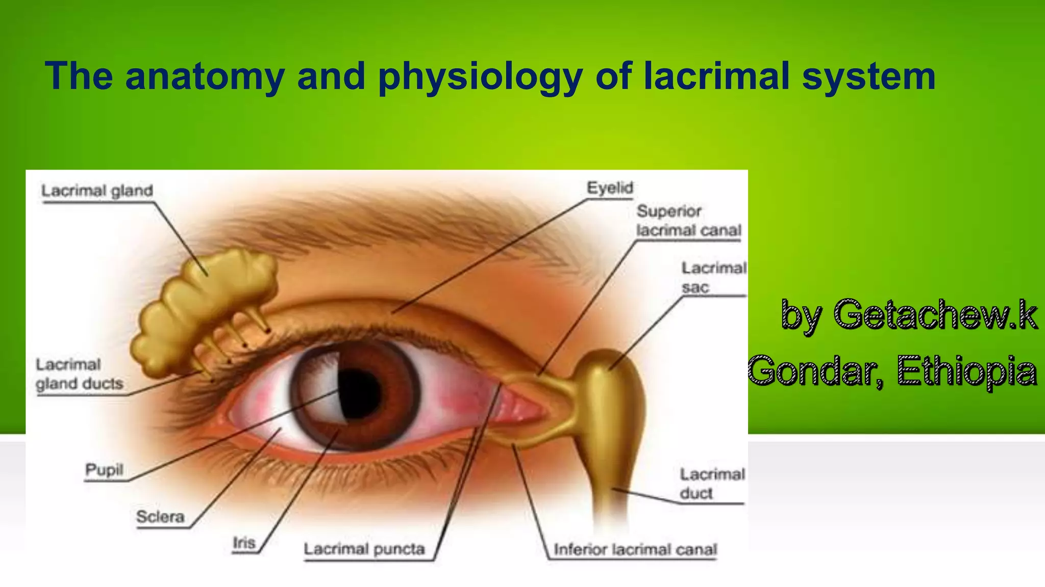
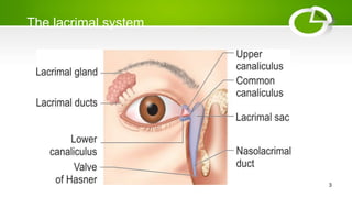
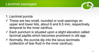
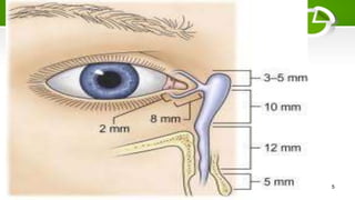
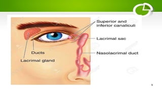
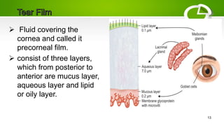
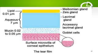
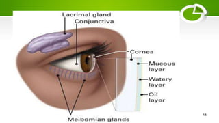


The lacrimal system comprises the lacrimal glands and passages, including puncta, canaliculi, lacrimal sac, and nasolacrimal duct, responsible for tear production and drainage. The gland secretes tears that form a multi-layered film to maintain the moisture and health of the cornea. Tears are continuously secreted and drained through an active pump mechanism involving the eyelids and facial muscles.